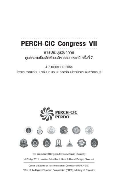
PERCH-CIC Congress VII
PERCH-CIC Congress VII
PERCH-CIC Congress VII

Create successful ePaper yourself
Turn your PDF publications into a flip-book with our unique Google optimized e-Paper software.
○ ○ ○ ○ ○ ○ ○ ○ ○ ○ ○ ○ ○ ○ ○ ○ ○ ○ ○ ○ ○ ○ ○ ○ ○ ○ ○ ○ ○ ○ ○ ○ ○ ○ ○ ○<br />
<strong>PERCH</strong>-<strong>CIC</strong> <strong>Congress</strong> <strong>VII</strong><br />
○ ○ ○ ○ ○ ○ ○ ○ ○ ○ ○ ○ ○ ○ ○ ○ ○ ○ ○ ○ ○ ○ ○ ○ ○ ○ ○ ○ ○ ○ ○ ○<br />
○ ○ ○ ○<br />
การประชุมวิชาการ<br />
ศูนยความเปนเลิศดานนวัตกรรมทางเคมี ครั้งที่ 7<br />
4-7 พฤษภาคม 2554<br />
โรงแรมจอมเทียน ปาลมบีช แอนด รีสอรท เมืองพัทยา จังหวัดชลบุรี<br />
The International <strong>Congress</strong> for Innovation in Chemistry<br />
4-7 May 2011, Jomtien Palm Beach Hotel & Resort Pattaya, Chonburi<br />
Center of Excellence for Innovation in Chemistry (<strong>PERCH</strong>-<strong>CIC</strong>)<br />
Office of the Higher Education Commission (OHEC), Ministry of Education
○ ○ ○ ○ ○ ○ ○ ○ ○ ○ ○ ○ ○ ○ ○ ○ ○ ○ ○ ○ ○ ○ ○ ○ ○ ○ ○ ○ ○ ○ ○ ○ ○ ○ ○ ○ ○ ○ ○ ○ ○ ○ ○ ○ ○ ○ ○ ○ ○ ○ ○ ○ ○ ○ ○ ○ ○ ○ ○ ○ ○ ○ ○ ○<br />
รวบรวมและเรียบเรียง<br />
ศูนยความเปนเลิศดานนวัตกรรมทางเคมี (<strong>PERCH</strong>-<strong>CIC</strong>)<br />
ISBN 978-974-11-1459-7<br />
จำนวนพิมพ 1,000 เลม<br />
เมษายน 2554
○ ○ ○ ○ ○ ○ ○ ○<br />
Contents<br />
○ ○ ○ ○ ○ ○<br />
○ ○<br />
Introductory Remarks.................................................................................. (5)<br />
Conference Schedule ................................................................................... (9)<br />
Presentation Schedule ............................................................................... (12)<br />
Special Lecture.................................................................................................. 1<br />
Plenary and Invited Lectures........................................................................ 3<br />
Abstracts for Oral Presentations<br />
- S1: Analytical Technologies................................................................. 43<br />
- S2: Innovation in Bioactive Natural Products ................................. 59<br />
- S3: Materials Science and Nanotechnology ................................... 77<br />
Abstracts for Poster Presentations<br />
- S1: Analytical Technologies................................................................. 97<br />
- S2: Innovation in Bioactive Natural Products and Synthesis ..... 179<br />
- S3: Materials Science and Nanotechnology ................................. 345<br />
List of Participants ....................................................................................... 483<br />
Keyword Index.............................................................................................. 501<br />
Author Index................................................................................................. 513<br />
Comments on <strong>PERCH</strong> <strong>Congress</strong> VI, May 3-6, 2009, Pattaya<br />
by Foreign Participants ...............................................................................523<br />
Sponsors.......................................................................................................... 533
○ ○ ○ ○ ○ ○ ○ ○ ○ ○ ○ ○ ○ ○ ○ ○ ○ ○ ○ ○ ○ ○ ○<br />
○ ○ ○ ○ ○ ○ ○ ○ ○ ○ ○ ○ ○ ○ ○ ○ ○ ○ ○ ○ ○ ○ ○<br />
Introductory Remarks<br />
The Center for Innovation in Chemistry (<strong>PERCH</strong>-<strong>CIC</strong>) was favorably evaluated by<br />
the 2010 Review Group appointed by The Office of the Higher Education Commission (OHEC).<br />
The Phase III operation of the Center for 2012-2017 has been approved. The three<br />
strategic and cutting-edge research areas: Analytical Technology, Innovation in Natural<br />
Products, and Materials Science and Nanochemistry together with the excellent track<br />
record of the Center will be the driving forces for the overall activities in the next five years.<br />
It will be a very challenging task in synchronizing the research excellence, innovation and<br />
human resources development for enhancing the crucial national strategies in four areas,<br />
i.e. environment and human security, food security, energy security and high value-added<br />
industries. <strong>PERCH</strong>-<strong>CIC</strong> is determined to attain excellence in all relevant categories. The<br />
Center has thirteen universities as its members, i.e. Mahidol (MU), Prince of Songkla (PSU),<br />
Khon Kaen (KKU), Chiang Mai (CMU), Kasetsart (KU), Ramkhamhaeng (RU), Burapha<br />
(BUU), Mahasarakham (MSU), Lampang Rajabhat (LPRU), Suratthani Rajabhat (SRU),<br />
Naresuan (NU), Ubon Rajathanee (UBU) and Bansomdejchaopraya Rajabhat (BSRU)<br />
Universities. The operational harmonization among all the members is crucial for the<br />
success of the Center in its effort to strengthen research excellence, innovation and<br />
research training, and to provide education opportunities in chemical sciences and related<br />
disciplines on a nation-wide basis. It is gratifying to be able to report that the member
(6)<br />
<strong>PERCH</strong>-<strong>CIC</strong> <strong>Congress</strong> <strong>VII</strong><br />
universities have contributed financially both in-kind and in-cash toward the research and<br />
education of the Center.<br />
The <strong>Congress</strong> series has become one of the milestone activities of the Center; it<br />
creates team spirit, allows researchers from the thirteen member universities to discuss and<br />
share their research results as well as their facilities and opportunities. The <strong>Congress</strong> has<br />
also become the most important international meeting in the chemical sciences in Thailand<br />
with the attendance of distinguished participants from abroad and from within Thailand.<br />
The general meeting at each <strong>Congress</strong> with the participation of staff, students and supporting<br />
personnel has been an extremely important forum for strategic planning of the Center.<br />
In its operation over approximately ten years, the Center has admitted a total of<br />
1,721 graduate students (1,329 M.Sc. and 392 Ph.D.), of which 772 have graduated (649<br />
M.Sc. and 123 Ph.D.). The members have published 870 papers in international journals<br />
together with 25 patents and 7 knowledge-based innovative products arising from the<br />
research results. The Center has invested heavily in the research facilities and infrastructure<br />
necessary for high-quality research and education in the chemical and pharmaceutical<br />
sciences. These facilities have allowed the faculty members to carry out state-of-the-art<br />
multidisciplinary research in the three strategic and cutting-edge research areas.<br />
The collaboration with industry has been further expanded. The main projects are<br />
as follows. Further development of the second generation products Plaitanoids TM and<br />
Lotusia TM with a group of companies led by Kovic Kate International (Thailand) Co., Ltd.<br />
and International Laboratory Corporation (ILC), respectively, are on-going. Chemical test<br />
kits for field use for trace analysis have been commercialized in co-operation with Higher<br />
Enterprise Co., Ltd. In addition, a spin-off company “TESTKIT INNOVATION” has been<br />
registered by Professor Yuwadee Shiowatana for the commercialization of the field test kits<br />
products. <strong>PERCH</strong>-<strong>CIC</strong> is one of the beneficiaries of the company. Researchers of the Center<br />
have been awarded 10 of The RGJ-Industry Ph.D. scholarships for research collaboration<br />
with industry; six of these are in Analytical Technologies, two in Materials Science and two<br />
in Innovation of Natural Products. Research collaboration with SCG Siam Cement Group<br />
is continuing in olefin polymerization catalysis research with the award of a grant worth<br />
approximately 25 million baht for five Ph.D. scholarships. An MOU has been signed with a<br />
food company Betagro on the development analytical methods for food products analysis.<br />
The collaboration with Bruker on the proteomics technology using high resolution NMR<br />
for food analysis is being initiated.<br />
The International <strong>PERCH</strong>-<strong>CIC</strong> <strong>Congress</strong> <strong>VII</strong>, having the theme “Chemistry,<br />
Environment and Society”, will offer an opportunity for students and faculty members of
4-7 May 2011 (7)<br />
the Center to present their research work both orally and by posters. Eleven countries are<br />
represented at this <strong>Congress</strong>. Plenary and invited speakers are drawn from noted researchers<br />
within and outside the Center. There are 12 plenary and 26 invited lectures plus 48 oral<br />
and 385 poster presentations. It is worth noting that the participants include 18 graduate<br />
students from Singapore, Taiwan and Belgium, together with 4 graduate students and<br />
faculty members from other institutions in Thailand that are non- members of the Center.<br />
An industrial meeting entitled “Chemistry for Innovation-Industry Forum” is organized in<br />
conjunction with the <strong>Congress</strong> to facilitate discussion between researchers and entrepreneurs<br />
about the utilization of research outcomes. There are 4 presentations from food, chemical,<br />
scientific equipment and petroleum industries together with 8 oral and 15 poster presentations<br />
from <strong>PERCH</strong>-<strong>CIC</strong> members. The total number of participants attending the <strong>Congress</strong> is 850.<br />
The Center has come a long way since its official inception in December 1999.<br />
I would like to quote a statement from the 2010 Review Group, which summarizes the<br />
achievement as follows.<br />
“The Center has a rigorous systematic administrative procedures which are<br />
flexible and conducive to achieving excellence, to enhancing the motivation and<br />
satisfaction of its members. The Center has exceeded its set targets of outputs. It is cost<br />
effective economic investment, especially in the areas of training high-quality human<br />
resources, the creation of new knowledge and new technology relevant to Thailand,<br />
comparing with the cost needed to acquire these resources from abroad. The Center has<br />
potential to attain a World Class Center of Excellence status in the three strategic and<br />
cutting-edge research areas, i.e. Analytical Technology, Innovation in Natural Products,<br />
and Materials Science”.<br />
I have to thank every member of the Center for making <strong>PERCH</strong>-<strong>CIC</strong> possible and<br />
for keeping the faith during the difficult path to realization of our vision of a World Class<br />
Center of Excellence in Chemistry.<br />
I would like to welcome all of you to this exciting <strong>Congress</strong>. I hope the experience<br />
gained during this event will be rewarding for your professional career.<br />
Finally, I wish the <strong>Congress</strong> every success.<br />
Vichai Reutrakul<br />
Director<br />
Center of Excellence for Innovation in Chemistry (<strong>PERCH</strong>-<strong>CIC</strong>)<br />
May 2011
○ ○ ○ ○ ○ ○ ○ ○ ○ ○ ○ ○ ○ ○ ○ ○ ○ ○ ○ ○ ○ ○ ○ ○ ○<br />
○ ○ ○ ○ ○ ○ ○ ○ ○ ○ ○ ○ ○ ○ ○ ○ ○ ○ ○ ○ ○ ○ ○ ○ ○<br />
Conference Schedule<br />
<strong>PERCH</strong>-<strong>CIC</strong> <strong>Congress</strong> <strong>VII</strong><br />
Theme: Chemistry, Environment and Society<br />
4-7 May 2011<br />
Jomtien Palm Beach Hotel & Resort Pattaya, Chonburi<br />
Wednesday, 4 May 2011<br />
15.00 - 18.30 Registration and Poster Set Up<br />
18.30 - 19.00 Opening Ceremony and Opening Address<br />
by Sumate Yamnoon,<br />
Secretary-General, Office of the Higher Education Commission<br />
19.00 - 19.30 SL-1: “Framework for Collaboration with Industry”<br />
by Wilaiporn Chetanachan, Director, Corporate Technology Office,<br />
The Siam Cement PLC, Thailand<br />
19.30 - 21.00 Cocktail / Welcome Party<br />
Thursday, 5 May 2011<br />
8.00 - 8.30 Registration<br />
8.30 - 9.10 PL-1: “Recent Advances in Hydrogen Storage Chemistry Research<br />
in the US DOE Metal Hydride Center of Excellence”<br />
by Lennie Klebanoff, Sandia National Laboratories, Livermore, USA.<br />
9.10 - 9.50 PL-2: “Discovery and Development of New Drugs from TCM<br />
Based on “System to System” Mode”<br />
by Guoan Luo, Tsinghua University, China
(10)<br />
<strong>PERCH</strong>-<strong>CIC</strong> <strong>Congress</strong> <strong>VII</strong><br />
9.50 - 10.10 Coffee Break<br />
10.10 - 10.50 PL-3: “Organic Field-effect Transistor/Memory Devices with<br />
Pentacene Films Embedding Metallic and Organic Charge<br />
Traps: Structure and Electric Bistability Study”<br />
by Yu-Tai Tao, Academia Sinica, Taiwan<br />
10.50 - 11.30 PL-4: “Some Medicinal and Mechanistic Aspects of Asymmetric<br />
Organocatalysis: From Stem-Cell Differentiation to<br />
Combined Organo/Biocatalysis”<br />
by Albrecht Berkessel, University of Cologne, Germany<br />
11.30 - 12.10 PL-5: “Last Development in Phosphinine Chemistry”<br />
by Franois Mathey, Nanyang Technological University, Singapore<br />
12.10 - 13.00 Lunch<br />
13.00 - 14.30 Poster Presentations<br />
S1: Analytical Technology<br />
S2: Innovation in Bioactive Natural Products<br />
S3: Materials Science and Nanotechnology<br />
14.30 - 15.00 Coffee Break<br />
15.00 - 17.20 Oral Presentations<br />
Session S1 S2A S2B S3<br />
Venue Marine I Marine II Marine IV Marine III<br />
S1-L1 S2A-L1 – S2A-L3 - S3-L1<br />
S1-O1 – S1-O4 S2A-O1 – S2A-O3 - S3-O1 – S3-O6<br />
15.00 - 17.20 Oral Presentations<br />
18.30 - 19.30 General Meeting of <strong>PERCH</strong>-<strong>CIC</strong><br />
by Vichai Reutrakul, <strong>PERCH</strong>-<strong>CIC</strong> Director<br />
Evening —Free—<br />
Friday, 6 May 2011<br />
8.30 - 10.10 Oral Presentations<br />
Session S1 S2A S2B S3<br />
Venue Marine I Marine II Marine IV Marine III<br />
S1-L2 S2A-L4 – S2A-L6 - S3-L2<br />
S1-O5 – S1-O8 S2A-O4 – S2A-O5 - S3-O7 – S3-O10<br />
10.10 - 10.30 Coffee Break<br />
10.30 - 12.10 Oral Presentations
4-7 May 2011 (11)<br />
Session S1 S2A S2B S3<br />
Venue Marine I Marine II Marine IV Marine III<br />
S1-L3 S2A-L7 – S2A-L9 -<br />
S1-O9 – S1-O12 S2A-O6 – S2A-O8 S2B-O1 – S2B-O4 S3-O11 – S3-O15<br />
12.10 - 13.00 Lunch<br />
13.00 - 14.30 Poster Presentations<br />
14.30 - 15.00 Coffee Break<br />
15.00 - 17.40 Oral Presentations<br />
Session S1 S2A S2B S3<br />
Venue Marine I Marine II Marine IV Marine III<br />
S1-L4 S2A-L10 – S2A-L11 S2B-L1 – S2B-L5 S3-L3 – S3-L5<br />
S1-O13 – S1-O16 S2A-O9 – S2A-O13 - S3-O16 – S3-O20<br />
18.30 Dinner / Recreation<br />
Saturday, 7 May 2011<br />
9.00 - 9.40 PL-6: “Electrochemical Synthesis of Organofluorine Compounds<br />
in Ionic Liquids Toward Green Sustainable Chemistry”<br />
by Toshio Fuchigami, Tokyo Institute of Technology, Japan<br />
9.40 - 10.20 PL-7: “Using NMR-based Methods to Assign the Stereochemistry<br />
of Natural Products”<br />
by Mary J. Garson, The University of Queensland, Australia<br />
10.20 - 10.40 Coffee Break<br />
10.40 - 11.20 PL-8: Asymmetric Formation of Quaternary Carbon Stereocenters<br />
by Sung Ho Kang, KAIST, Korea<br />
11.20 - 12.00 PL-9: “Cycloaddition Reactions of Masked o-Benzoquinones with<br />
Cyclopentadiene and Furans and Their Synthetic Applications”<br />
by Chun-Chen Liao, Chung Yuan Christian University, Taiwan<br />
12.00 - 13.00 Lunch<br />
13.00 - 13.40 PL-10: Materials based on Bowl-shaped Polynuclear Aromatics<br />
by Jay S. Siegel, University of Zurich, Switzerland<br />
13.40 - 14.20 PL-11: “Development of Natural Products to More Potent Bioactives”<br />
by Zhu-Jun Yao, Chinese Academy of Sciences, China<br />
14.20 - 15.20 Presentation of Awards and Closing Ceremony
Presentation Schedule<br />
Thursday, 5 May 2011<br />
Session S1 - Analytical Technology: Analytical Method Development<br />
Oral Presentations (Venue: Marine I)<br />
Chairpersons: J. Sabine Becker / Proespichaya Kanatharana<br />
15.00 - 15.20 S1-L1 High-Throughput Natural Products Chemistry for Drug Discovery: Is it Possible Mark O’Neil-Johnson<br />
15.20 - 15.40 S1-O1 Enhancement of Fluorescence Quenching Ability of DPPHby Colloidal Nanocrystalline Quantum เตือนใจ นอยพา Tuanjai Noipa<br />
Dot in Aqueous Micelle<br />
15.40 - 16.00 S1-O2 Photon Flux Densitometry of Agricultural Area in Thailand: The Study Through Ground Truth ศุภโชค อุปาลี Suphachock Upalee<br />
Remote Sensing<br />
16.20 - 16.40 S1-O3 High Performance Liquid Chromatographic Analysis of Penicillin Residues in Milk Samples After ชัญภักต คูคูสมุทร Chunyapuk Kukusamude<br />
Mixed Micelle-cloud Point Extraction<br />
16.40 - 17.00 S1-O4 Development of Simultaneously Carbaryl, Dimethoate and Fenvalerate Residual Extraction in วชิราภรณ เขียวมั่ง Wachiraporn Kheowmung<br />
Tangerine (Sai Nam Pung) by Using SPE Technique and Detection with HPLC and LC/MS<br />
Session S2 - Innovation in Bioactive Natural Products: Synthetic Methodologies and Organic Materials<br />
Oral Presentations (Venue: Marine II)<br />
Chairpersons: Rungnapha Saeeng / Yaowapa Sukpondma<br />
15.00 - 15.20 S2A-L1 Palladium(II)-catalyzed Ortho Arylation of 2-Phenoxypyridines and 2-Phenaminopyridines with Potassium Aryltrifluoroborates Ming-Jung Wu<br />
15.20 - 15.40 S2A-L2 Synthesis, and Biological Evaluation of 2-Phenylbenzothiazoles and Bis(benzylidene-benzenamine)-1-disulfides as Anticancer Agents Jeh-Jeng Wang<br />
15.40 - 16.00 S2A-L3 Transition Metal-catalyzed C–H Bond Functionalizations for Sustainable Synthesis Lutz Ackermann<br />
16.00 - 16.20 S2A-O1 Electroluminescent and Photophysical Properties of Metallotetraazaporphyrin Complexes Anurach Poloek<br />
16.20 - 16.40 S2A-O2 Synthesis and Characterization of Novel Oligothiophenes Bearing Dialkylaniline End-capped for OFETs ดวงรัชนีกร เหมือนมาตย Duangratchaneekorn Muenmart<br />
16.40 - 17.00 S2A-O3 Asymmetric Synthesis of gem-Difluoro-1-azabicyclic Compounds วัชราภรณ ทาหาร Watcharabhorn Thaharn<br />
Session S3 - Materials Science & Nanotechnology: Catalysis<br />
Oral Presentations (Venue: Marine III)<br />
Chairpersons: Ekasith Somsook / Chomjai Suksai<br />
15.00 - 15.20 S3-L1 Design of Vanadium Complex Catalysts for Efficient Olefin Insertion/Metathesis Polymerization Kotohiro Nomura<br />
15.20 - 15.40 S3-O1 Modified Ceria-based Catalyst for Water Gas Shift Reaction: Influence of Preparation Methods กิ่งแกว ฉายากุล Kingkaew Chayakul<br />
on the Role of Added Metal<br />
(12)<br />
<strong>PERCH</strong>-<strong>CIC</strong> <strong>Congress</strong> <strong>VII</strong>
15.40 - 16.00 S3-O2 Synthesize, Characterize and Kinetic Studies of the Ring-opening Bulk Polymerization of มานิตา ดุมกลาง Manita Dumklang<br />
ε-Caprolactone Using Novel Soluble Tin(II) Alkoxide Initiators<br />
16.00 - 16.20 S3-O3 Graphene Based Titanium Dioxide Composites for Water Treatment ณุตตรา หลาสกุล Nuttara Lasakul<br />
16.20 - 16.40 S3-O4 Photocatalytic Activity of TiO 2<br />
-immobilized Sheets Prepared by Different Methods ชวาลย ศรีวงษ Chaval Sriwong<br />
16.40 - 17.00 S3-O5 Serendipitous Discovery of a Phospherene-Phosphindole Rearrangement Duanghathai Panichakul<br />
17.00 - 17.20 S3-O6 Photochemistry of Iodine Solution and Titanium Oxalate Complex ธานินทร ทัดระเบียบ Tanin Tudrabiab<br />
Friday, 6 May 2011<br />
Session S1 - Analytical Technology: Plasma Spectrochemistry and Sample Preparation<br />
Oral Presentations: (Venue: Marine I)<br />
Chairpersons: Pakawadee Sutthivaiyakit / Supalax Srijaranai<br />
8.30 - 8.50 S1-L2 Progress and Trends in Elemental Imaging and Metallomics of Biological Tissue from Micrometer to Nanometer Scale J. Sabine Becker<br />
8.50 - 9.10 S1-O5 Gel Electrophoresis with Laser Ablation Inductively Coupled Plasma Mass Spectrometry for อุษารัตน คำทับทิม Usarat Kumtabtim<br />
Investigation of Metal Binding Rice Protein<br />
9.10 - 9.30 S1-O6 Inductively Coupled Plasma Mass Spectrometry for Trace Elements and Speciation Analysis นพรัตน วรพลาวุฒิ Nopparat Vorapalawut<br />
in Petroleum Matrices<br />
9.30 - 9.50 S1-O7 Development of Eggshell Membranes as Solid Phase Adsorbent for Preconcentration of Cd(II), ธนวัฒน ดวงคำ Tanawat Duangkum<br />
Pb(II) and Ni(II) Coupled with Flame Atomic Absorption Spectrometry<br />
9.50 - 10.10 S1-O8 Preparation and Characterization of Molecularly Imprinted Polymer for Coumaphos ปรัตถกร เอี่ยมอราม Paratthakorn Iamaram<br />
10.10 - 10.30 Coffee Break<br />
Session S1 - Analytical Technology: Flow-based and Microfluidic Technology<br />
Oral Presentations: (Venue: Marine I)<br />
Chairpersons: Peter C. Hauser / Jaroon Jakmunee<br />
10.30 - 10.50 S1-L3 Chemical Analysis Using Reagents and Materials Around Ourselves: Green Chemical Analysis-Green Innovation เกตุ กรุดพันธ Kate Grudpan<br />
10.50 - 11.10 S1-O9 Determination of Glucosamine by Sequential Injection Chromatography (SIC) with Fluorescence Detector อาทิตย บุญมา Arthit Bunma<br />
11.10 - 11.30 S1-O10 Chemiluminescence Microfluidic Device for Monitoring of Nitrite in Water นันทนภัส ลายทิพย Nunnapus Laitip<br />
11.30 - 11.50 S1-O11 Direct Analysis of Urine for Evaluation of Iodine Deficiency Disorder จิรายุ สิตานุรักษ Jirayu Sitanurak<br />
11.50 - 12.10 S1-O12 Size Characterization of Selenium Nanoparticles Prepared from Various Synthesis Conditions พรวิลาส เอ็มเอ็ม Pornwilard M-M<br />
12.10 - 13.00 Lunch<br />
13.00 - 14.30 Poster Presentations<br />
14.30 - 15.00 Coffee Break<br />
4-7 May 2011 (13)
Session S1 - Analytical Technology: Sensors and Sensing Technology<br />
Oral Presentations: (Venue: Marine I)<br />
Chairpersons: Kate Grudpan / Duangjai Nacapricha<br />
15.00 - 15.20 S1-L4 Contactless Conductivity Detection for Microseparation Techniques Peter C. Hauser<br />
15.20 - 15.40 S1-O13 Capillary Electrophoresis (CE) with Capacitively Coupled Contactless Conductivity Detection ฐิติรัตน แมนทิม Thitirat Mantim<br />
(C 4 D) for Chiral Separation of Stimulants in Urine Samples<br />
15.40 - 16.00 S1-O14 Detection of White Spot Syndrome Virus in Shrimp Pond Water by a Highly Sensitive Capacitive Biosensor สาลูมา สมานหมาน Saluma Samanman<br />
16.00 - 16.20 S1-O15 Ru/Rh-Carbon Fiber Microelectrode Biosensor for Sensitive Detection of แ-Ketoglutarate สุจิตรา ภูระหงษ Sujittra Poorahong<br />
16.20 - 16.40 S1-O16 Cost-Effective Formaldehyde Sensing Device for Industry Applications โอภาส บุญเกิด Opas Bunkoed<br />
(14)<br />
Session S2A - Innovation in Bioactive Natural Products: Synthetic Methodologies and Synthesis of Bioactive Compounds<br />
Oral Presentations (Venue: Marine II)<br />
Chairpersons: Boonsong Kongkathip / Vachiraporn Ajavakom<br />
8.30 - 8.50 S2A-L4 Fun with Organic Electrochemistry Istvn E. Mark<br />
8.50 - 9.10 S2A-L5 The Development of New Organocatalysts for Enantioselective C-C Bond Formation through Michael Reaction of Aldehydes to Nitroolefins Biing-Jiun Uang<br />
9.10 - 9.30 S2A-L6 Is There a General Rule to Predict the Regiochemical Pathways in the Ring Opening Reactions of 2-Substituted Aziridines Hyun-Joon Ha<br />
9.30 - 9.50 S2A-O4 Total Synthesis of Tamiflu and Tamiphosphor from D-Ribose ภาวิณี วิเชียรนุกูล Pawinee Wichienukul<br />
9.50 - 10.10 S2A-O5 Synthesis of the Disaccharide Moiety of an Anticancer OSW-1 จันจิรา รุจิรวณิช Janjira Rujirawanich<br />
10.10 - 10.30 Coffee Break<br />
Session S2A - Innovation in Bioactive Natural Products: Isolation and Synthesis<br />
Oral Presentations (Venue: Marine II)<br />
Chairpersons: Somyote Sutthivaiyakit / Kwanjai Kanokmedhakul<br />
10.30 - 10.50 S2A-L7 New Chemical Scaffold Starting Points for Targeting Inflammation Preferred, Novel and Neglected Scaffolds in a Drug Discovery Program Mark O’Neil-Johnson<br />
10.50 - 11.10 S2A-L8 Stemona Alkaloids and Derivatives with Potential Agricultural and Medicinal Applications Stephen G. Pyne<br />
11.10 - 11.30 S2A-L9 Total Syntheses of Structurally Complex Polycyclic Alkaloids Hidetoshi Tokuyama<br />
11.30 - 11.50 S2A-O6 Phthalide and 2,5-Dihydroxyphenyl Butanone Derivatives from the Endophytic Fungus อุบลทา สมมารถ Ubonta Sommart<br />
Microsphaeropsis arundinis PSU-G18<br />
11.50 - 12.10 S2A-O7 Clauraila A-D, Cytotoxic Carbazole Alkaloids from the Roots of Clausena harmandiana อุไรวรรณ สงเสียง Uraiwan Songsiang<br />
12.10 - 12.30 S2A-O8 New Cyclopeptide Alkaloids with Antiplasmodial and Antimycobacterial Activities from the ณัฏฐกัลย ลมเชย Natthakaln Lomchoey<br />
Root of Ziziphus mauritiana Lam.<br />
<strong>PERCH</strong>-<strong>CIC</strong> <strong>Congress</strong> <strong>VII</strong>
12.30 - 13.00 Lunch<br />
13.00 - 14.30 Poster Presentations<br />
14.30 - 15.00 Coffee Break<br />
Session S2A - Innovation in Bioactive Natural Products: Organic Sensors and Synthesis<br />
Oral Presentations (Venue: Marine II)<br />
Chairpersons: Palangpon Kongsaeree / Uthai Sakee<br />
15.00 - 15.20 S2A-L10 “Reactive” Fluorescent Probes for Heavy Metal Species Kyo Han Ahn<br />
15.20 - 15.40 S2A-L11 Development of Molecular Probes for Imaging of Gene Expression and Bacteria Functions Bengang Xing<br />
15.40 - 16.00 S2A-O9 PhI(OAc) 2<br />
-mediated Iminobromination for Synthesis of Bromomethyl Cyclic Imine Starting Stephen Sanjaya<br />
from Alkenyl Carbonitriles and Grignard Reagents<br />
16.00 - 16.20 S2A-O10 Palladium- and Gold-catalyzed Hydroamination of C-(Tetra-O-acetyl-β-D-galactopyranosyl)allene เฉลียวศักดิ์ คำวงษ Chaleowsak Khamwong<br />
16.20 - 16.40 S2A-O11 Synthetic Development and Mechanistic Study on Pd(II)-catalyzed Cyclization of Enediynes to Chen-Chau Chen<br />
Benzo[a]carbazoles<br />
16.40 - 17.00 S2A-O12 Synthesis and Nucleic Acid Binding Properties of Novel Pyrrolidinyl Peptide Nucleic Acid นิษานาถ รีนับถือ Nisanath Reenabthue<br />
Carrying 3-Aminopyrrolidine-4-carboxylic Acid Spacer<br />
17.00 - 17.20 S2A-O13 Ring-Closing Metathesis of Vinyl Bromides: Construction of Tricyclic Compounds พจนีย พันดอกรักษ Potchanee Pandokrak<br />
Session S2B - Innovation in Bioactive Natural Products: Biological Activities & Pharmaceuticals Sciences<br />
Oral Presentations (Venue: Marine IV)<br />
Chairpersons: Klaokwan Srisook / Nanteetip Limpeanchob<br />
10.30 - 10.50 S2B-O1 Investigation of Anti-androgenic Compounds from Curcuma aeruginosa Roxb. หนึ่งฤทัย สุพรม Nungruthai Suphrom<br />
10.50 - 11.10 S2B-O2 The Efficacy of Cryptolepis buchanani Oil Formulation versus Indomethacin Solution in Symptomatic ณัฐิยา หาญประเสริฐพงษ Nutthiya Hanprasertpong<br />
Treatment of Osteoarthritis of Knee<br />
11.10 - 11.30 S2B-O3 An ELISA Using Monoclonal Antibody against Shatavarin IV for Determination of Steroidal ณัฏธยานภรณ เริ่มยินดี Nattayaporn Reamyindee<br />
Saponins in Asparagus racemosus Roots<br />
11.30 - 11.50 S2B-O4 Effect of Green Tea Extract on Human Fibroblast and Type I Collagen Production วุฒิชัย วิสุทธิพรต Wudtichai Wisuitiprot<br />
12.10 - 13.00 Lunch<br />
13.00 - 14.30 Poster Presentations<br />
14.30 - 15.00 Coffee Break<br />
4-7 May 2011 (15)
Session S2 - Innovation in Bioactive Natural Products: Bioorganic and Chemical Biology<br />
Oral Presentations (Venue: Marine IV)<br />
Chairpersons: Boon-ek Yingyongnarongkul / Pitak Chuawong<br />
15.00 - 15.20 S2B-L1 Synthesis of Dierctly Linked Azo-Pyrrole-Polyamide Capable of DNA Photocleavage and Mixed Sequence Recognition Chi Wi Ong<br />
15.20 - 15.40 S2B-L2 Effect of Highly Fluorinated Amino Acids on Protein Secondary Structure Stability Richard P. Cheng<br />
15.40 - 16.00 S2B-L3 Methylation Effects on Charge Transport in Duplex DNA Fangwei Shao<br />
16.00 - 16.20 S2B-L4 RXR Ligand Discovery from Natural Sources: Crystal Structure Analysis with Biological Function Assay Xu Shen<br />
16.20 - 16.40 S2B-L5 Unraveling the Structural Energetics of Protein Cages and Their Application to Nanomaterials Brendan P. Orner<br />
(16)<br />
Session S3 - Materials Science & Nanotechnology: Altermative Energy<br />
Oral Presentations (Venue: Marine III)<br />
Chairpersons: Vinich Promarak / Metha Rutnakornpituk<br />
8.30 - 8.50 S3-L2 High Performance Solution Processable Small Molecular Host Material SimCP2 for Blue and White Phosphorescence OLEDs Chin-Ti Chen<br />
8.50 - 9.10 S3-O7 Porphyrin Dyes for Dye Solar Cells (DSCs) : The Effect of meso- Substituted π-Conjugation กนกกรณ ศิริทิพย Kanokkorn Sirithip<br />
9.10 - 9.30 S3-O8 Novel Carbazole-Fluorene based D-π-A Conjugated Organic Dyes as Dye-Sensitizer in ธนิสร ยาขันทิพย Thanisorn Yakhanthip<br />
Dye-sensitized Solar Cells: DFT/TDDFT<br />
9.30 - 9.50 S3-O9 D-π-A Type Organic Dyes Bearing Carbazole-Triphenylamine as Donor for DSC ธนิกา ขันอาสา Tanika Khanasa<br />
9.50 - 10.10 S3-O10 Molecular Simulations of Ion Sputter Coating on Nafion and Carbon Models as Alternative Catalyst จันทรฉาย ยานะ Janchai Yana<br />
in PEMFC Application<br />
10.10 - 10.30 Coffee Break<br />
Session S3 - Materials Science & Nanotechnology: Materials Science<br />
Oral Presentations (Venue: Marine III)<br />
Chairpersons: Thapanee Sarakonsri / Jonggol Tantirungrotechai<br />
10.30 - 10.50 S3-O11 Cationic Modification of Zeolite NaY for Ethylene Adsorption ณภสินธุ พัฒนากุล Nopbhasinthu Patdhanagul<br />
10.50 - 11.10 S3-O12 Structural, Electronic and Gas Adsorption Properties of Metal Doped Single Wall Carbon Nanotube: ฌาณุกรณ ทับทิมใส Chanukorn Tabtimsai<br />
A Theoretical Study<br />
11.10 - 11.30 S3-O13 Preparation of Sn-carbon Composites Used as Anode Materials for Li-ion Battery กัญญาพร อาจผักปง Kanyaporn Adpakpang<br />
11.30 - 11.50 S3-O14 Effect of Phase Transformation on the Luminescent Properties of the Eu 3+ Doped Yttria-Stabilized Zirconia Crystals รังสิต ลันดา Rungsit Lunda<br />
11.50 - 12.10 S3-O15 Ultrafast Vibrational Relaxation Dynamics of Carbonyl Stretching Modes in Os 3<br />
(CO) 12<br />
Suxia Yan<br />
12.10 - 13.00 Lunch<br />
13.00 - 14.30 Poster Presentations<br />
14.30 - 15.00 Coffee Break<br />
<strong>PERCH</strong>-<strong>CIC</strong> <strong>Congress</strong> <strong>VII</strong>
Session S3 - Materials Science & Nanotechnology: Catalysis & Materials Science<br />
Oral Presentations (Venue: Marine III)<br />
Chairpersons: Winita Punyodom / Kampanart Chayajarus<br />
15.00 - 15.20 S3-L3 Chiral Ar-M Complexes Mediated Asymmetric Synthesis of Functionalized Chiral Phosphines Pak-Hing Leung<br />
15.20 - 15.40 S3-L4 Matrix Effect in the quasi-Homogeneous Nanogold Catalyst Hidehiro Sakurai<br />
15.40 - 16.00 S3-L5 Amazing Chemistry of Silanols: to Next Generation Materials Masafumi Unno<br />
16.00 - 16.20 S3-O16 Photocatalytic Activity of Flower-like ZnO Derived by a D-Gluclose-assisted Sonochemical Method อัษฎาวุธ ศรีขาว Assadawoot Srikhaow<br />
16.20 - 16.40 S3-O17 MRI-visible Polymeric Micelles for Cancer Cell Detection แมน ธีระศิลป Man Theerasilp<br />
16.40 - 17.00 S3-O18 Synthesis and Characterization of Biocompatible Poly(2-hydroxyethyl methacrylate)-chitosan สมเกียรติ เจนจบ Somkieath Jenjob<br />
Core-shell Hydrogel Particles<br />
17.00 - 17.20 S3-O19 NR-g-PVA/zeolite-g-PAA Mixed Matrix Membrane (MMM) for Dehydration Pervaporation ฤทธเนศ วัฒนวิบูลยกิจ Ridhaned Wattanawiboonkid<br />
of Water-ethanol Mixtures<br />
17.20 - 17.40 S3-O20 Effect of Porous 3-Dimensional Scaffolds of Poly(L-lactide-co-ε-caprolactone) on the บุณฑริกา เทพสุคนธ Boontharika Thapsukhon<br />
Biocompatibility of Mesenchymal Stem Cells<br />
4-7 May 2011 (17)
Poster Presentations<br />
Session S1 - Analytical Technology<br />
Coordinators: Prapin Wilairat / Panote Thavarungkul / Sunanta Wangkarn / Somchai Lapanantnoppakhun / Waret Veerasai / Apinya Navakhun / Rattikan Chantiwas / Kanchana Uraisin<br />
Pitchaya Mungkornasawakul / Chongdee Thammakhet / Warakorn Limbut / Apon Numnuam<br />
S1-P1 Fabrication of a Home-made Spectrophotometric Detector for Flow Injection System อภิเดช ทองโสม Apidech Thongsom<br />
S1-P2 A Compact Flow Injection Colorimetric Detection for Determination of Ethanol in Distilled Liquor วัญเพ็ญ คงเพ็ชร Wanpen Khongpet<br />
S1-P3 Determination of Salinity in Seawater with a Reagent-free Flow Injection System ปยวรรณ พันสี Piyawan Phansi<br />
S1-P4 A Simple Flow Injection Analysis Spectrophotometric Method for Determination of Trace Nitrite in Water Samples ปวีรลดา ประเสริฐบุญใหญ Paweelada Prasertboonyai<br />
S1-P5 Determination of Ammonia by Reverse Flow Injection Analysis นพวรรณ ทับขัน Nopphawun Thunkhun<br />
S1-P6 Comparison of the Efficiency of Gas Collection Units for Determination of Nitrogen Dioxide by Flow Injection Analysis นพดล มะโนเย็น Noppadon Manoyen<br />
S1-P7 Application of Cds Quantum Dots as Luminescent Probes for Arsenic Determination by Gas-Diffusion Flow Injection Analysis ณัฏฐธยาน บุตรวงศ Nutthaya Butwong<br />
S1-P8 Development of a Flow Injection-capillary Electrophoresis System ธรารัตน มูลตะ Thararat Moonta<br />
S1-P9 Sequential Injection Spectrophotometric Determination of Aluminium(III) Using Quercetin พจนีย หนอฝ น<br />
Poachanee Norfun<br />
S1-P10 Spectrophotometric Sequential Injection Analysis for Determination of Phenol in Water ธนากร เปลื้องกลาง Thanakorn Pluangklang<br />
S1-P11 Determination of Orthophosphate by Cross Injection Analysis ศศินันท จรรยา Sasinan Janya<br />
S1-P12 Possibility Study of Continuous-Flow Sequential Extraction and Determination of Phosphorus in Soil ศิรินารถ ปรีชา Sirinart Preecha<br />
S1-P13 Monolith-based Immobilized Enzyme Micro-reactor for Determination of Organophosphorus Pesticides ภารวี รัตนกิจ Parawee Rattanakit<br />
S1-P14 Monolithic Materials for a Microfluidic System วิสาขะ ชุนหกรณ Visakha Chunhakorn<br />
S1-P15 Rapid Fabrication of PMMA Chips Using mini-CNC Machine Modified with Laser Diode ณรภัทษ รัญนุรักษ Narabhat Rannurags<br />
S1-P16 Low Cost Lithography and Sputtering Process for Electrochemical-based Microfluidic Device Fabrication จันทรเพ็ญ ครุวรรณ Chanpen Karuwan<br />
S1-P17 Surface Plasmon Resonance Immunosensor Based on Immobilized Antibody on Electropolymerized Poly(o-phenylenediamine) ศุภมาศ ยอดบุตร Supamas Yodbutr<br />
S1-P18 Surface Plasmon Resonance Immunosensor for Melioidosis Detection สุภาพร ดาวัลย Supaporn Dawan<br />
S1-P19 Detection of Total Bacteria Using Surface Plasmon Resonance Affinity Biosensor จงจิตร จันตรา Jongjit Jantr<br />
S1-P20 Capacitive DNA Sensor Using Immobilized PNA สุพรรณี สันเกาะ Supannee Sankoh<br />
S1-P21 Capacitive Immunosensor Using para-Phenylenediamine Modified Electrode for HSA Detection อรวรรณ ทิพยมณี Orawan Thipmanee<br />
S1-P22 Multilayer Modified Electrode for Label-free Impedimetric Immunosensing of Small Molecule กชพร จุลสัตย Kochaporn Chullasat<br />
S1-P23 Preparation of Magnetic Iron Oxide Particles by Co-precipitation ชรินรัตน ศิริธรรม Charinrat Siritham<br />
S1-P24 Utilization of Metal Oxide Semiconductor as Sensor for Determination of Ethanol อำนาจ เรืองชัยวัตร Amnat Ruangchaiwat<br />
S1-P25 Simple and Direct Determination of Ethanol in Gasohol by Raman Spectrometry พูนทวี แซเตีย Phoonthawee Saetear<br />
S1-P26 Sensitivity Enhancement of Glucose Fluorescence Sensor Based on Alizarin-Boronic Acid Complex in Aqueous Micelle เกษรินทร งามดี Kessarin Ngamdee<br />
S1-P27 Development of Oxygen Biosensor Based on Entrapping of Laccase on the Nanocomposite Film of Carbon Nanotubes-Chitosan ยุวากร เสนศรี Yuwakorn Sensri<br />
S1-P28 Electrochemical Analysis of Enrofloxacin Using Boron-doped Diamond Electrode Applied to Homemade Flow through Cell ไพโรจน จันทรหอม Phyroajne Janhom<br />
S1-P29 Bismuth Film Modified Electrode For Tetracycline Detection จารุวรรณ แซลอ Jaruwan Saelor<br />
(18)<br />
<strong>PERCH</strong>-<strong>CIC</strong> <strong>Congress</strong> <strong>VII</strong>
S1-P30 A Membraneless Unit for Measuring Calcium Carbonate with pH-ISFET Detection via Vaporization of CO 2<br />
พันธุวดี วัฒนสิน Panwadee Wattanasin<br />
S1-P31 Cadmium Ionophore Based Tripodal Aniline Calix[4]arene Derivatives for Fabrication of Polymeric Membrane Cd-ISE อุทศาวดี คำจุมพล Uisawadee Khamjumphol<br />
S1-P32 Optimization of the Integrated Pulsed Amperometric Waveform for b-Lactam Analysis สุธาสินี บุญเชียงมา Suthasinee Boonchiangma<br />
S1-P33 Determination of Vitamin B 12<br />
by Voltammetry ปยพร มังสังคำ Piyaporn Mangsangkam<br />
S1-P34 Flow Injection Anodic Stripping Voltammetric Method for Determination of Cadmium and Lead วนิตา ปาวสกุล Wanita Powsakul<br />
S1-P35 Cathodic Stripping Voltammetric Method for Determination of Some Inorganic Arsenic Species สุทิศา เงินเรืองโรจน Suthisa Ngoenruangrote<br />
S1-P36 Study of Trehalose Immobilization on Si Surface ภาติยา ภาสกนธ Patiya Pasakon<br />
S1-P37 XANES Investigations of the Final Product of Ni(II) Chelate with Nitrogen Dioxide อภิชัย ทองธำรงรัตน Apichai Thongtomroungrat<br />
S1-P38 Development of Home-made Digestion Apparatus for Trace Elements Analysis in Biological Samples รสสุคนธ สิทธิพรต Rossukon Sittipout<br />
S1-P39 Solid Phase Extraction for Preconcentration and Determination of Lead in Environmental Sample by Flame Atomic Absorption Spectrometry สุรียรัตน แสงอุทัย Sureerat Sanguthai<br />
S1-P40 Improving of the Calcium and Magnesium Separation Method from Dolomite วลัยลักษณ สงรักษ Walailak Songrak<br />
S1-P41 The Study on Bioaccessibity of Cd and Pb in Oyster and Seabass Samples Using Physiologically Based Extraction Test สุพัตศา ศรีรักษา Supatsa Sriraksa<br />
S1-P42 Speciation Analysis of Antimony And Arsenic in Chilli Pepper and Tomato Samples by Continuous Flow Hydride นิตยา ทาหาร Nitaya Thaharn<br />
Generation Atomic Absorption Spectrometry<br />
S1-P43 Determination of Heavy Metals in Atmospheric PM10 Samples by ICP-OES จตุพร ชัยชนะ Jatuporn Chaichana<br />
S1-P44 Development of a Vapour Generation-Inductively Coupled Plasma Optical Emission Spectrometry ประดับ มีสวัสดิ์ Pradup Mesawat<br />
(VG-ICP-OES) for Determination of Sulfur and Halogen<br />
S1-P45 Analysis of Multielement in Thai Rice by Using Inductively Coupled Plasma Optical Emission Spectroscopy (ICP-OES) สิริพร ยศบรรเทิง Siriporn Yodbuntung<br />
S1-P46 Application of Laser Ablation ICP-MS for Shrimp Samples ประชา เจียเจษฎากุล Pracha Cheajesadagul<br />
S1-P47 Fractional Extraction of Organosulfur Compounds in Garlic Clove and Effect of Some Heavy Metals on their Antioxidant Capacity จันทรทิวา ทรงสังขาร Jantiwa Songsungkan<br />
S1-P48 Extraction of Tetrabromobisphenol-A in Polystyrene by Ultrasonic Extraction Method อนุรักษ จันทรแกว Anurak Chankaew<br />
S1-P49 Cryogel: Sorbent for Sample Preparation กนกรัตน เจริญพรภักดี Kanokrat Charoenpornpukdee<br />
S1-P50 On-line Preconcentration of Xylene and Styrene with a Tubetrap จินดาพร แซลิ่ม Jindaporn Saelim<br />
S1-P51 Determination of Absorption Efficiency for VOCs by Methanol and Acetonitrile and Analysis by GC-FID เอกชัย สิงหคำ Eakachai Singcom<br />
S1-P52 Determination of Lauric acid in Virgin Coconut Oil by Gas Chromatography อะไอเสาะ ตึงเงาะ Aisah Tenngah<br />
S1-P53 Application of an Automated Headspace-Gas Chromatographic (HS-GC) Technique with a Nitrogen-Phosphorous Detector (NPD) พิทยาพร บุญทาคำ Pittayaporn Boontakham<br />
for the Determination of an Aroma Compound in Rice Leaves<br />
S1-P54 Optimization of Micro-Hydrodistillation for Some Aroma Compounds in Citronella Grass Leaf and Herbal Incense Priorto Analysis by GC ธนาวัฒน จูมแพง Thanawat Jumepaeng<br />
S1-P55 Survey of Acrylamide Contamination in Fried-fruit Snacks from Nong Mon Market, Chonburi Using GC-MS Technique พงษพันธ คำทอง Phongphan Komthong<br />
S1-P56 Possibility of Purifying the Spermatogenic Cells in Rat Using Gravitational Split-flow Thin Cell Fractionation นันทิยา เภาทอง Nanthiya Paothong<br />
S1-P57 Determination of Amino Acids and Biogenic Amines Using High Performance Liquid Chromatography จารุวรรณ ดรเถื่อน Jaruwan Donthuan<br />
S1-P58 Determination of Fluoroquinolone Antibiotics Using Solid-Phase Extraction and High Performance Liquid Chromatography นิทัชชา พลเกง Nithachcha Phonkeng<br />
S1-P59 Determination of Organophosphorus Pesticides in Fruits by Cloud-Point Extraction Followed by HPLC-DAD เกษริน สีบุญเรือง Ketsarin Seebunrueng<br />
4-7 May 2011 (19)
S1-P60 On-Line Solid-Phase Extraction Using Sequential Injection Bead Injection Lab-On-Valve and HPLC for จิตรลดา วิชาผง Jitlada Vichapong<br />
Determination of Carbamate Insecticides<br />
S1-P61 Sample Preparation and Determination of Pyrethroid Insecticides Using Solid-phase Extraction นิยม วงศา Niyom Wongsa<br />
Combined with High Performance Liquid Chromatography<br />
S1-P62 Comparison of Two Extraction Methods for Determination of Benzoic Acid, Sorbic Acid and สุวารุณีย แกวคงสุข Suwarunee Kaeokhongsuk<br />
Salicylic Acid in Some Dried Fruits by High Performance Liquid Chromatography<br />
S1-P63 Flow Field-Flow Fractionation for Characterization of Macromolecules in Pineapple Juices เกวลีน ทวมกลาง Kawaleen Thuamklang<br />
S1-P64 Field-Flow Fractionation for Investigation of Protein-Nanoparticles Association พนิดา วิมุกติวรรณ Panida Wimuktiwan<br />
S1-P65 Application of Field-Flow Fractionation for Sub-Micrometer Particles ระเบียบ สุวรรณเพ็ชร Rabiab Suwanpetch<br />
S1-P66 Possibility of Purifying the Spermatogenic Cells in Rat Using Gravitational Split-Flow Thin Cell Fractionation นันทิยา เปาทอง Nanthiya Paothong<br />
S1-P67 Determination of Iron Using 1,2-Dimethyl-3-hydroxypyrid-4-one as Reagent ไกรวิณี ประเกาพันธ Kraivinee Pragourpun<br />
S1-P68 Spectrophotometric Method for Determination of Vitamin B 12<br />
Using Nitroso-R-salt นงคราญ ดวงสิน Nongkran Duangsin<br />
S1-P69 Application Of UV-Digestion for the Determination of Orthophosphate in Natural Rubber Latex เบญญานันท พันธวงศ Benyanan Panwong<br />
S1-P70 Determination of Copper Contents in Natural Rubber Latex by Visible Spectrophotometry ขวัญนภา รัตนเดชา Khwannapha Rattanadaecha<br />
S1-P71 Spectrophotometric Method for Determination of Phosphate in Concentrated Latex จรรยาภรณ ศิริกาญจน Janyaporn Sirikarn<br />
S1-P72 Digital Image-based Colorimetry for Protein Assay in Natural Rubber นนทวัฒน บางเอี่ยม Nontawat Bang-iam<br />
S1-P73 A Green Analytical Chemistry for Determination of Curcuminoids ประภัสสร เวียงนนท Prapussorn Wiengnon<br />
S1-P74 Screening Method for Determination of Nitrosamines in Baby Nipples สุจรรยา จิตรหลัง Sujanya Jitlang<br />
S1-P75 Spectrofluorometric Determination of Hydrogen Peroxide ลักษณากรณ ศิริมุสิกะ Laksanaporn Sirimusika<br />
S1-P76 Some Chemometrics Approaches for the Assay of Each Component in Ternary Mixture ระพีพรรณ ฐิลานันท Rapeephan Thilanan<br />
S1-P77 Chemical Interactions Between β-Carotene and Chitosan by Coagulation and Flocculation: สุภลักษณ คงศรี Supalak Kongsri<br />
A Study on Light Scattering Intensity and Infrared Spectrum<br />
S1-P78 Using Physical Property for Ore Dressing กิตติทัต ทานทา Kittitat Tanta<br />
S1-P79 Production and Determination of Total Reducing Sugars from Cassava Stem by Hydrolysis with Diluted Sulfuric Acid เสรี มหาวิชัด Seri Mahavichad<br />
S1-P80 Preparation and Characterization of Activated Carbon from Oil Palm Shell ไพจิตร กลับศรี Paijit Klubsri<br />
S1-P81 Extraction and Separation of Fulvic Acid from Leonardite กนิษฐา แสงศร Kanitha Sangsorn<br />
S1-P82 In Vitro Release and Characterization of PLGA Fiber as Dexamethasone Carrier ขวัญชนก วนะวนานนท Kwanchanok Wanawananon<br />
Session S2 - Innovation in Bioactive Natural Products<br />
Coordinators: Vatcharin Rukachaisirikul / Somdej Kanokmedhakul / Ngampong Kongkathip / Shuleewan Rajviroongit / Chatchanok Karalai / Thitima Rukachaisirikul / Sirirat Chancharunee /<br />
Korakot Navakun / Wanchai Pluempanupat / Arisara Issaree / Darunee Soorukram / Natthinee Anantachoke<br />
S2-P1 Benzophenone, Chromone, Cyclohexene and Dibenzyl Ether Derivatives from the Mangrove-derived ศรัณยู ใคลคลาย Saranyoo Klaiklay<br />
Fungus Pestalotiopsis sp. PSU-MA69<br />
(20)<br />
<strong>PERCH</strong>-<strong>CIC</strong> <strong>Congress</strong> <strong>VII</strong>
S2-P2 Benzopyranone, Xanthone and Anthraquinone Derivatives from the Sea Fan-derived Fungus Penicillium sp. PSU-F51 นันทพงศ ขำทอง Nanthaphong Khamthong<br />
S2-P3 Cytotoxic Lasiodiplodin Derivatives from the Fungus Syncephalastrum racemosum มงคล บัวใหญรักษา Mongkol Buayairaksa<br />
S2-P4 Pyranone Derivatives from the Seagrass-derived Fungus Polyporales PSU-ES83 ศิริกานต กันนัย Sirikan Kannai<br />
S2-P5 Biologically Active Cytochalasins from the Endophytic Fungus Phomopsis sp. CGLA B8 ธวัชชัย ทองคงแกว Tawahchai Thongkongkaew<br />
S2-P6 Cyclohexenone, Mellein, Pyrone and Quinone Derivatives from the Endophytic Fungus Xylaria sp. PSU-G30 สาธิต บัวดำ Sathit Buadam<br />
S2-P7 Tetralone, Isocoumarin, δ-Lactone and Naphthol Derivatives from the Seagrass-derived Fungus Xylariales PSU-ES163 จิราพร อรุณพาณิชเลิศ Jiraporn Arunpanichlert<br />
S2-P8 Microbial Transformation of Neoandrographolide by Aspergillus Species ทิพวรรณ ดวงสงค Tippawan Duandsong<br />
S2-P9 Bioassay-guided Isolation of Cytotoxic Compounds from Allamanda cathartica. ปสุตา กีรติยาพงษ Pasuta Keeratiyapong<br />
S2-P10 Chemical Constituents from Rhizomes of Alpinia purpurata ณัฐพัชร สังเฉวก Natthapat Sungchawek<br />
S2-P11 Chemical Constituents and Biological Activities of the Leaves of Agapetes hosseana พรพนา กรวงษวาล Pornpana Kornwongwan<br />
S2-P12 Chemical Constituents from the Roots of Hybrid between Artocarpus heterophyllus and Artocarpus integer กนกวรรณ โตะดี Kanogwan Tohdee<br />
S2-P13 Chemical Constituents and Biological Activities of Amomum uliginosum Rhizome วรรณพร ชะเต Wannaporn Chatea<br />
S2-P14 Polymethoxyflavones from the Leaves of Citrus reticulata Blanco (cv. Som Cho Kun) บุสรอ ยะลาแป Busro Yalapae<br />
S2-P15 Chemical Constituents from the Roots of Citrus hystrix ยุรนันท ศรีสุด Yuranan Srisud<br />
S2-P16 Chemical Constituents from the Roots of Citrus reticulata Blanco (Som Cho Khun) ณัฐทกาณ หวันลาโสะ Nutthakran Wanlaso<br />
S2-P17 1-O-Isopropyl-β-D-glucoside Conjugates from Citrus hystrix Fruit Peels จุฑามณี อยูขวัญ Juthamanee Youkwan<br />
S2-P18 Chemical Constituents from Clausena excavata and Their Biological Activities ถวนันท ศรีพิสุทธิ์ Tawanun Sripisut<br />
S2-P19 Chemical Constituents from Clausena lansium วิษณุ มณีรัตน Wisanu Maneerat<br />
S2-P20 Chemical Constituents and Biological Activities from the Roots of Decaschistia parviflora นิคม วงศา Nikhom Wongsa<br />
S2-P21 Isolation and Structure Identification of Bioactive Compounds from Leaves and Twigs of Diospyros ranongensis (Ebenaceae) ศริญารัชจ ธนสารสุรพงศ Sariyarach Thanasansurapong<br />
S2-P22 Biflavonoid and Flavonoid Derivatives from the Roots of Ellipanthus tomentosus Kurz var. tomentosus จรินทร ศรประสิทธิ์ Jarinthon Sonprasit<br />
S2-P23 Chemical Constituents from Etlingera littoralis Rhizomes โชติกา จีระพงศ Chotika Jeerapong<br />
S2-P24 Xanthones from the Twig of Garcinia cowa Roxb. นภาพร เจริญรัมย Napaporn Charoenram<br />
S2-P25 Chemical Constituents from the Flowers of Garcinia speciosa วิรวรรณ อนุอินทร Wirawan Anuin<br />
S2-P26 Xanthones from the Root of Garcinia fusca Pierre จันทรนรินทร นนทะขาม Jannarin Nontakham<br />
S2-P27 Labdane-type Diterpenes from the Flowers of Hedychium coronarium กานตชิษชนก สารสุข Ganchitchanuk Sarasuk<br />
S2-P28 Chemical Constituents from the Twigs of Homalium tomentosum ฐิตา ยอดสวัสดิ์ Thita Yodsawad<br />
S2-P29 Sesquiterpene Alkaloids from Maytenus mekongensis ธิติมา หลินหะตระกูล Thitima Lhinhatrakool<br />
S2-P30 Chemical Constituents from the Stems of Grammatophyllum speciosum Blume อรวรรณ เกษแกว Orawan Geskeaw<br />
S2-P31 C-7 Oxygenated Coumarins from the Fruits of Micromelum minutum รัศมี เหล็กพรม Ratsami Lekphrom<br />
S2-P32 Chemical Constituents from Murraya koenigii Stems ชลพิสุทธิ์ ตันตาปกุล Cholpisut Tantapakul<br />
S2-P33 Diterpenoids from the Roots of Premna obtusifolia อับดุลวาหาบ สาและ Abdulwahab Salae<br />
S2-P34 Chemical Constituents from the Leaves of Premna pyramidata ครองขวัญ มนตประสาธน Khrongkwan Monprasart<br />
4-7 May 2011 (21)
S2-P35 Bioactive Alkaloids from the Tuber of Stephania venosa (Blume) Spreng สำอาง หมอกขุนทด Samang Mogkhunto<br />
S2-P36 Diterpenoids from the Roots of Trigonostemon reidioides ประภากร แคมจันทึก Parphakorn Kaemchantuek<br />
S2-P37 Chemical Constituents from Dichloromethane Extract of Vernonia scandens Twigs อนุรักษ ใชยลังกา Anuruk Chailungka<br />
S2-P38 Phenolic Alkanones from Zingiber officinale Rhizomes with Cytotoxic Activity Against Cancer Cell Lines อนันต อธิพรชัย Anan Athipornchai<br />
S2-P39 Anticancer Compounds in Bran of Black Rice Cultivar Riceberry พนาวัลย สุทธิอาภรณ Panawan Suttiarporn<br />
S2-P40 Total Phenolic Content and DPPH Radical Scavenging Activity of Rice Bran Extract from Colored and Non-colored Thai Rice Cultivars กมลชนก ชากุทน Kamonchanok Chakuton<br />
S2-P41 The Evaluation of Phenolic Compounds from Corn and Ground Nut Husks by Alkaline Hydrolysis and Solid State Fermentation กีรติ ตันเรือน Keerati Tanruean<br />
S2-P42 Protein Engineering of OsBADH1 from Rice (Oryza sativa) for Substrate Specificity กุลธิดา เจียมสมบุญ Kultida Jiamsomboon<br />
S2-P43 The Total Phenolic Contents and Their Antioxidant Activity from Leaves in Different Growth Stage of Thai Glutinous Rice Cultivars จิราภรณ กระแสเทพ Jirapora Krasaetep<br />
S2-P44 Volatile Metabolite Profile of the Brown Planthopper Resistance Thai Rice Varieties cv. Supan Buri กฤษดา ปติจะ Kitsada Pitija<br />
S2-P45 Investigation of Metabolites and Precursors Involved in Biosynthetic Pathway of an Aroma วัชรพงษ ชุมพลศรี Watcharapong Chumpolsri<br />
Compound, 2-Acetyl-1-pyrroline (2AP), in Rice<br />
S2-P46 Rice Vinegar Production by Using Solid State Fermentation พีรนาฏ ใจมาลัย Peranart Jaimalai<br />
S2-P47 Chlorophyll-A, B, Total Carotenoids Contents and Phytochemical Screening of Gynura procumbens Extract นิวัฒน แกวสีจันทร Niwat Kaewseejan<br />
S2-P48 Total Phenol and Anthocyanidin Content of Andidesma thwaitesianum Mll. Arg. Seeds and Marcs Extract ทนงศักดิ์ ราชโยธา Tanongsak Rachyotha<br />
S2-P49 Black Pepper and Piperine Regulate Cholesterol Transporter Proteins อัจฉราภรณ ดวงใจ Acharaporn Duangjai<br />
S2-P50 Phosphodiesterase 5 Inhibitors from Kaempferia parviflora ประภาพรรณ เต็มกิจถาวร Prapapan Temkitthawon<br />
S2-P51 Anti-inflammatory Effect of Pluchea indica Less. Leaf Extracts in Macrophage ดวงนภา บัวพูล Doungnapa Buapool<br />
S2-P52 Analgesic, Anti-inflammatory Activities and Acute Toxicity of Alpinia purpurata Rhizome Extract เนตรฤทัย หมายชม Nedruthai Maichom<br />
S2-P53 Anti-inflammatory Activity of Some Selected Plants in the Zingiberaceae Family in RAW264.7 Cells ธีรทัศน สุดสาย Teeratad Sudsai<br />
S2-P54 Potential for Acrylamide Biodegradation of Kluyvera ascorbata Isolated from Wastewater in Thailand อุทุมพร ธัญญเจริญ Uthumporn Thanyacharoen<br />
S2-P55 Anti-allergic Activity of Some Selected Plants in the Genus Boesenbergia and Kaempferia ฟามีรา มะดากะ Fameera Madaka<br />
S2-P56 Effects of Piper betle Leaf and Areca catechu Nut Extracts Against Bacterium Causing Malodor อิงดาว รอดเลี้ยง Ingdao Rodleang<br />
S2-P57 Evaluation of Antifungal Activity of Asparagus racemosus Willd. Root Extracts จุรัญญา ออนลอม Churanya Onlom<br />
S2-P58 Antioxidant Activity and Cytotoxicity to Human Skin Cell of Artocarpus incisus’s Heartwood Extract ขวัญจิต อิสระสุข Khwunjit Itsarasook<br />
S2-P59 In vitro Anti-oxidant and Anti-tyrosinase Activities of the Leaf Extracts from Amomum biflorum Jack. เยาวลักษณ เจริญสุข Yaowalak Charoensuk<br />
S2-P60 Free Radical Scavenging Activity of Hesperethusa crenulata (Roxb.)’s Bark Extract and Cytotoxicity ปวีณา อมรนพรัตนกุล Paveena Amornnopparattanakul<br />
of the Extract to Human Skin Fibroblast<br />
S2-P61 Biological Activities Study of Terminalia Chebula Retzius, Terminalia bellirica and Rafflesia kerrii Meijer Extracts วิจิตรา นิตยใจพรหม Wijittra Nittayajaiprom<br />
S2-P62 Study of Immunomodulatory Activity of β-Glucan from Wild Mushroom พรทิพา มีพยุง Phonthipa Meephayoong<br />
S2-P63 Involvement of Heme Oxygenase-1 Expression in Inhibitory Effect on Nitric Oxide Production of Etlingera paviena มัลลิกา ปาละโชติ Mullika Palachot<br />
(Pierre ex Gagnep.) R.M.Sm. Rhizome Extract<br />
S2-P64 Lutein and Vitamin E Inhibit UV-B Induced Oxidative Stress in Retinal Epithelial Cells สาธิต เอี่ยมจงจันทร Sathid Aimjongjun<br />
(22)<br />
<strong>PERCH</strong>-<strong>CIC</strong> <strong>Congress</strong> <strong>VII</strong>
S2-P65 Effect of Vernonia cinerea Less. Extracts on Nicotine-induced Withdrawal Symptoms in Mice พัทธชัย ปนนาค Pattachai Pinnak<br />
S2-P66 Effects of Thai Edible Plants on Lipofuscin and Reactive Oxygen Species (ROS) Formation in Caenorhabditis elegans ขวัญตา แกวนรินทร Khwanta Kaewnarin<br />
S2-P67 Hypocholesterolemic Effect of Sericin and Its Effect on Liver Protein Expression ขนิษฐพร ไตรศรัทธ Kanittaporn Trisat<br />
S2-P68 Sericin Consumption Suppresses Development and Progression of Colon Tumorigenesis in 1,2-Dimethylhydrazine–treated Rats วราภรณ แกวคอน Waraporn Kaewkon<br />
S2-P69 Effect Of Lipopolysaccharide and Interleukin-1β on Hyaluronan Synthase Gene Expression and Hyaluronan Synthesis นวรัตน วิริยะเขษม Nawarat Viriyakhasem<br />
S2-P70 Immunostaining Chromatographic Determination of Bacopaside I and Its Application in Metabolic Studies in Rat Model สนธยา สุขยิ่ง Sontaya Sookying<br />
S2-P71 Effects of Curcuminoids on Ethanol Induced Toxicity in HepG2 Cells and Rats รัตติยา ทองรุง Ruttiya Thongrung<br />
S2-P72 Antimetastatic Effect of Isomorellinol in Human Cholangiocarcinoma KKU-M156 Cells ฐิติพร สหกุลบุญญรักษ Thitiporn Sahagunboonyarak<br />
S2-P73 Induction of Cell Cycle Arrest by Rhinacanthin-C in Human Cholangiocarcinoma Cell Lines วิศนุกร พุทธบาล Wissanukorn Puthabaln<br />
S2-P74 Effect of Cyclopentanone–anthracene Adducts on Activity of Cytochrome P450 ลัดดาวัลย พจนพรหมมณี Laddawan Potprommanee<br />
S2-P75 Identification of Endophytic Actinomycete by 16S rRNA Sequence จุฬาลักษณ แตงอ่ำ Julaluck Tang-um<br />
S2-P76 Endophytic Fungi from Rhodomyrtus tomentosa (Ait.) Hassk. Which Produced Antimicrobial Substances จุฑาทิพย จีนแกวเปยม Juthatip Jeenkeawpieam<br />
S2-P77 Callus Production from Drumstick Trees (Moringa oleifera Lam.) as an Alternative Source of Enzymes and Secondary Metabolites ปฐมา อุดมสันติ Patama Udomsanti<br />
S2-P78 Screening for Antimicrobial Substance Producing Actinomycetes from Soil สุนันทา สวัสดี Sunanta Sawasdee<br />
S2-P79 Proteomic Analyses of Rat Liver Proteins Affected by “Trikatu” A Thai Herbal Formulation ชาติชาย ไชยชนะ Chartchai Chaichana<br />
S2-P80 Proteome Analysis of Murine Macrophage Cell in Response to EAMA จิรภา จันทิมาลย Jirapa Chantiman<br />
S2-P81 Proteomic Study of Proteins Involved in 2-Acetyl-1-pyrroline Biosynthesis in Isogenic Rice Seedlings อภิญญา วงศเปย Aphinya Wongpia<br />
S2-P82 Molecular Cloning of Alpha-amylase Inhibitor from Kaw Dok Mali 105 Indica Rice นัฐวุธ พุมศิลา Natthawut Poomsila<br />
S2-P83 Development of Heamorrhagic Septicaemia Vaccine Using Microencapsulation Technique ภัทรนภา นิ่มตระกูล Pataranapa Nimtrakul<br />
S2-P84 Change in Biochemical Composition of Spermatozoa from Cryopreserved Seabass (Lates calcarifer) milt. พัชรี คลายวัฒนะ Patcharee Klaiwattana<br />
S2-P85 Development of Facial Cream Containing Pomegranate Peel Extract Loaded Nanostructure Lipid Carriers (NLCs) ณุกัญญา ตกตน Nukanya Tokton<br />
S2-P86 Potential of Broussonetia papyrifera Leaf Extract as a Whitening Agent สุรัติวดี ทั่งมั่งมี Suradwadee Thungmungmee<br />
S2-P87 Potential of Thai Herbal Extracts for Application in Cosmetics as Skin Anti-aging สวรรยา ยาแกว Swanya Yakaew<br />
S2-P88 Development of Hydrogel Patch Containing Breadfruit’s Heartwood Extract for Application in Skin Lightening จุฑาทิพย ขวัญแกว Jutatip Kwankaew<br />
S2-P89 Whitening Activities of Bio-actives, Extracted from Stem of Mulberry (Morus alba L.) with Different Solvents ชินดนัย ใยระยา Shindanai Yairaya<br />
S2-P90 Skin Wound Healing Promoting Effect of Some Thai Medicinal Plant Extracts on ex-vivo Porcine Skin Wound Healing Model รัชนี คำลือ Ratchanee Kumlue<br />
S2-P91 Development of Nanoemulsion Containing Asparagus racemosus Extract Complexation ธรรมนูญ รุงสังข Tammanoon Rungsang<br />
S2-P92 Development of Chitosan Nanoparticles Containing GABA อรรถวิช ภักดีอาษา Atthawith PAkdee-asa<br />
S2-P93 Preparation of Solid Lipid Nanoparticles Containing Asparagus racemosus Extract จิรศิต อินทร Jirasit Inthorn<br />
S2-P94 Chitosan-dextran Sulfate Nanoparticles for Lutein Delivery: Preliminary Preparation and Characterization วรรณฉัตร ใจยะสัน Wanachat Chaiyasan<br />
S2-P95 The Relation between UV Absorption and Color of Sericin Extracts from Polyvoltine (Nang-Noi), Bivoltine (SW1), สุพัฒศร เชื้อลี Supatsorn Chuelee<br />
Eri (Philosamia Cynthhia ricini) and Fagara (Attacus atlas Linn.) Silk Cocoons<br />
S2-P96 Development of Sericin Nanoparticles Using Water in Silicone Emulsion Technique ฐาปนา อรชุน Thapana Orachun<br />
4-7 May 2011 (23)
S2-P97 Thiourea-base Charge Neutral Host Molecule with Methanol รัฏฐา หนูราช Rattha Noorat<br />
S2-P98 The Deacylation of Aminoacyl-tRNAs: Mechanistic Study Using Linear Free Energy Relationship ณิชาภา ชนะวังเมือง Nichapa Chanawungmuang<br />
S2-P99 Development of Cell-based Model for NFAT Nuclear-cytoplasm Translocation for Bioactive Immuno-modulator Screening นิคม นาคสุพรรณ Nikhom Naksupan<br />
S2-P100 Mapping Molecular Evolution of the Non-discriminating Aspartyl-tRNA Synthetase (ND-AspRS) from Helicobacter pylori วิโรจน ลิขิตตระกูลวงศ Wirot Likittrakulwong<br />
S2-P101 Conformation Analysis of 8-Hydroxy Germacrene B Using Variable-temperature NMR Spectroscopy จักรินทร ศรีวิไล Jukkarin Srivilai<br />
S2-P102 An Effort toward Structural Study and Domain Communication in the Non-discriminating Aspartyl-tRNA Synthetase (ND-AspRS) พิชญดา เฟองฟูลอย Pitchayada Fuengfuloy<br />
from the Human Pathogen Helicobacter pylori<br />
S2-P103 Efficacy Immobilization of Peptide Nucleic Acid on Functionalized Magnetite Nanoparticles for DNA Sequencing กฤษฎา ตันกันยา Kritsada Tankanya<br />
S2-P104 A Coumarin-based Ratiometric and Selective Fluorescent Probe for Magnesium Ions กนกอร เวชกรณ Kanokorn Wechakorn<br />
S2-P105 Design of Novel Compounds as Potential DPP-4 Inhibitors ไชยยันต ทาวมะลิ Chaiyun Taomali<br />
S2-P106 Molecular Design of Anticancer Drug Derivatives of Microtubule Inhibitors by Molecular Docking and ADMET Methods พิรดา สุดประเสริฐ Pirada Sudprasert<br />
S2-P107 Synthesis of an Acid Labile Reagent for the Purification of Specific tRNAs สุวิมล สืบคา Suwimon Suebka<br />
S2-P108 Synthesis and Inhibitory Evaluation of Substrate Analogs for the AsptRNA(Asn)/Glu-tRNA(Gln) กฤษณา กลิ่นจันทร Krisana Klinchan<br />
Amidotransferase (Asp/GluADT) from Helicobacter pylori<br />
S2-P109 Multifunctional Polar Head Cholesterol-based Cationic Lipids for DNA Delivery น้ำฝน เบาทองคำ Namfon Baowthongkum<br />
S2-P110 The Electrostatic Polarization is Essential to Differentiate the Helical Propensity in Polyalanine Mutants Wei Caiyi<br />
S2-P111 Synthesis and Antibacterial Activity of Natural Amide Analogues Using Reusable Linker ณัฐธิสา นิยมธรรม Nattisa Niyomtham<br />
S2-P112 Silica Supported Base in Organic Synthesis ปริญทิพย รัตนบุรี Parintip Rattanaburi<br />
S2-P113 Enzyme Responsive Multifunctional Magnetic Nanoparticles for Tumor Intracellular Drug Delivery and Imaging Yanmei Yang<br />
S2-P114 Synthesis and of Cationic Lipids with Ether Spacer between Cationic Head and Linker for DNA Delivery ขนิษฐา รุงวิทยวทัญู Khanitha Roongwitwathunyoo<br />
S2-P115 Synthesis of Cationic Lipids Having Triterpene as a Hydrophobic Tail วิศิษฎ แซจิ๋ว Wisith Saejew<br />
S2-P116 Effect of Arginine Side Chain Length and Charge on Helix Formation and Capping Energetics, Cell Penetration, and RNA Recognition Cheng-Hsun Wu<br />
S2-P117 Reduced Ecdysteroid Analogue with Plant Growth Regulating Activity ศศิธร กลิ่นสาคร Sasithorn Klinsakorn<br />
S2-P118 Chemically Modified Analogues of 3-Hydroxyflavanones, Khonklonginols ศานิตย ทองเนตร Sanit Thongnest<br />
S2-P119 Synthesis and Cytotoxicity of 2-Deoxy-2-iodo-α-manopyranosyl Glycosides ญาดา ศิริจันทร Yada Sirichan<br />
S2-P120 Synthesis of Bioreductive Anticancer Agents of Zerumbone Derivatives from Zingiber zerumbet Smith สิริพิศ พิศชวนชม Siripit Pitchuanchom<br />
S2-P121 Synthesis and Cytotoxicity of Fmoc-Aeg-Artemisinin-OtBu Oligomers in HT-29 Cells สุพรรณี โพธิ์ทองคำ Supannee Phothonkam<br />
S2-P122 Synthesis of Anti-cancer Hydroxynaphthoquinone Derivatives วันทนีย แพงศรี Wanthani Paengsri<br />
S2-P123 Synthesis of Eugenol Derivatives for Anasthetic Test in Aquatic Animals ฐิติพงษ คำเคน Thitiphong Khumkhen<br />
S2-P124 Synthesis and Biological Activity Evaluation of Fluvastatin Derivatives ธัญญะ รักษกิจการ Thanya Rukkijak<br />
S2-P125 Synthesis of Capsaicin Analogues as a Novel Anti-pain Agent ธเนศ เหลารบ Thanet Laorob<br />
S2-P126 Synthesis of Capsiate Analogues as a Novel Anti-Pain Agent ปรินธร เอี่ยมสะอาด Parintorn Eiamsa-ard<br />
S2-P127 Synthesis and Cytotoxic Activity Against NCI-H187 Cell Line of (1E,4E,6E)-Heptatrien-3-one Analogues of Curcuminoids ทิพวรรณ จูประจบ Thipphawan Chuprajob<br />
(24)<br />
<strong>PERCH</strong>-<strong>CIC</strong> <strong>Congress</strong> <strong>VII</strong>
S2-P128 Efficient Synthesis of Anti-tumor Activity of Pyrrolo[2,1-c][1,4]benzodiazepines Chung-Yu Chen<br />
S2-P129 A New Synthetic Approach to Oseltamivir Phosphate from D-Mannose ณัฐวัชร เชื้อนพรัตน Nutthawat Chuanopparat<br />
S2-P130 Synthesis of Tamiflu from D-Glucose สุนิสา อัคคะรัสมิโย Sunisa Akkarasamiyo<br />
S2-P131 Plant Growth Regulating Action of 5α-Ecdysteroid Analogue ดรุณี เจนการ Darunee Chenkan<br />
S2-P132 Synthesis of Cyclic Imides from Simple Diols Jian Zhang<br />
S2-P133 Synthetic Strategy to α-gem-Difluoromethylenated Bicyclic Compounds ธีรชัย ภูนิรันดร Teerachai Punirun<br />
S2-P134 Enantioselective Total Synthesis of (+)-Eudesmadiene-12,6-olide Yu-Yu Chou<br />
S2-P135 Fluoride-catalyzed Addition of PhSCF 2<br />
TMS to Succinic Anhydrides: Synthetic Approach to gem-Difluoromethylated γ-Lactams วรรณภา ไพรครบุรี Vannapha Pharikronburee<br />
S2-P136 Microwave-accelerated Preparation of 5-Ethoxy-N,N-dialkyl-[α,α,β,β-H 4<br />
]- and [α,α,β,β-D 4<br />
]-Tryptamines รัชนก เธียรวาริช Ruchanok Tearavarich<br />
S2-P137 Morita–Baylis–Hillman Reaction of Chiral Polyhydroxylated Cyclopentenones ชลธิชา มะสุใส Chonticha Masusai<br />
S2-P138 Camphorquinone-mediated Carbon-Sulfur Bond Formation by Oxidation-reduction Condensation ปรินทร เต็มญารศิลป Parinthorn Temyarasilp<br />
S2-P139 Synthesis of Vinyl Sulfones by the Reaction of Aryl Sulfinates with Alkenes and Alkynes แพรวพรรณ กาศรุณ Praewpan Katrun<br />
S2-P140 Molecular Iodine Mediated the Reaction of Aryl Sulfinates with Alkenes and Alkynes: Synthesis of Vinyl Sulfones ทัศพร แสวงผล Tassaporn Sawangphon<br />
S2-P141 Difluorophenylsulfanylmethane: A New Alternative Electrophilic Formylating Agent of Aromatic Compounds in HFIP โสภณัฐ คงศรีประพันธุ Sopanat Kongsriprapan<br />
S2-P142 Efficient Synthesis of 2,3-Dihydro-1H-benzo[b]azepines and 2-Vinyl Indolines via Gold Catalyzed Dewi Susanti<br />
Cyclization of 2-Tosylaminophenyl Cyclopropyl Methanols<br />
S2-P143 Rhodium-catalyzed Homocoupling of (1-Acyloxyvinyl)silanes: Synthesis of 1,3-Diene-2,3-diyl Diesters and Their Derivatives Yanni Yue<br />
S2-P144 Palladium(0) Mediated Heck-type Reactions of (Bromodifluoromethylsulfonyl)benzene นคินทร สุรพานิช Nakin Surapanich<br />
S2-P145 Synthesis of Pterosin A via Suzuki Cross-Coupling Reactions Shao-Chian Hsu<br />
S2-P146 Synthesis of Catalysts for Biodiesel Production สิริพร คงนิยาย Siriporn Kongniyai<br />
S2-P147 A New Fluorine-containing Chiral Derivatizing Agent for Determination of Absolute Configuration of Secondary Alcohols กุลวดี ดลโสภณ Kulvadee Dolsophon<br />
S2-P148 Poly(3-hexylthiophene)-based High-performance Space-charge-limited Transistor with Well-ordered Nanoporous Aluminum Base Electrode Kun-Yang Wu<br />
S2-P149 The Reactivity of N-Heterocyclic Carbene Palladium Complexes with Osmium Clusters Liu Yu<br />
S2-P150 [3+2] Cycloaddition on Carbohydrate Templates: Stereoselective Synthesis of Pyrrolidines Shuting Cai<br />
S2-P151 Application of Benzotriazoles as Mild and Stable Esterifying Agents พัทจารี เทียบแสน Pattajaree Teabsaen<br />
S2-P152 Benzoin Condensation Catalyzed by N,N-Dimethylbenzimidazolium Iodide in Aqueous Medium วิจิตรา แวงดงบัง Wijitra Waengdongbung<br />
S2-P153 Study, Development and Application of a Novel Multi-component Reaction in Heterocyclic Chemistry Florian Schevenels<br />
S2-P154 Synthesis and Characterization of D-π-A Dyes Having Oligothiophene π-Conjugated Bridges for DSCs นิตยา จันทะสิงห Nittaya Janthasing<br />
S2-P155 Synthesis Toward Conjugated Thiophene-quinone Systems เดือนเพ็ญ อุนเจริญ Duenpen Unjaroen<br />
S2-P156 Carbazole-Thianapthene Derivatives as Emitting Materials for OLED ศักดิ์ระวี พันสาย Sakravee Punsay<br />
S2-P157 An Efficient Synthesis of Dinaphthothiophene Derivatives การุณย สาดออน Karoon Sadorn<br />
S2-P158 Synthesis and Characterization of Bulky D-D-π-A Type Organic Dyes for DSCs เทอดเกียรติ แกวเพวง Teadkait Kaewpuang<br />
S2-P159 Pyrene Derivatives: Synthesis and Their Photo- , Electrochemical and Electroluminescence Properties กิจณิชญ เนรานนท Kitjanit Neranon<br />
4-7 May 2011 (25)
S2-P160 Synthesis and Characterization of N-Coumarin Derivatives for OLEDs ทิติยา สุนนทนาม Thitiya Sunonnam<br />
S2-P161 An Alternative Non-photochemical Approach Toward [9]-Heterohelicenebisquinone นันทิยา บุญบำรุง Nantiya Bunbamrug<br />
S2-P162 Carbazole Dendronized O-Coumarins as Electroluminescent Materials for OLEDs สิรินทรา พจนาโสภา Sirintra Potjanasopa<br />
S2-P163 Synthesis and Characterization of D-π-A Dyes Having Carbazole Dendrons as Donor for DSCs อมรรัตน แทงทอง A-monrat Thangthong<br />
S2-P164 D-π-A Type Organic Dyes with Oligo(phenyl fluorene) as π-Conjugation for DSSCs ปาลิตา คชประดิษฐ Palita Kochpradist<br />
S2-P165 Analysis of Organic Components in Bio-oils from Tea Waste by Gas Chromatography-Mass Spectrometry เรือนทรัพย เจริญผล Rueansap Charoenphon<br />
Session S3 - Materials Science and Nanotechnology<br />
Coordinators: Sumpun Wongnawa / Sujittra Youngme / Ruangsri Watanesk / Apisit Songsasen / Apinpus Rujiwatra / Chanaiporn Danvirutai / Kanidtha Hansongnern / Sunan Saikrasun<br />
Supranee Kaewpirom / Siwaporn Meejoo Amith / Sayant Saengsuwan / Taweesak Sudyoadsuk<br />
S3-P1 Fabrication and Characterization of Heterogeneous Catalyst CaO Supported on Silica for Biodiesel Pilot Plan ภาวิณี พงษวัน Pawinee Pongwan<br />
S3-P2 Ligninolytic Enzymes Secreted by Lentinus polychrous Lv. and their Potential Application for Black Liquor from Paper Industry Decolorization วิภาวดี บุดดา Wipawadee Budda<br />
S3-P3 Photocatalytic Degradation of Acid Orange by Using ZnO and Mn-doped ZnO ณัตติยา เรืองทิพย Nattiya Reungtip<br />
S3-P4 Treatment of Methylene Blue by Titanium Dioxide Supported Charcoal from Corncob (TiO 2<br />
/C) รัศณีญา ทับปลา Rassaniya Tabpla<br />
S3-P5 Preparation and Characterization of N-S Co-doped Titanium Dioxide Photocatalyst วัลลภา จิตตเจียรนัย Wanlapa Chitchiaranai<br />
S3-P6 Screening and Characterization of Fungal Lipase from Soil ปรีดาพร ยศเมฆ Preedaporn Yotmek<br />
S3-P7 Screening and Characterization of Bacterial Lipase from Soil สุธาสินี นาคนอย Suthasinee Naknoi<br />
S3-P8 Chemical Analysis in Virgin Coconut Oil สุนีย ชินโคตร Sunee Chinkort<br />
S3-P9 Corundum Alumina Supported Strontium as Heterogeneous Catalysts for Biodiesel Production ศิต บุญชูชวย Zit Boonchuchauy<br />
S3-P10 Atom Transfer Radical Polymerization Catalyzed by Cu/Bidentate Ligand with Ferrocene Moiety อุดมชัย เทวะเศกสรรค Udomchai Tewasekson<br />
S3-P11 Copper Complexes Featuring Tripodal “Click” Ligand นงลักษณ ขุนโอษฐ Nonglak Khunoad<br />
S3-P12 Copper-catalyzed ATRP of MMA Supported by Click-substituted Tripodal Ligands เพิ่มพูน ใหมโสภา Purmpoon Maisopa<br />
S3-P13 Synthesis and Characterization of Calcium Incorporated Mesoporous Silica ภารวี ธนานุภาพไพศาล Pharawee Thananupappaisal<br />
S3-P14 Synthesis, Characterization and Photocatalytic Activity of Nitrogen and Iron(III) Co-doped TiO2 ภัทรศยา ทรงคำ Patsaya Songkhum<br />
S3-P15 Development of Asymmetric Catalysts for Diethylzinc Addition Reaction ปณณพัฒน โชติมงคลทรัพย Pannapat Chotmongkolsap<br />
S3-P16 Quality Assessment of Biodiesel Produced with SrO/Al2O3 Solid Base Catalyst อธิตยา อารีย Atitaya Aree<br />
S3-P17 Synthesis of Complexes Supported by Nitrogen-based Tetradentate Ligands Featuring “Click Reaction” กรรณิกา สิทธิสุวรรณกุล Kannika Sitthisuwannakul<br />
S3-P18 Synthesis and Characterization of the Single-site N2O2 Tin(II) Complexes for Ring-opening Polymerization of Lactide ศิริวรรณ พราบรรณ Siriwan Praban<br />
S3-P19 Transesterification of Vegetable Oils from Different Sources Catalyzed by Catalysts Prepared by Using Vermicelli as a Template ศศิณิฎา คงแชมดี Sasinida Khongchamdee<br />
S3-P20 Aerobic Oxidation of Benzyl Alcohol Catalyzed by Ferricinium Doped-MnO2 อัจจนา ขำทิพย Achjana Khamthip<br />
S3-P21 Synthesis of Stereo-block Polylactide ศรีสุดา ปาทำมา Srisuda Patamma<br />
S3-P22 Synthesis and Characterization of Tin(II) Complexes for the Polymerization of Cyclic Esters ปาริชาติ ภิรมจิตรผอง Parichat Piromjitpong<br />
S3-P23 Synthesis and Characterizations of Tin(II) Complexes Containing 2-Iminopyrrolyl Ligands ศดานันท เกิดโพชา Sadanan Kerdpocha<br />
(26)<br />
<strong>PERCH</strong>-<strong>CIC</strong> <strong>Congress</strong> <strong>VII</strong>
S3-P24 Catalytic Activity of Gold Nanoparticles on C-C Bond Forming in the Suzuki-Miyaura Cross-coupling Reaction พัชรินทร แกวมาธิ Patcharin Kaewmati<br />
S3-P25 Degradation of Dye Using M(II)-Titanium Compound Photocatalyst เพชรดาว กาเราะ Petdaw Karoh<br />
S3-P26 Photocatalytic Degradation of Dye by TiO2 Doped with Some Anions สุธาทิพย เจียรศิริ Sutatip Jiansiri<br />
S3-P27 Photocatalytic Activities of ZnO Thin Films Prepared by Sol-gel Dip-coating Method กนกวรรณ ทองสุริวงศ Kanokwan Thongsuriwong<br />
S3-P28 Photocatalytically Active Amorphous TiO2 Doped with M(III) Ions สุพัฒน บุตรดี Supat Buddee<br />
S3-P29 Photocatalytic Degradation of Dye Using Ag – TiO2 Catalyst ฮาซัน ดอปอ Hasan Dopo<br />
S3-P30 Kinetic Studies of Polyesterification of D-Lactic Acid อนุชิต แถมสุข Anuchit Thaemsuk<br />
S3-P31 Theoretical Study of Ring-opening Polymerization of ε–Caprolactone Initiated by Tin(II) Alkoxides ชาญชัย สัตยนนท Chanchai Sattayanon<br />
S3-P32 Kinetic Studies of the Titanium(IV) Alkoxide-initiated Bulk Ring-opening Polymerization วิจิตรา มีเหลือ Wijitra Meelua<br />
of ε-Caprolactone by Differential Scanning Calorimetry<br />
S3-P33 A New Recipe for Studying the Chemical Oscillation in the Belousov-Zhabotinsky Reaction ฐิติกานต สมบูรณ Titikan Somboon<br />
S3-P34 Turbulence Pattern in the Belousov-Zhabotinsky Reaction ศุภรินทร อนุพงศ Suparinthon Anupong<br />
S3-P35 Effect of Large Particle TiO 2<br />
Content in Light Scattering Layer on the Efficiency of Dye-sensitized Solar Cell สมภพ มรดา Somphop Morada<br />
S3-P36 Theoretical Investigation of Carbazole-fluorene Derivatives for Dye-sensitized Solar Cells (DSCs): DFT and TD-DFT Study เยาวรัตน สุระโคตร Yaowarat Surakhot<br />
S3-P37 Optimization of Multilayer TiO 2<br />
Electrodes for Dye-sensitized Solar Cells (DSSCs) ธีรวัชร เนาวนนท Theerawat Naowanon<br />
S3-P38 Preparation of Novel Gel Polyelectrolyte for Dye-sensitized Solar Cells ฐิตาพร ขวัญประชาธรรม Thitaporn Kwanprachatham<br />
S3-P39 The Preparation Pt-based Ternary Catalysts Support on Treated Carbon N115 by Reflux Method for PEMFC ศิวัช ตั้งประเสริฐ Siwat Thungprasert<br />
S3-P40 Structural Study of Iron-arrowroot Starch and Iron-cassava Starch in the Solution by EXAFS อัจชลีญา จิณเสน Atchaleeya Jinasan<br />
S3-P41 (Fe, Sb, Cu or Zn)-doped Tin Dioxide Thin Films Deposited by Spray Pyrolysis: Doping Influence จำเนียร พุฒพันธ Jumnain Putpan<br />
on the Structural and Optical Properties of Film<br />
S3-P42 Preparation and Characterization of Iron Doped Tin Oxide Thin Films by Dip-coating Technique ประพันธ เคนทาว Praphan Kenthao<br />
S3-P43 Preparation of Fluorine Doped SnO 2<br />
Nanoparticle by Sol-gel Method พงศธร ทองกระสี Pongsathorn Thongkasee<br />
S3-P44 Improvement of the Properties of Fluidized Bed Combustion Fly Ash-geopolymer with Aluminum Hydroxide ศิวานันท ไทยวิชญเจริญ Siwanant Thaiwitcharoen<br />
S3-P45 The Effect of Microwave Curing on the Properties of Fly–ash Geopolymer สมภพ แตบวนฮวด Sompop Taebuanhuad<br />
S3-P46 Adsorption Kinetics of Extracted Dye from Artocarpus heterophyllus onto Cotton ชไมพร อินชู Chamaiporn Inchoo<br />
S3-P47 Preparation of Meso-tetraphenylporphyrin and Its Derivatives as Fluorescent Sensing for Determination of Metal Cation เธียรกุล กังวานวงษ Tienkul Kangwanwong<br />
S3-P48 Anion Recognitions of Disubstituted Isophthalamide-base Anion Receptors: Experimental and Theoretical Studies เรณู สวางศรี Ranu Sawangsri<br />
S3-P49 Structural, Electronic and Gas Adsorption Properties of Transition Metal Doped Boron Nitride Nanotubes: A Theoretical Study ศราวุฒิ ตนตะภา Sarawut Tontapha<br />
S3-P50 A Theoretical Study of Anion Recognition Based on bis-Thiourea Derivative Receptor วันดี รักไร Wandee Rakrai<br />
S3-P51 Adsorption and Thermodynamic of Reactive Dyes onto Chitosan ยุวรัตน อินทรเชื้อ Yuwarat Inchue<br />
S3-P52 DFT Calculations of Ferrocenylbenzoic Acid and Its Derivatives ประทานพร ชวนประสิทธิ์ Pratanphorn Chuanprasit<br />
S3-P53 Effects of Solid Lipid to Drug Distribution in Lipid Nanoparticles นัฐกาญจน ระหงษ Natthakarn Rahong<br />
S3-P54 Nanoparticulate Matters in the Atmospheric Environment of Songkhla,Thailand ไพจิตรา ชัยชะนะ Paichittra Chaichana<br />
4-7 May 2011 (27)
S3-P55 Synthesis, Characterization and Crystal Structures of Mononuclear Mixed-ligand Silver(I) Complexes of Triphenylphosphine and Acetylthiourea ปยะพงษ จันทรมาศ Piyapong Jantaramas<br />
S3-P56 Synthesis, Spectroscopic and Structural Characterization of Binuclear Copper(I) Complexes Containing Phenylthiourea ฤทัยรัตน นิ่มทอง Ruthairat Nimthong<br />
and Bis(diphenylphosphino)methane Bridges<br />
S3-P57 Synthesis, Characterization and Crystal Structure of Dinuclear Copper Complex with 2,22 -Bipyridine and N-Cyanoaniline วัฒนา เรืองวุฒิ Wattana Ruangwut<br />
S3-P58 Study on The Complex Formation between Curcumin with Some Metal Ions ชไมภรณ เฉลิมวรรณ Chamaiporn Chalermwan<br />
S3-P59 Interaction of Cu(II) Ion with Malva Nut Fiber ณาฏวดี เอี่ยวเจริญ Natwadee Eowjarern<br />
S3-P60 Synthesis and Characterization of [Ru(dcbpy) 2<br />
(azine)](PF 6<br />
) 2<br />
(dcbpy = 4,4'-Dicarboxy-2,2'-bipyridine, azine = 2-(phenylazo)pyrazine) นุดา หวาหลำ Nuda Walam<br />
S3-P61 Synthesis and Characterization of [Ru(dcbpy) 2<br />
(3aazpy)](PF 6<br />
) 2<br />
, (dcbpy = 4,4'-Dicarboxy-2,2'-bipyridine,3aazpy = 3-amino-2-(phenylazo)pyridine) ทัศนีย โรมินทร Thassanee Romin<br />
S3-P62 Synthesis, Characterization and Electrochemistry of a Ru(II)-azoimine Complex: A Light Harvesting วาสนา รื่นเริง Wasana Runrueng<br />
Dye Promising Application in Dye Sensitized Solar Cell<br />
S3-P63 Study of Interaction between Some Metal Ions and Malva Nut กนกกาญจน เกตุเกิดเกลา Kahnokkan Kedkoedklao<br />
S3-P64 Surface Properties of ZnO Thin Films Prepared by Sol-gel Method พัชรี จงนวกิจ Patcharee Jongnavakit<br />
S3-P65 Synthesis and Characterization of the Ruthenium(II) Complex Containing 4-4'-Dicarboxy-2,2'-bipyridine and 2,6-(Diphenylazo)pyridine สุกานตภิรมย ศิริธรรม Sukanpirom siritam<br />
S3-P66 Synthesis and Characterization of the Ruthenium(II) Complex Containing 4,4’-Dicarboxy-2,2’-bipyridine and ภราดร วารีศรี Paradorn Wareesri<br />
4-Methyl-2-(phenylazo)pyridine Ligands<br />
S3-P67 Synthesis and Characterization of [Ru(dcbpy) 2<br />
(5mazpy)](PF 6<br />
) 2<br />
(dcbpy = 4,4'-dicarboxy-2,2'-bipyridine, 5mazpy = นันทนัช ยาทิพย Nanthanach Yathip<br />
5-methyl-2-(phenylazo)pyridine)<br />
S3-P68 Synthesis and Characterization of the [Ru(dcbpy) 2<br />
(bsazpy)](PF 6<br />
) 2<br />
Complex (dcbpy = 4,4'-Dicarboxy-2,2'-bipyridine, อลิษา บือราเฮง Alisa Buerahenga<br />
bsazpy = 2-(phenylazo)benzothizole)<br />
S3-P69 Synthesis, Characterizations and Fluorescence Properties of Chalcones and Heteroaryl Chalcone Derivatives ธวัลรัตน กอบเกียรติถวิล Thawanrat Kobkeatthawin<br />
S3-P70 Synthesis and Effect of Substituent Groups on Fluorescent Properties of New Hydrazone Derivatives พัชราภรณ จันทรศรีสวางวงค Patcharaporn Jansrisewangwong<br />
S3-P71 The Effect of Substituted Groups and Solvent Polarity on Fluorescence Property of Nicotinonitrile Derivatives ฐิติพร สุวรรณวงศ Thitipone Suwunwong<br />
S3-P72 Effect of 4-Substituent on Aromatic Moiety of 4-[(4-Substituted benzylidene)amino]benzenesulfonic Acid Derivatives ภูมิศักดิ์ เรือนวาส Pumsak Ruanwas<br />
to Their Fluorescent Properties<br />
S3-P73 Synthesis and Magnetic Properties of MFe 2<br />
O 4<br />
(M = Co, Ni, Cu and Zn) Ceramics Prepared by Sol-gel Process รวิพงษ วงศนิล Rawiphong Wongnin<br />
S3-P74 Synthesis and Characterization of Thiocyanate-Bridge Co II –Cu II Bimetallic Complex: [CoCu(2,2'-bpy) 2<br />
(SCN) 2<br />
Cl 2<br />
] สิทธิชัย พานจำนงค Sittichai Phanjamnong<br />
S3-P75 Rapid and Energy Saving Route Synthesis and Characterization of Copper Hydroxyphosphat สายฝน กัลยากุล Saifon Kullyakool<br />
S3-P76 Effects of Calcining Temperature on Structural and Dielectric Properties of LiAlNiO Giant Dielectric Materials ชุลีรัตน พันธุยาง Chulirat Phanyang<br />
S3-P77 Synthesis and Characterization of Binuclear Copper (II) with 1,10-phenanthroline Ligand นิตยา ทุยเวียง Nittaya Thuyweang<br />
S3-P78 DNA Purine Base-binding to (N-(Methylpyridin-2-yl)-amidino-O-methylurea)copper(II) Chloride รมยระวี ประทุมเวียง Romrawee Pratumwieng<br />
S3-P79 Formation of CuO-Montmorillonite Hybrid ณัฐพร ขำชู Nuttaporn Khumchoo<br />
S3-P80 Magneto-structural Correlations in Ferromagnetic Triply Bridged Dinuclear Cu(II) Compounds นันทวัฒน วรรณฤทธิ์ Nanthawat Wannarit<br />
Containing Carboxylato and Hydroxo Bridges<br />
(28)<br />
<strong>PERCH</strong>-<strong>CIC</strong> <strong>Congress</strong> <strong>VII</strong>
S3-P81 Study of Physical Properties of Li 0.30<br />
Cr 0.02<br />
Ni 0.68<br />
O Powders Prepared by Sol-gel Process with Varying Cross-linking Agents บัวหลัน คุมไพทูลย Bualan Khumpaitool<br />
S3-P82 Thermal Dehydration Kinetics of LiZnPO 4<br />
H 2<br />
O นันทวัท ผลจันทร Nantawat Phonchan<br />
S3-P83 The Studies of Dehydration Mechanism of Potassium Nickel Phosphate Monohydrate พิทยากรณ นอยทรงค Pittayagorn Noisong<br />
S3-P84 Carbazole Dendronized Triphenylamines as High Tg Amorphous Hole-transporting Materials for OLEDs ปรีชา มูลสิน Preecha Moonsin<br />
S3-P85 Hydrogen-bonding Supramolecular Assembly of cis-Diaquabis(1,10-phenanthroline-K 2<br />
N,N’) nickel(II) Nitrate โยธิน ฉิมอุปละ Yothin Chimupala<br />
S3-P86 CTAB-assisted Hydrothermal Synthesis of Tungsten Oxide Microflowers อรนุช ยะยาเปา Oranuch Yayapao<br />
S3-P87 Luminescence of Uniform LaPO 4<br />
Nanorods Synthesized by a Facial Microwave Method หนึ่งฤทัย เอกธรรมทัศน Nuengruethai Ekthammathat<br />
S3-P88 Synthesis of α-MoO 3<br />
Nanobelts by Facile Hydrothermal Method and Its Optical Properties หทัย สินอิ่ม Hathai Sinaim<br />
S3-P89 Degradation Behaviors of Eri Silk Fibroin Exposure to Protease Enzymes ชุลีรัตน วงศณรัตน Chuleerat Wongnarat<br />
S3-P90 Effect of Silk Concentration on Morphology of Electrospun Nanofibers คัทลียา ครองยุติ Kuttaleeya Krongyut<br />
S3-P91 Preparation and Characterization of Porous and Crosslinked Styrene Based Copolymers พรพรรณ สีลาผอง Pornpun Seelaphong<br />
S3-P92 Adsorption of Cu 2+ Ions on Chelating Glycidyl Methacrylate Copolymer Micro-beads นิตยา เตยหลา Nittaya toeyla<br />
S3-P93 Curing Characteristics of UV-curable Coating based on Urethane Acrylate Oligomer ดาภะวัลย คุณวงค Dapawan Kunwong<br />
S3-P94 Synthesis and Modification of Polymeric Adsorbent Microspheres Containing Epoxy Groups for Cd(II) Removal วิภาวดี ทองนาค Wiphawadee Thongnak<br />
S3-P95 Preparation and Characterization of Biodegradable PLA/starch Blends ศศิประภา นราพันธ Sasiprapa Naraphan<br />
S3-P96 Surface Modification of Magnetite Nanoparticle with External Stimuli-responsive Polymer via ATRP and Click Reaction ภาวิณี เทียมดี Pawinee Theamdee<br />
S3-P97 Polysiloxane-poly(ethylene glycol) Methyl Ether Methacrylate Amphiphilic Block Copolymer บัณฑิต ทองออน Bandit Thong-On<br />
Prepared via Atom Transfer Radical Polymerization<br />
S3-P98 Sorbitol Derivatives for Using as a Nucleating Agent and Their Effect on Mechanical Properties of Polypropylene Fiber ธนิตา สุทธะตั้ง Thanita Sutthatang<br />
S3-P99 Synthesis of Poly(N-isopropylacrylamide)-grafted Carboxymethyl Chitosan นันทรักษ รอดเกตุ Nantharak Rodkate<br />
S3-P100 Synthesis of Polyelectrolyte-grafted Magnetite Nanoparticle พัชรินทร กันหาเขียว Patcharin Kanhakeaw<br />
S3-P101 Influences of Cassava Starch on the Mechanical Properties and Degradation Behavior of LDPE/impact Modifier Blends สุนันท วิจิตขจี Sunan Wichitkhachee<br />
S3-P102 The Depletion of the Lubricant Films After Being Heated by a Laser Using Gas Chromatography–Mass Spectroscopy สุทธิลักษณ สุขทนารักษ Suttilak Suktanarak<br />
S3-P103 Magnetite Nanoparticles Stabilized with Polydimethylsiloxane Brush ศิรประภา มีรอด Siraprapa Meerod<br />
S3-P104 Effects of Chemical Linkers on Color Switching Behaviors and Fluorescence Properties of ชนิตา ขนันทอง Chanita Khanantong<br />
Polydiacetylene Assemblies Prepared from Diamidodiacetylene Monomers<br />
S3-P105 Improvement of Adhesion between High Density Polyethylene Fiber and Cementitious Matrix ฐาปณี วงศปรีดี Thapanee Wongpredee<br />
S3-P106 Rheological, Dynamic Mechanical Properties and Bound Rubber of NBR Filled with Precipitated Silica and TESPT มนุเชศวร นิลวงษ Manuchet Nillawong<br />
S3-P107 Chitosan/Poly(hydroxyethyl methacrylate) Polymer Particles as Drug Delivery Carriers ณัฏฐชิสา มหัทธนาดุลย Natshisa Mahattanadul<br />
S3-P108 Formulation Development for the Production of Packaging Film from Poly(L-lactide) Modified with Cellulose Acetate Butyrate พิมภนิจภา กันทาดง Pimpanitpa Kunthadong<br />
S3-P109 Preparation of Plai Oil Nanoemulsion Using Chitosan-SDS Complex as an Emulsifying Agent กรรญาพร พุมดวง Kunyaporn Pumduang<br />
S3-P110 Synthesis of Quaternized Chitosan Nanoparticles for Gene Delivery Application ดลพวรรณ ตันวิลัย Dolphawan Tanwilai<br />
S3-P111 Adhesion Improvement of Natural Rubber/Carboxylated Nitrile Rubber (XNBR) Interface by Blending XNBR with Polychloroprene สุรชัย ศิริพัฒน Surachai Siripat<br />
4-7 May 2011 (29)
S3-P112 Modification of Natural Rubber to Improve Compatibility between Rubber and Filler Phases ดวงฤทัย ศรีนุน Duangruthai Srinun<br />
S3-P113 The Study of Functionalized Natural Rubber by UV Radiation as a Continuous Process บงกช กุลจิตร Bongkot Kulajit<br />
S3-P114 Nanostructure of Protein-lipid membrane of Natural Rubber Particle from Hevea Brasiliensis by Atomic Force Microscopy รัฐภูมิ กาละ Ruttapoom Kalah<br />
S3-P115 Rheological Study for Predicting Molecular Characteristics of Polyethylene Melts ยอดปรารถนา สมณะ Yodpradthana Samana<br />
S3-P116 A Study of Reinforcement in Fluoroelastomer (FKM) สุพัฒน เจริญศิลป Supat Charoensilp<br />
S3-P117 Preparation of Polyethylene Porous Membrane วิสรินทร นาควัชระ Wisarin Nakwatchara<br />
S3-P118 Enhancement of NBR-CB Interaction by the Use of Functionalized Liquid Polybutadiene กรรณิกา หัตถะปะนิตย Kannika Hatthapanit<br />
S3-P119 Preparation and Electrical Properties of PVA/CNT Films อำนวย เลิศพันธวงศ Amnuey Lertpuntawong<br />
S3-P120 Preparation and Study of Polypropylene/Macca Charcoal Composite ๛bers ชาติชาย สรฤทยางกูร Chadchai Sorarutayangkoor<br />
S3-P121 Preparation and Characterization of Biodegradable Nanoparticles of Amphiphilic Diblock Copolymer Blends for Drug Delivery วิชุดา นันทกาศรี Wichuda Nanthakasri<br />
S3-P122 Effect of Silk Surface Modification Using Atmospheric Pressure Plasma Treatment on Adsorption กัลยาณี บุญหลา Kanlayanee Boonla<br />
Kinetics and Thermodynamics of Lac Dyeing on Silk<br />
S3-P123 Uniaxially Oriented Short PALF Reinforced PP Composites: Fibre Content and Mechanical Properties นันทยา เกงเขตรกิจ Nanthaya Kengkhetkit<br />
S3-P124 Preparation of Injectable Polymeric Implants from Tri-component Copolymers of D,L-Lactide, วรรณรงค คำเหลา Wannarong Khamlao<br />
Epsilon-caprolactone, and Poly(ethylene glycol)<br />
S3-P125 Versatile PLGA-PEG-PLGA Rods as Drug Delivery Systems for Cancer Chemotherapy ธารารัตน จันเลน Tararat Chanlen<br />
S3-P126 UV-induced Grafting of Poly(acrylic Acid)(PAA) onto Silica Nanoparticles ประภาสินี จิระสุโข Prapasinee Jirasukho<br />
S3-P127 Properties Characterization of Silk Fibroin-Rice Starch Blend Film by Differential Scanning อนุชา รักสันติ Anucha Racksanti<br />
Calorimetry and Fourier Transform Infrared Spectroscopy<br />
S3-P128 An Approach on Blending Silk Fibroin with Polymers for Eco-friendly Food Packaging Films ภูษิตา คูชัยภูมิ Pusita Kuchaiyaphum<br />
S3-P129 Adsorption Behavior of Low Molecular Weight Sodium Polyacrylate on Ceramic Glaz อภิรดี เทิดพุทธคุณ Apiradee Terdputtakun<br />
S3-P130 Preparation and Characterization of Natural Rubber with Soft Nanomatrix Structure รัชนีวรรณ ศุทธางกรู Ratchaniwan Sutthangkul<br />
S3-P131 Preparation of Composite Macca charcoal/PET Fibers วรรณิตา เลิศชีวานันท Wannita Lerdcheewanan<br />
S3-P132 Study of Stability of PNADNA and PNAPNA Duplexes Using Molecular Dynamics Simulations วัชราภรณ ไชยทุม Watcharaporn Chaiyatoom<br />
S3-P133 Computational Alanine Scanning and Binding Free Energy for Predicting Mutations of scFv Anti-p17 พันธุทิพย ถือเงิน Panthip Tue-ngeun<br />
S3-P134 Molecular Simulation of HIV-1 p17 Epitope Adsorbed on Polystyrene ภาคภูมิ ตาอินทร Pakpoom Ta-intorn<br />
S3-P135 Molecular Docking Studies of Peptide Based Inhibitors of an American Foulbrood Metalloprotease from Paenibacillus larvae ศศิประภา ครองแดง Sasiprapa Krondang<br />
S3-P136 Removal of Crystal Violet Dye by Adsorption on Titanium Dioxide พิมพาภรณ ศรีปรางค Pimpaporn Sriprang<br />
S3-P137 Synthesis and Characterization of Bis(amidinate) Tin(II) Complexes for Ring-opening Polymerization of ε-Caprolactone วิภาวี ธำรงพัฒนารักษ Wipavee Thumrongpatanaraks<br />
(30)<br />
<strong>PERCH</strong>-<strong>CIC</strong> <strong>Congress</strong> <strong>VII</strong>
Abstract<br />
Special Lectures
SL-1<br />
Wilaiporn Chetanachan<br />
The Siam Cement PLC, Thailand<br />
Framework for Collaboration with Industry
Abstracts<br />
Plenary and<br />
Invited Lectures
PL-1<br />
Recent Advances in Hydrogen Storage Chemistry<br />
Research in the US DOE Metal Hydride Center of Excellence<br />
Lennie Klebanoff<br />
Director, Metal Hydride Center of Excellence, Sandia National Laboratories, Livermore, CA 94551, USA.<br />
For the past 5 years, the U.S. Department of Energy (DOE) Office of Energy Efficiency and<br />
Renewable Energy (EERE) has funded three materials research centers devoted to finding a solidstate<br />
hydrogen storage material that can enable hydrogen-powered light duty vehicles. One of these<br />
Centers was the Metal Hydride Center of Excellence (MHCoE), led by Sandia National Laboratories.<br />
The purpose of the MHCoE has been to conduct highly collaborative and multi-disciplinary applied<br />
R&D to develop new reversible hydrogen storage materials with high hydrogen capacity and excellent<br />
volumetric and gravimetric efficiencies. The MHCoE, consisting of 6 US National Laboratories, 10<br />
universities and several companies, combined two broad areas: mechanisms and modeling (which<br />
provided a theoretically driven basis for pursuing new materials) and materials development (in which<br />
new materials were synthesized and characterized). The MHCoE work has focused on modifying the<br />
thermodynamics of hydrogen release through chemical modification, finding new high-capacity<br />
amide and borohydride compounds containing hydrogen, improving the hydrogen absorption<br />
properties of metal hydrides and exploring the effects of nanoconfinement on hydrogen storage<br />
chemistry. Selected results from these areas will be presented. In addition, directions for future<br />
hydrogen storage chemical research will be discussed.<br />
Lennie Klebanoff<br />
Research field: materials chemistry, surface science, hydrogen storage, interactions of<br />
radiation with matter, analysis of fuel cell systems
PL-2<br />
Discovery and Development of New Drugs from TCM Based on<br />
“System to System” mode<br />
Guoan Luo and Yiming Wang<br />
Modern Research Center for Traditional Chinese Medicine, Tsinghua University, Beijing 100084, China.<br />
(luoga@tsinghua.edu.cn)<br />
The conventional drug (western medicine) is screened based on single entity and its interaction<br />
with single target, representing as ‘point to point’ (P2P) mode. However, its R&D productivity has<br />
experienced decades of decline with the greatly increased cost and lengthened time. Some of those<br />
problems of single-target-based screening may be overcome with the proposal of systems biology<br />
which believe that the body system is a holistic well-organized system composed of ordered networks<br />
including genes, proteins, metabolites, and so on. The network pharmacology based on the<br />
development of systems biology may represent an interaction mode of single (or multiple) point and<br />
biological system (point to system, P2S).<br />
TCM has not been fully accepted by mainstream medicine whereas it has a long history of clinical<br />
practice in China and beyond China. Besides of the complex nature of the formulae, as well as a lack<br />
of stringent quality control, the main obstacles of understand may be attributed to the fact that TCM is<br />
compound medicine with a treatment concept quite different with the “P2P” mode of western<br />
medicine. The TCM is a complex system, so that the treatment of TCM represents the interaction of<br />
both complex systems.<br />
In the past study we have introduced approaches of “omics” and systems biology so that a novel<br />
approach Chemomics was proposed to study the composition of a chemome (e.g. a TCM formula) and<br />
the correlation between its change and biological effect. Chemomics provides a new “omics”<br />
approach to develop “modernized composite medicine” (MCM), where “the phytochemical<br />
composition of a herbal formula with demonstrated clinical efficacy is regarded as a global chemome,<br />
which can be simplified successively through bioactivity-guided screening to achieve an optimized<br />
chemomome with minimal phytochemical composition for further drug development, while<br />
maintaining its curative effect for a specific disease”.<br />
Here we present a mode of “system to system” (S2S) by integrating Chemomics and systems<br />
biology which is so called Integrative System Biology approach to study the interaction of drug<br />
system and biological system. Different from western conventional mode by screening lead<br />
compound based on the selected target (P2P mode), the presented methodology (S2S) is advantaged<br />
for TCM formula with proven clinical efficacy, characterizing the chemical composition and their<br />
relationship of the TCM drug system by means of Chemomics, characterizing the response of the<br />
biological system by means of Systems Biology, providing a comprehensive approach for<br />
understanding the interaction of both systems.<br />
Guoan Luo<br />
Research field: research and development of new drugs, focusing on Capillary<br />
Electrophoresis(CE) and it’s application technology, Bio-MS, and Bio-electrochemistry
PL-3<br />
Organic Field-effect Transistor/Memory Devices with Pentacene<br />
Films Embedding Metallic and Organic Charge Traps: Structure and<br />
Electric Bistability Study<br />
Yu-Tai Tao<br />
Institute of Chemistry, Academia Sinica, Taipei, Taiwan.<br />
Pentacene films are widely used as the channel material in the fabrication of field-effect<br />
transistor. The crystallinity and packing orientation of the pentacene film are critical to the<br />
performance of the devices. Carrier traps within the films are commonly suggested to be one reason<br />
for degrading performances. However, traps can also be utilized to influence or even switch the<br />
device between different conductivities, allowing the fabrication of a memory device. In this work, we<br />
purposely introduced various metallic or organic moieties at the organic semiconductor/insulating<br />
dielectrics interface as carrier traps and assessed their effects on the crystallinity and morphology on<br />
the pentacene films deposited on top. Transistor properties as well as electrical bistability as a<br />
function of the gate bias were studied. Strategy to improve memory window, on/off ratio will be<br />
discussed.<br />
Selected References:<br />
1. Tseng, C. W.; Tao, Y. T. J. Am. Chem. Soc., 2009, 131, 12441.<br />
2. Tseng, C. W.; Tao, Y. T. ACS Appl. Mater. & Interf., 2010, 2, 3231.<br />
Yu-Tai Tao<br />
Research field: organic materials chemistry and surface chemistry, structure of<br />
self-assembled molecular thin films, development of organic electronic materials, including<br />
electroluminescent materials and devices, organic thin film transistor materials and devices,<br />
organic memory devices
PL-4<br />
Some Medicinal and Mechanistic Aspects of Asymmetric<br />
Organocatalysis: From Stem-Cell Differentiation to Combined<br />
Organo/Biocatalysis<br />
Albrecht Berkessel<br />
Department of Chemistry, University of Cologne, Greinstraße 4, 50939 Cologne, Germany. (www.berkessel.de)<br />
Over the last decade, organocatalysis has secured its role as the "third pillar" of asymmetric<br />
catalytic methodology (besides metal-based and enzymatic/biocatalytic methods). As evidenced by a<br />
wealth of publications, the search for new organocatalysts and new enantioselective transformations<br />
accounts for the majority of the work in this flourishing field of chemical research. In this context, our<br />
laboratory has, for example, contributed by providing access to the novel and enantiopure diamine<br />
building blocks DIANANE (endo,endo-2,5-diaminonorbornane) 1 , IPDA (isophorone diamine) 2,3 , and<br />
monoprotected cis-DACH (1,2-diaminocyclohexane) 4 . Catalysts derived from these building blocks<br />
perform excellently e.g. in asymmetric Morita-Baylis-Hillman 3 and aldol reactions 4 . Both<br />
organocatalysis and enzymatic catalysis were used for the (dynamic) kinetic resolution of oxazinones,<br />
affording enantiopure 2 - and 3 -amino acids 5,6 .<br />
The lecture will focus on two (as yet) less elaborated facets of organocatalysis: (i) The potential<br />
of organocatalysts to interact with biological systems, and (ii) the combination of organocatalytic<br />
transformations with other catalytic methodology, e.g. metal- or bio-catalysis.<br />
Ad (i): Screening of organocatalyst libraries has indentified compounds that affect stem-cell<br />
differentation, inducing significantly increased formation of cardiomyocytes. 7<br />
Ad (ii): Stereochemically uniform 1,3-diols are important synthetic building blocks. A method was<br />
elaborated which establishes one center of chirality by asymmetric aldol reaction, and the second one<br />
by enzymatic ketone reduction. As the result, each one of the four diol stereoisomers can be obtained<br />
in > 98 % ee by simple combination of the appropriate organo- and biocatalyst. 8 The success of the<br />
method is based on mechanistic studies which revealed unexpected and "contra-intuitive" effects of<br />
e.g. catalyst loading on enantioselectivity. 9<br />
Selected References:<br />
1. Berkessel, A.; Schröder, M.; Sklorz, C. A.; Tabanella, S.; Vogl, N.; Lex, J.; Neudörfl, J.-M. J. Org. Chem.,<br />
2004, 69, 3050-3056.<br />
2. Berkessel, A.; Roland, K.; Schröder, M.; Neudörfl, J.-M.; Lex, J. J. Org. Chem., 2006, 71, 9312-9318.<br />
3. Berkessel, A.; Roland, K.; Neudörfl, J.-M. Org. Lett., 2006, 8, 4195-4198.<br />
4. Berkessel, A.; Ong, M.-C.; Nachi, M.; Neudörfl, J.-M. ChemCatChem, 2010, 2, 1215-1218.<br />
5. Berkessel, A.; Cleemann, F.; Mukherjee, S. Angew. Chem. Int. Ed., 2005, 44, 7466-7469.<br />
6. Berkessel, A.; Jurkiewicz, I. Mohan, R. ChemCatChem, 2011, 3, 313-330.<br />
7. Berkessel, A.; Seelig, B.; Schwengberg, S.; Hescheler, J.; Sachinidis, A. ChemBioChem, 2010, 11, 208-217.<br />
8. Baer, K.; Kraußer, M.; Burda, E.; Hummel, W.; Berkessel, A.; Gröger, H. Angew. Chem. Int. Ed., 2009, 48,<br />
9355-9358.<br />
9. Rulli, G.; Duangdee, N.; Baer, K.; Hummel, W.; Berkessel, A.; Gröger, H. Angew. Chem. Int. Ed., 2011,<br />
50, in press.<br />
Albrecht Berkessel<br />
Research field: metal-, organo- and biocatalysis, mechanistic studies, medicinal<br />
chemistry, combinatorial chemistry
PL-5<br />
Last Developments in Phosphinine Chemistry<br />
François Mathey<br />
Nanyang Technological University, CBC-SPMS, 21 Nanyang Link, Singapore. (fmathey@ntu.edu.sg)<br />
During these last two years, we have described three new methods for the synthesis of<br />
phosphinines. The first route relies on the oxidation of the bridge of the readily accessible 1-<br />
phosphanorbornadienes, followed by the elimination of formol and the aromatisation of the resulting<br />
6-membered rings. [1] The second route involves the decomposition of 1-diazoalkylphosphole<br />
sulfides. [2] It proceeds through intermediate phosphinine sulfides. The third route starts by the [4+2]<br />
cycloaddition of furans with methylenechlorophosphine as stabilized by pentacarbonyltungsten. [3] This<br />
last method produces 2-phosphaphenols whose chemistry has been investigated.<br />
Me<br />
Ph<br />
P<br />
Me<br />
R 2<br />
R 1<br />
Me<br />
Me R 1<br />
Me<br />
Me<br />
R<br />
Ph P R 2 P<br />
S C(N2 )CO 2 Et R P CO 2 Et<br />
Me<br />
Me<br />
H 2 C P<br />
Cl<br />
W(CO) 5<br />
O<br />
0 o C, 2h HO P<br />
Selected References:<br />
1. Wang, H.; Li, C.: Geng, D.; Chen, H.; Duan, Z.; Mathey, F. Chem. Eur. J., 2010, 16, 10659-10661.<br />
2. Chen, H.; Li, J.; Wang, H.; Liu, H.; Duan, Z.; Mathey, F. Eur. J. Inorg. Chem., 2011, in press.<br />
3. Mao, Y.: Mathey, F. Org. Lett., 2010, 12, 3384-3385.<br />
François Mathey<br />
Research field: organophosphorus chemistry, transition metal chemistry, homogeneous<br />
catalysis
PL-6<br />
Electrochemical Synthesis of Organofluorine Compounds in Ionic<br />
Liquids: Toward Green Sustainable Chemistry<br />
Toshio Fuchigami<br />
Department of Electronic Chemistry, Tokyo Institute of Technology, 4259 G1-2 Nagatsuta, Midori-ku, Yokohama 226-8502,<br />
Japan. (fuchi@echem.titech.ac.jp)<br />
In sharp contrast to widely used anhydrous HF, ionic liquid HF salts like Et 3 N-nHF and Et 4 NFnHF<br />
are stable enough and easy to use. We have developed highly selective electrochemical<br />
fluorination of organic compounds in organic solvents containing ionic HF salts. Volatile organic<br />
solvents (VOC)s are flammable and not always safe from health and environmental perspectives.<br />
Therefore, we have developed VOC-free selective electrochemical fluorination of organic molecules<br />
and conducting polymers in ionic liquid HF salts.<br />
1) Mediatory Use of Task-Specific Ionic Liquid for Selective Anodic Fluorination: A taskspecific<br />
ionic liquid having iodoarene was prepared and it was successfully used as a mediator for<br />
anodic fluorination and fluorodesulfurization ion ionic liquid fluoride salts (Scheme 1). 1,2) Both the<br />
mediator and ionic liquid fluoride salt were repeatedly used for the fluorination.<br />
2) Selective Anodic Fluorination of Polymer in Ionic Liquid: Although it is quite difficult to<br />
carry out "Polymer Electrochemical Reaction", we have achieved selective anodic<br />
fluorodesulfurization of poly(fluorene) derivatives in ionic HF salt as shown in Scheme 2. 3)<br />
3) Selective Anodic Fluorination Under Ultrasonication”: High viscosity of ionic liquids cause<br />
extremely slow mass transport of substrates to the surface of electrode, which results in low<br />
fluorination efficiency. However, we found that ultrasonication markedly increased the yield and<br />
changed product- and stereoselectivity (Scheme 3). 4)<br />
In addition, “Organic Synthesis like Prins Cyclization in Ionic Liquids” will be mentioned<br />
(Scheme 4). 5)<br />
-2e, 2F -<br />
Iodobenzene<br />
with IL moiety<br />
IF 2<br />
N<br />
N S EWG Et 3 N-3HF<br />
EWG = COOEt, COMe, CN<br />
I<br />
N F<br />
N S EWG<br />
without mediator: 0 - 39%<br />
with mediator: 65 - 87%<br />
Octyl<br />
Octyl<br />
Anode<br />
S<br />
S<br />
n<br />
-2e, +2F<br />
in Et 4 NF-5HF<br />
Octyl<br />
Octyl<br />
Anode<br />
F<br />
F<br />
n<br />
(Scheme 1) (Scheme 2)<br />
Selected References:<br />
1. Sawamura, T.; Kuribayashi, S,; Inagi, S,; Fuchigami, T. Org. Lett., 2010, 12, 644.<br />
2. Sawamura, T.; Kuribayashi, S,; Inagi, S,; Fuchigami, T. Adv. Synth. Catal., 2010, 352, 2757.<br />
3. Inagi, S.; Hayashi, S.; Fuchigami, T. Chem. Commun., 2009, 1718.<br />
4. Sunaga, T.; Atobe, M.; Inagi, S.; Fuchigami, T. Chem. Commun., 2009, 956.<br />
5. Kishi, Y.; Nagura, H.; Inagi, S.; Fuchigami, T. Chem. Commun., 2008, 3809.<br />
Toshio Fuchigami<br />
Research field: organofluorine electrochemistry, new electrolytic systems toward green<br />
sustainable chemistry
PL-7<br />
Using NMR-based Methods to Assign the Stereochemistry of Natural<br />
Products<br />
Ken W. L. Yong, James J. De Voss and Mary J. Garson<br />
School of Chemistry and Molecular Biosciences, The University of Queensland, Brisbane QLD 4072, Australia.<br />
Natural product researchers, armed with a suite of 2D NMR methods and knowledge of<br />
biosynthetic pathways, can usually deduce the structure of a complex natural product without<br />
difficulty. The more complex task of assigning stereochemistry (that is relative and absolute<br />
configuration) presents more of a challenge.<br />
R<br />
R<br />
24 23<br />
7<br />
O<br />
S R<br />
R<br />
O<br />
(1) R = H; plakortolide K O 2<br />
H<br />
(3) R = OH; plakortolide M<br />
S<br />
O O<br />
(2) R = H; plakortolide L H<br />
(4) R = OH; plakortolide N<br />
(7) plakortone L<br />
S<br />
O<br />
H<br />
S<br />
O<br />
O<br />
O<br />
O<br />
O<br />
R<br />
(5) seco-plakortolide K<br />
(6) seco-plakortolide L<br />
O<br />
X<br />
(8) R = H or OH<br />
(9) X = OH or OOH<br />
OH HO<br />
H<br />
OH HO<br />
H<br />
O O<br />
H<br />
O O<br />
H<br />
This talk focuses primarily on the metabolite-rich extract of the Australian sponge Plakinastrella<br />
clathrata Kirkpatrick, 1900. 1 Structural elucidation of new plakortolides (eg. 1 - 4), seco-plakortolides<br />
(eg. 5-6), and plakortones (eg. 7), including relative configurational assignment, was based on<br />
extensive spectroscopic analysis, while the absolute configurations were deduced from Mosher ester<br />
analysis. Diastereomeric sets of plakortolides differed in configuration at C-3/C-4 rather than at C-6, a<br />
stereochemical result which suggested a biosynthesis involving cyclization of a 6-hydroperoxydienoic<br />
acid intermediate. During the isolation work, some seco-plakortolides converted under mild<br />
conditions into plakortones with full retention of configuration, an observation that enabled us to<br />
correct errors in the published literature on plakortolide metabolites. Other metabolites isolated<br />
include side chain-truncated plakortolide metabolites (eg. 8-9), including some examples with the<br />
very rare hydroperoxy functionality.<br />
Selected Reference:<br />
1. Yong, K. W. L.; De Voss, J. J.; Hooper, J. N. A.; Garson, M. J. “The configurational assignment of cyclic<br />
peroxy metabolites provides an insight into their biosynthesis: new plakortolides, seco-plakortolides, and<br />
plakortones from the Australian marine sponge Plakinastrella clathrata Kirkpatrick, 1900.,” J. Nat. Prod.,<br />
article publication date January 24 2011, DOI: 10.1021/np100620x.<br />
Mary J. Garson<br />
Research field: marine natural products<br />
O<br />
O<br />
O<br />
O<br />
O<br />
O<br />
O<br />
O
PL-8<br />
Asymmetric Formation of Quaternary Carbon Stereocenters<br />
Sung Ho Kang<br />
Department of Chemistry, KAIST, Daejeon 305-701, Korea. (shkang@kaist.ac.kr)<br />
Enantioselective construction of quaternary carbon centers belongs to one of the most challenging<br />
functionalizations in organic synthesis. The most prevalent centers comprise hydroxyl and amino<br />
functional groups, and all-carbon substituents, which correspond to tertiary alcohols, tert-alkylamines<br />
and all-carbon-substituted methanes, respectively. In particular, they are intriguing because they are<br />
found in a variety of biologically potent natural and artificial compounds. Much effort has been<br />
devoted to exploiting their stereoselective preparation due to their synthetic values as building blocks.<br />
Chiral tertiary alcohols have been formed by epoxidation and dihydroxylation of alkenes, and<br />
nucleophilic addition reactions to ketones such as cyanide addition, aldol condensation and alkylzinc<br />
addition. tert-Alkylamines have been built asymmetrically through alkylation of alanine and glycine<br />
esters, allylic alkylation of azlactones, and cyanide addition of ketoimines. All-carbon-substituted<br />
methanes have been synthesized enantioselectively via Lewis acid-mediated Diels-Alder<br />
cycloaddition, Rh-promoted C-H bond insertion and cyclization, amino acid-activated aldol<br />
cyclization, Cu-catalyzed tosylation, Pd-induced coupling reactions and PTC-delivered<br />
transesterification.<br />
If the chiral quaternary building blocks can be generated from meso stereoisomers by asymmetric<br />
desymmetrization, the synthetic route is conceivably useful and versatile. Although the<br />
desymmetrization has been sometimes implemented to resolve meso compounds, their prostereogenic<br />
centers are mostly tertiary but rarely quaternary. It is of remarkable value to extend the<br />
desymmetrization method to their enantioselective synthesis. In this presentation, we will discuss<br />
the desymmetrization approaches to the chiral quaternary carbons 1-3,5 and their synthetic application to<br />
some of the physiologically significant compounds. 4<br />
Selected References:<br />
1. Jung, B.; Kang, S. H. Proc. Nat. Acad. Sci., 2007, 104, 1471.<br />
2. Jung, B.; Hong, M. S.; Kang, S. H. Angew. Chem. Int. Ed., 2007, 46, 2616.<br />
3. Hong, M. S.; Kim, T. W.; Jung, B.; Kang, S. H. Chem. Eur. J., 2008, 14, 3290.<br />
4. Kim, H. C.; Kang, S. H. Angew. Chem. Int. Ed., 2009, 48, 1827.<br />
5. Lee, J. Y.; You, Y. S.; Kang, S. H. J. Am. Chem. Soc., 2011, 133, 1772.
Sung Ho Kang<br />
Seoul National University, Korea, Chemistry, B.S. 1972<br />
Seoul National University, Korea, Organic Chemistry, M.Sc. 1977<br />
University of Texas at Austin, USA, Organic Chemistry, Ph.D. 1982<br />
KAIST, Korea, Professor, Department of Chemistry, 1985–present; Chairman,<br />
Department of Chemistry, 1994–1995; Dean, College of Natural Sciences and<br />
Director, Natural Science Research Institute, 2003–2004<br />
Director, School of Molecular Science (BK21 project), 2006–2009<br />
Research field: synthesis of natural products, development of asymmetric<br />
reactions
PL-9<br />
Cycloaddition Reactions of Masked o-Benzoquinones with<br />
Cyclopentadiene and Furans and Their Synthetic Applications<br />
Chun-Chen Liao<br />
Department of Chemistry, Chung Yuan Christian University, Chungli 32023, Taiwan. (ccliao@cycu.edu.tw)<br />
Masked o-benzoquinones (MOBs), which are 6,6-dialkoxy-2,4- cyclochexadienones and easily<br />
prepared from oxidative dearomatization of 2-alkoxyphenols, are one of the most readily accessible<br />
types of 2,4-cyclochexadienones with immense synthetic potentials. We have examined the Diels-<br />
Alder reactions of MOBs with various dienophiles inter- and intramolecularly to construct<br />
bicyclo[2.2.2]oct-5-en-2-ones with high regio- and stereoselectivites in good yields, 1 and recently<br />
some asymmetric modes of the Diels-Alder reactions of MOBs. 2 We have also applied these<br />
methodologies to the constructions of structures of various skeletons, and to the total syntheses of<br />
several natural products. 1,3 In this lecture, the cycloaddition reactions of MOBs with cyclopentadiene<br />
and furans, and their synthetic applications will be presented. 4<br />
Selected References:<br />
1. (a) Liao, C.-C.; Peddinti, R. K. Acc. Chem. Res., 2002, 856. (b) Liao, C.-C. Pure Appl. Chem., 2005, 77, 1221.<br />
2. (a) Lu, P.-H.; Yang, C.-S.; Devendar, B.; Liao, C.-C. Org. Lett., 2010, 12, 2642. (b) Luo, S.-Y.; Jang,Y.-J.;<br />
Liu, J.-Y.; Chu, C.-S.; Liao, C.-C.; Hung, S.-C. Angew. Chem. Intl. Ed. Engl., 2008, 47, 8082.<br />
3. (a) Kao, S.-Y.; Chuang, G. J.; Chittimalla, S. K.; Liao, C.-C. J. Org. Chem., 2009, 74,1632. (b) Chang, C.-<br />
P.; Chen, C.-H.; Chuang, G. J.; Liao, C.-C. Tetrahedron Lett., 2009, 50, 3414. (c) Kao, T.-C.; Chuang, G. J.;<br />
Liao, C.-C. Angew. Chem. Intl. Ed. Engl., 2008, 47, 7325. (d) Lu, Y.-B.; Lee, T.-H.; Liu, W.-C.; Chuang, G.<br />
J.; Liao, C.-C. Chem. Asian J., 2008, 3, 1422. (e) Hsu, D.-S.; Liao C.-C. Org. Lett., 2007, 9, 4563. (f) Shiao,<br />
H.-Y.; H.-P. Hsieh; Liao, C.-C. Org. Lett., 2008, 10, 449.<br />
4. (a) Hsu, D.-H.; Chou, Y.-Y.; Tung, Y.-S.; Liao, C.-C. Chem. Eur. J., 2010, 16, 3121. (b) Chou, Y.-Y.;<br />
Peddinti, R. K.; Liao, C.-C. Org. Lett., 2003, 5, 1637. (c) Yen, C.-F.; Liao, C.-C. Angew. Chem. Intl. Ed.<br />
Engl., 2002, 41, 4090.<br />
Chun-Chen Liao<br />
Research field: photochemical reactions, total syntheses of natural products
PL-10<br />
Jay S. Siegel<br />
Materials Based on Bowl-shaped Polynuclear Aromatics<br />
Organic Chemistry Institute, University of Zurich, Winterthurerstrasse 190, Zurich CH-8006 Switzerland.<br />
The formula C 10n H 10 includes a series of bowl-shaped carbon-rich structures where in corannulene<br />
(1) represents n=2. Derivatives of class n=2 continue to lead to many interesting materials. 2<br />
Sym-pentakisarylalkynylcorannulenes offer a different type of materials application in the area of<br />
oriented solids and liquid crystals with efficient fluorescence activity. Decachlorocorannulene (2)<br />
serves as the starting material for many decasubstituted derivatives like decamethylcorannulene (3)<br />
with modified structure and physical properties.<br />
Among other isomers, the class of n=4 (C 40 H 10 ) is represented by decaethynylcorannulene (4) and<br />
the simplest capped nanotube (5). A simple bond energy estimate for the energetics of 4 compared<br />
to 5 results in a remarkable value of 300-400 kcal/mol in favor of 2, and leads one to a family of<br />
derivative of 3 as high energy yet an "inert" materials.<br />
From 2, decapentynylcorannulene (6) can be prepared with the hope of making decaalkyl versions<br />
of 2. Such an approach would open a solution phase method to the synthesis of mono-disperse<br />
single-walled carbon nanotubes. Alternatively, cognates of 6 could form the base of new discotic<br />
liquid crystals. Thio derivatives like 7 drastically alter the electrochemistry of the corannulene core,<br />
while maintaining the possibility for liquid crystal behavior. In general, recent advances in the<br />
chemical synthesis of molecular bowls now makes investigation of their materials properties possible.<br />
Cl<br />
Cl<br />
H 3 C<br />
CH 3<br />
Cl<br />
Cl<br />
H 3 C<br />
CH 3<br />
Cl<br />
Cl<br />
H 3 C<br />
CH 3<br />
Cl<br />
Cl<br />
Cl<br />
Cl<br />
H 3 C<br />
CH 3<br />
H 3 C CH 3<br />
1 2 3<br />
4<br />
S<br />
S<br />
S<br />
S<br />
S<br />
S<br />
S<br />
S<br />
S<br />
S<br />
5<br />
6<br />
Jay S. Siegel<br />
Princeton University, USA, Ph.D.<br />
ETH Zurich, Swiss Universites Fellow<br />
University of Louis Pasteur, Strasbourg, NSF-CNRS postdoctoral fellow<br />
University of California, San Diego, Assistant Professor, 1986; Associate Professor,<br />
1992; Professor, 1996<br />
University of Zurich, Professor and co-director of the Organic chemistry institute of<br />
the and Director of its laboratory for process chemistry research (LPF), 2003; Dean<br />
of Studies and Head of the Research Council for the Faculty of Sciences, present<br />
Research field: Synthesis and stereochemistry of designed supramolecular<br />
architectures<br />
7
PL-11<br />
Zhu-Jun Yao<br />
Development of Natural Products to More Potent Bioactives<br />
Nanjing National Laboratory of Microstructures, Nanjing University School of Chemistry and Chemical Engineering, 22<br />
Hankou Road, Nanjing 210093, China; Shanghai Institute of Organic Chemistry, Chinese Academy of Sciences, 345<br />
Lingling Road, Shanghai 200032, China. (yaoz@sioc.ac.cn)<br />
Naturally occurring compounds have attracted wide attentions from various fields including<br />
chemistry, biology, medicine and materials. Though a very small component is shared in total organic<br />
compound number, the bioactive natural products and their derivatives gain extremely high priority in<br />
sciences. In the cross fields of chemistry and biology, many bioactive natural products have been<br />
employed as the powerful probes in biological investigations; and some have been regarded as the<br />
privileged scaffold of further innovations leading to successful drugs for human healthcare.<br />
In the past decade, we have attempted quite a number of syntheses of bioactive natural products<br />
and some have been accomplished. Using the methodologies established in these syntheses, we<br />
continuously explored further chemistry innovations for new molecular tools/probes of biological<br />
investigation, and created new chemical entities for the drug discovery. These experiences also clearly<br />
show that bioactive natural products are a bonanza of scientific discovery, which constantly provides<br />
us new challenges to initiate new ideas and protocols leading to new discoveries. In this lecture,<br />
couple examples in our research projects will be illustrated to address the importance of bioactive<br />
natural products.<br />
Zhu-Jun Yao<br />
Fudan University, China, B.Sc. 1990<br />
Shanghai Institute of Organic Chemistry (SIOC), China, Ph.D. 1995<br />
NIH, USA, NIH Visiting fellow, 1995-1998<br />
SIOC, Professor and Principle Investigator, 1999-2007<br />
Nanjing University, China, Cyrus Tang Professor, 2008<br />
Research field: chemical biology of natural products
S1-L1<br />
High-Throughput Natural Products Chemistry for Drug Discovery:<br />
Is It Possible<br />
Mark O’Neil-Johnson, a Gary Eldridge a and Oskar Schett b<br />
a Sequoia Sciences, Inc. 1912 Innerbelt Business Center Drive Saint Louis, MO 63119, USA. (moj@sequoiasciences.com)<br />
b Bruker Biospin AG, Industriestrasse 26, CH-8117 Faellanden. (Oskar.Schett@bruker.ch)<br />
Sequoia Sciences identifies novel chemistry from its library of structurally diverse small molecules<br />
isolated from plants. The proprietary design of this library allows for the biological screening of these<br />
compounds at optimal HTS concentrations without non-drug-like interferences. Sequoia built this<br />
analytical process such that rapid isolation and structure elucidation of active compounds could be<br />
accomplished. Using the extremely sensitive TCI MicroCryoProbe, structure elucidation of active<br />
compounds is completed on samples of limited mass. Relative to synthetic library HTS, the question<br />
remains, is it really high-throughput natural products chemistry Can the rate limiting step, structure<br />
elucidation process, be a higher throughput process<br />
The scientific strategy and hardware that Sequoia employs in order to rapidly uncover the chemical<br />
diversity contained in plant natural products will be outlined. This presentation will expand upon the<br />
ground breaking cryoprobe introduced by Bruker BioSpin, the TCI 1.7mm MicroCryoProbe. For the<br />
structure elucidation process, this advanced capillary scale NMR cryoprobe acquires complete NMR<br />
data sets on micrograms (10 micrograms) of material. The Bruker TCI 1.7mm MicroCryoProbe has<br />
now extended the high-throughput process to include NMR data acquisition.<br />
This talk presents Sequoia’s analytical approach to extraction, purification and isolation of plant<br />
biomass. Examples showing the sample loading and data acquisition ease of the 1.7mm probe will be<br />
detailed. Data will be presented on biologically active compounds isolated from preparative HPLC<br />
fractions from Sequoia’s library. Sequoia’s inclusion of the TCI 1.7mm MicroCryoProbe compliments<br />
its current platform technologies for high-throughput natural products research for drug discovery.<br />
Mark O’Neil-Johnson<br />
Research field: drug discovery
S1-L2<br />
Progress and Trends in Elemental Imaging and Metallomics of<br />
Biological Tissue from Micrometer to Nanometer Scale<br />
J. Sabine Becker<br />
Research Centre Jülich, Germany. (www.brainmet.com)<br />
In recent years, there has been a growing interest in studying elemental distribution (imaging) in<br />
biological and especially in clinical tissues. Metal ions such as Cu, Zn, Fe, Na, Ca, K, Co, Mn, Mg<br />
and others required for cell development, differentiation, function and survival, play an important role<br />
in all biological processes. About one third of all proteins are believed to require a metal cofactor,<br />
usually a transition metal, such as copper, iron or zinc. In most neurodegenerative diseases, abnormal<br />
metal deposition has been observed within the brain [e.g. in Alzheimer’s, Parkinson’s or Wilson<br />
diseases]. Laser ablation inductively coupled plasma mass spectrometry (LA-ICP-MS) with spatial<br />
resolution on the 12 – 160 µm scale has been successfully applied as a powerful imaging (mapping)<br />
technique to produce quantitative images of detailed regionally specific element distributions in thin<br />
tissue sections of biological and clinical samples.<br />
Bioimaging of essential metals (e.g., Cu, Zn, Fe, Mn) and toxic metals (e.g., Pb) in human brain<br />
tissue on human hippocampus, cortex and brain hemisphere and on rat and mouse brain tissue (e.g.,<br />
aging studies, Parkinson's diseased brain, Alzheimer's diseased studies and investigation of brain<br />
infarct and tumour growth) from micrometre to nanometre scale will be discussed.<br />
To improve the spatial resolution of LA-ICP-MS we created a new experimental arrangement<br />
using a laser microdissection system (LMD) for tissue ablation coupled together with a sensitive<br />
quadrupole-based inductively coupled plasma mass spectrometer (ICP-QMS, XSeries 2, Thermo<br />
Scientific, Bremen) for the analysis of ablated material. First experiments by LMD-ICP-MS on brain<br />
tissues at low µm scale will be presented. This novel LMD-ICP-MS technique with sub-cellular<br />
spatial resolution down to the 300 nm will open up the field of quantitative imaging to study the metal<br />
distribution in tissue, single cells and cell organelles.<br />
Selected References: (http://www.brainmet.de/publikationen/)<br />
1. Becker, J. S. Inorganic Mass Spectrometry, Principles and Applications, Wiley & Sons, Chichester, 2007.<br />
2. Becker, J. S. Intern. J. Mass Spectrom., 2010, 289, 65–75.<br />
3. Becker, J. S.; et al. Mass Spectrom. Rev., 2010, 29, 156–175, Trends in Anal. Chem., 2010, 29, 966–978.<br />
J. Sabine Becker<br />
Research field: LA-ICP-MS
S1-L3<br />
Kate Grudpan<br />
Chemical Analysis Using Reagents and Materials around<br />
Ourselves: Green Chemical Analysis-Green Innovation<br />
Department of Chemistry and and Center for Innovation in Chemistry, Faculty of Science and Center of Excellence for<br />
Innovation in Analytical Science and Technology, Chiang Mai University, Chiang Mai 50200, Thailand.<br />
(kgrudpan@gmail.com)<br />
This presentation will discuss aspects in novel approaches in modern chemical analysis by using reagents and<br />
materials around ourselves including simple aqueous clude extracts of natural reagents such guava leaf or tea<br />
for iron determination; soap bubbles for gas determination. Modification of soap bubbles even could be possible<br />
for chiral separation.<br />
This is possible by incorporating some flow based analysis techniques. This leads to novel approaches in green<br />
analytical chemistry .It would be parts of green innovation.<br />
Some specific example cases will be discussed.<br />
Selected References:<br />
1. Wongwilaia, W.; Lapanantnoppakhuna, S.; Grudpan, S.; Grudpan, K. Webcam camera as a detector for a<br />
simple lab-on-chip time based approach. Talanta, 2010, 81, 1137–1141.<br />
2. Grudpan, K.; Kradtap Hartwell, S.; Lapanantnoppakhun, S.; McKelvie I. The case for the use of unrefined<br />
natural reagents in analytical chemistry—A green chemical perspective. Anal. Methods, 2010, 2, 1651–1661.<br />
3. Kanyanee, T.; Borst, W. L.; Jakmunee, J; Grudpan, K.; Li, J.; Dasgupta, P. K. Soap Bubbles in Analytical<br />
Chemistry-Conductometric Determination of Sub-Parts Per Million Levels of Sulfur Dioxide with a Soap<br />
Bubble. Anal. Chem., 2006, 78, 2786–2793.<br />
4. Kanyanee, T.; Jakmunee, J.; Grudpan, K.; Dasgupta, P. K. Doped Soap Membranes Selectively Permeate a<br />
Chiral Isomer. J. Am. Chem. Soc., 2010, 132, 18045–18047.<br />
Kate Grudpan<br />
Research filed: Analytical technology
S1-L4<br />
Contactless Conductivity Detection for<br />
Microseparation Techniques<br />
Worapan Pormsila, a,b Thitirat Mantim, a,c Thanh Duc Mai, a,d Hong Heng See, a, e Marko Stojkovic, a<br />
Benjamin Bomastyk a and Peter C. Hauser a<br />
a Department of Chemistry, University of Basel, Spitalstrasse 51, 4056 Basel, Switzerland.<br />
b Department of Chemistry, Rajamangala University of Technology Krungthep, 2 Nanglingi, Sathorn, 10120 Bangkok,<br />
Thailand.<br />
c Department of Chemistry and Center of Excellence for Innovation in Chemistry, Faculty of Science, Mahidol University,<br />
Rama VI Rd, Bangkok 10400, Thailand.<br />
d Centre for Environmental Technology and Sustainable Development (CETASD), Hanoi University of Science, Nguyen Trai<br />
Street 334, Hanoi, Viet Nam.<br />
e Ibnu Sina Institute for Fundamental Science Studies, Universiti Teknologi Malaysia, 81310 UTM Skudai, Johor, Malaysia.<br />
Capacitively coupled contactless conductivity detection (C 4 D) has gained considerable popularity<br />
over the last few years for quantification in electrophoresis (CE) carried out in conventional<br />
capillaries as well as with lab-on-chip devices. The method allows the facile detection of all charged<br />
inorganic as well as organic and biochemical analytes with good sensitivity, but of particular interest<br />
are those species which are not readily accessible by optical means. The detectors have been<br />
thoroughly investigated and a comprehensive understanding of the fundamental properties has been<br />
reached. Commercial devices which have been optimized in accordance to these findings are now<br />
available and can be retrofitted to existing instruments.<br />
A range of projects based on C 4 D are carried out in our laboratory. Applications in conventional<br />
capillary electrophoresis as well as chip based separations are developed.<br />
These include the separation of cationic and anionic enantiomers, the determination of native<br />
inorganic and organic blood electrolytes, the clinical analysis of illicit drugs as well as therapeutic<br />
drug monitoring (TDM). The monitoring of enzymatic reactions is also investigated, including the<br />
study of acetylcholinesterase inhibitors, and the electrophoretically mediated microanalysis (EMMA)<br />
approach.<br />
Field portable CE-instruments have been designed and are further refined. An automated system<br />
based on a sequential injection analysis (SIA) mainfold has been developed for unattended monitoring<br />
operations in environmental or process analysis. This system also allows the introduction of a<br />
hydrodynamic flow. This is a useful additional parameter for optimisation of separation and analysis<br />
time which is only possible when using narrow capillaries in combination with a C 4 D.<br />
Contactless conductivity detection is furthermore suitable for detection in chromatographic<br />
separations. The combination with capillary electrochromatography (CEC) using miniature<br />
monolithic columns is investigated as well as the detection in miniaturized pumped HPLC and ionchromatography.<br />
Peter C. Hauser<br />
Research field: analytical applications, including portable CE, lab-on-a-chip, with detection<br />
by gas sensor, contactless conductivity and micro plasma
S2A-L1<br />
Palladium(II)-catalyzed Ortho Arylation of 2-Phenoxypyridines and<br />
2-Phenaminopyridines with Potassium Aryltrifluoroborates<br />
Ming-Jung Wu<br />
Department of Chemistry, National Sun Yat-sen University, Kaohsiung, Taiwan. (mijuwu@faculty.nsysu.edu.tw)<br />
The palladium-catalyzed synthesis of 2-arylphenols and carbazoles via carbon-hydrogen (C-H)<br />
bond activation is described. Treatment of 2-phenoxypyridines 1 with two and half equivalents of<br />
potassium aryltrifluoroborates 2 and 10 mol % of Pd(OAc) 2 in the presence of two equivalents of<br />
Ag 2 CO 3 and one equivalent of p-benzoquinone (BQ), four equivalents of DMSO and with (or without)<br />
H 2 O at 130-140 o C for 48 h in CH 2 Cl 2 gave the ortho arylated 2-phenoxypyridines 3 in modest to<br />
excellent yields. The investigation of kinetic isotope effect (k H /k D ) is determined to be 5.25, which<br />
indicates that C-H bond cleavage occurs in the rate-determining step. One of the arylated compound,<br />
2-(4'-nitrobiphenyl-2-yloxy)pyridine, was treated with methyl trifluoromethanesulfonate and<br />
subsequently sodium methoxide to give the 2-(4-nitrophenyl)phenol (4a) in 79% yield to demonstrate<br />
the pyridine is a removable directing group.<br />
cat. Pd(OAc)<br />
R<br />
2<br />
N<br />
N<br />
Ag 2 CO 3 , BQ<br />
+<br />
O<br />
O<br />
130-140 o C, 48 h<br />
up to 95% yield<br />
BF 3 K<br />
H<br />
R<br />
1 2 3<br />
C-H bond activation (k H /k D = 5.25)<br />
(a) MeOTf/dried<br />
Toluene, 100 o C, 2 h<br />
(b) Na/MeOH, reflux,<br />
15 min O 2 N<br />
HO<br />
4a, 79%<br />
On the other hand, a novel one-pot synthesis for 9-(pyridin-2-yl)-9H-carbazoles (6) by the<br />
reaction of N-phenylpyridin-2-amines 5 with potassium aryltrifluoroborates 2 using Pd(OAc) 2 as the<br />
catalyst is presented. For instance, reaction of 5 with two equivalents of 2a (R = H) under the<br />
described reaction conditions gave carbazole 6a in 80% yield along with 7a in 10% yield. The<br />
investigation of kinetic isotope effect (k H /k D ) for the first C-H activation/C-C bond formation step is<br />
determined to be 2.14, and that of the second C-H activation/C-N bond formation step is 1.18. On the<br />
basis of KIE analysis, it might indicate that first C-H activation should undergo direct C-H bond<br />
cleavage, and second step should be via electrophilic aromatic substitution.<br />
N<br />
HN<br />
H<br />
5<br />
+<br />
R<br />
2a<br />
BF 3 K<br />
10 mol % Pd(OAc) 2<br />
AgOAc (3 equiv)<br />
BQ (1 equiv)<br />
DMSO (4 equiv)<br />
1,4-dioxane<br />
130-140 o C, 48 h<br />
R<br />
HN<br />
N<br />
7a<br />
+<br />
R<br />
N<br />
6a<br />
N<br />
Ming-Jung Wu<br />
Pennsylvania State University, USA, Postdoctoral fellow, 1987-1989<br />
Smith Kline & Beecham Pharmaceuticals, Research Fellow, 1989-1990<br />
Kaohsiung Medical University, Associate Professor, 1990-1994; Professor, 1994-2008<br />
National Sun Yat-sen University, Professor, 2008-prsent<br />
Research field: transition metal catalyzed organic reactions, design and synthesis of new<br />
antitumor agents, total synthesis of natural products
S2A-L2<br />
Synthesis, and Biological Evaluation of 2-Phenylbenzothiazoles and<br />
Bis(benzylidene-benzenamine)-1-disulfides as Anticancer Agents<br />
Jeh-Jeng Wang, a<br />
Wan-Ping Hu b and Jaya Kishore Vandavasi a<br />
Department of Medicinal and Applied Chemistry, b Department of Biotechnology, College of Life Sciences, Kaohsiung<br />
Medical University, Kaohsiung, Taiwan. (jjwang@kmu.edu.tw)<br />
Photodynamic therapy (PDT) employing exogenous photosensitizers is currently being approved<br />
for treatment of non-melanoma skin cancers such as basal cell carcinoma (BCC) and Bowen’s disease.<br />
2-phenyl-benzothiazoles (6), a group of chromophoric structure, absorb light in the UVA (315-400<br />
nm) whereupon it becomes phototoxic through the production of reactive oxygen species (ROS). In<br />
this presentation, we report a synthesis of 6 from phenylthiobenzamides (4) mediated by oxidative<br />
reagents. 1,2 While reaction of 4 with DDQ formed bis[benzylidene-benzenamine]-1-disulfides (7) in<br />
high yields. The biological activities of compound 6 and 7 will also be reported. 3<br />
Selected References:<br />
1.<br />
Hu, W. P.; Yu, H. S.; Chen, Y. R.; Tsai, Y. M.; Chen, Y. K.; Liao, C. C.; Chang, L. S.; Wang, J. J. Bioorg.<br />
Med. Chem., 2008, 16, 5295-5302.<br />
2. Hu, W. P.; Chen, Y. K.; Liao, C. C.; Yu, H. S.; Tsai, Y. M.; Huang, S. M.; Tsai, F. Y.; Shen, H. C.; Chang, L.<br />
S.; Wang, J. J. Bioorg. Med. Chem., 2010, 18, 6197-6207.<br />
3. Lo, W. S.; Hu, W. P.; Lo, H. P.; Chen, C. Y.; Kao, C. L.; Vandavasi, J. K.; Wang, J. J. Org. Lett., 2010, 12, 5570.<br />
Jeh-Jeng Wang ( 王 志 鉦 )<br />
b. 1956 in Taichung, Taiwan<br />
National Chung-Hsiung University, Taiwan, B.S. 1979<br />
The Ohio State University, USA, Ph.D. 1989 (Prof. H. G. Floss)<br />
University of Texas at Austin, USA, Postdoc. 1989-1991 (Prof. L. Hurley)<br />
Koahsiung Medical University, Taiwan, Associate Professor, 1991; Professor,<br />
2001-present; Chairman, Department of Biotechnology, Koahsiung Medical University,<br />
2003-4; Dean, College of Life Sciences, Kaohsiung Medical University; 2006-present<br />
Research field: drug design and synthesis, new synthetic methodologies, and biological<br />
chemistry
S2A-L3<br />
Transition Metal-catalyzed C–H Bond Functionalizations for<br />
Sustainable Synthesis<br />
Lutz Ackermann<br />
Institute for Organic and Biomolecular Chemistry, Georg-August-University Göttingen, Tammannstrasse 2, 37077<br />
Göttingen, Germany.<br />
Transition metal-catalyzed cross-coupling reactions between organic halides and organometallic<br />
reagents are among the most important tools for C(sp 2 )–C(sp 2 ) bond formations (a). The<br />
corresponding organometallic nucleophilic starting materials are, however, often not commercially<br />
available, and lead to the formation of undesired side-products. Therefore, focus has shifted in recent<br />
years to the development of catalytic direct arylations through C–H bond cleavages as economically<br />
attractive alternatives (b). [1]<br />
The use of ruthenium, palladium or copper catalysts for efficient direct arylations of (hetero)arenes<br />
will be presented. Particularly, the development of generally applicable C–H bond functionalizations<br />
with aryl tosylates, [2] and mesylates [2b,c] as well as unactivated alkyl halides bearing β-hydrogens [3] as<br />
inexpensive electrophilic substrates will be discussed.<br />
M X<br />
(a) conventional cross‐coupling<br />
H<br />
R 2<br />
R 1 R 1 cat. [TM],-MX<br />
R 2<br />
X, cat. [TM]<br />
R 1<br />
R 2<br />
(b)directarylation<br />
Keywords: C–H bond functionalization, catalysis mechanism, palladium, ruthenium<br />
Selected References:<br />
1. (a) Ackerman, L; Vicente, R.; Kapdi, A. Angew. Chem. Int. Ed., 2009, 48, 9792–9826. (b) Ackermann, L.<br />
Modern Arylation Methods, Wiley-VCH, Weinheim, 2009. (c) Alberico, D.; Scott, M. E.; Lautens, M.<br />
Chem. Rev., 2007, 107, 174–238.<br />
2. (a) Ackermann, L.; Althammer, A.; Born, R. Angew. Chem. Int. Ed., 2006, 45, 2619–2622. (b) Ackermann,<br />
L.; Althammer A.; Fenner, S. Angew. Chem. Int. Ed., 2009, 48, 201–204. (c) Ackermann, L.; Fenner, S.<br />
Chem. Commun., 2011, 47, 430–432.<br />
3. (a) Ackermann, L. Chem. Commun., 2010, 46, 4866–4877. (b) Ackermann, L.; Novák, P.; Vicente, R.;<br />
Hofmann, N. Angew. Chem. Int. Ed., 2009, 48, 6045–6048.<br />
Lutz Ackermann<br />
b 1972 in Kiel, Germany<br />
Max-Planck-Institut für Kohlenforschung, Mühlheim/Ruhr, Germany, Ph.D. 2001<br />
Georg-August-University Göttingen, Germany, Organic Chemistry, Professor since 2007<br />
Research field: C–H bond functionalizations, air-stable preligands for cross coupling<br />
chemistry, syntheses of biologically active compounds
S2A-L4<br />
Fun with Organic Electrochemistry<br />
István E. Markó<br />
Université catholique de Louvain, Bâtiment Lavoisier, Place Louis Pasteur 1, B-1348 Louvain-la-Neuve, Belgium.<br />
Over the years, chemists have been faced with growing ecological concerns and atom economical<br />
reactions. Beautiful multi-component reactions have been discovered and cascade processes have<br />
been developed to increase the efficiency of our synthetic transformations. In this regard, the use of<br />
electrochemistry in the selective reduction or oxidation of various functions is particularly important.<br />
Indeed, electricity is the cheapest and less polluting source of electrons and, by adjusting the reaction<br />
parameters properly, high chemoselectivity can be observed.<br />
In this lecture, the selective removal of a hydroxyl group under electrochemical and<br />
metal-promoted conditions will be presented. Subsequently, the exquisite selectivity displayed by<br />
allylic benzoates will then be discussed in the context of their alpha versus gamma allylation reaction.<br />
Reduction of hydroxyl functions<br />
SmI 2 , THF, HMPA or<br />
O<br />
O<br />
R<br />
C graphite electrodes<br />
DMF, Bu 4 NBr<br />
O<br />
OH<br />
+ H<br />
R<br />
Alpha versus gamma allylations<br />
R<br />
OH<br />
R 2<br />
R 1<br />
Cgr-Pt<br />
DMF<br />
O<br />
R O Ar<br />
+<br />
O<br />
R 2<br />
R 1<br />
SmI 2<br />
HMPA<br />
R<br />
OH<br />
R 2<br />
R 1<br />
alpha adduct<br />
gamma adduct<br />
Selected References:<br />
1. Lam, K.; Markó, I. E. Org. Lett., 2008, 10, 2773-2776.<br />
2. Lam, K., Markó, I. E. Chem. Commun., 2009, 95-97.<br />
3. Lam, K., Markó, I. E. Org. Lett., 2009, 11, 2752-2755,<br />
4. Lam, K., Markó, I. E. Tetrahedron, 2009, 65, 10930-10940.<br />
5. Lam, K., Markó, I. E. Org. Lett., 2011, 13, 406-409.<br />
István E. Markó<br />
Research field: short, efficient and stereocontrolled total synthesis of natural products,<br />
development of new methodologies based on multiple bonds and rings formation,<br />
asymmetric catalysis with and without metals, new organometallic reagents, anionic<br />
polycyclisation reactions, electroorganic synthesis
A2A-L5<br />
The Development of New Organocatalysts for Enantioselective C-C<br />
Bond Formation through Michael Reaction of Aldehydes to<br />
Nitroolefins<br />
Yu-Cheng Yeh, Yu-Han Tsao and Biing-Jiun Uang<br />
Department of Chemistry, National Tsing Hua University, Hsinchu, Taiwan. (bjuang@mx.nthu.edu.tw)<br />
In recent years, organocatalysis has received considerable attention as one of the most important<br />
carbon-carbon bond formation reactions in organic synthesis. Much effort has been devoted toward developing<br />
efficient organocatalysts for the asymmetric synthesis of enantiopure molecules under mild, metal-free, nontoxic,<br />
environmentally benign conditions.<br />
Since the great significances of all-carbon quaternary centers in organic synthesis, unmet challenges of<br />
developing efficient organocatalysts for the synthesis of -nitrocarbonyl compounds possess all-carbon<br />
quaternary center has attracted much interests from chemists. In addition, there were some -aminoacids with<br />
-substitution that possess interesting biological activity. For example, (S)-baclofen 1 (an antispastic agent),<br />
(R)-pregabalin 2 (an anticonvusant), (S)-rolipram 3 (an antidepressant). It is envisaged that this class of<br />
molecules could be synthesized from the Michael addition of acetate to -nitroolefins.<br />
Cl<br />
O<br />
O<br />
O<br />
O<br />
HO<br />
NH 2<br />
HO<br />
NH 2<br />
O<br />
N<br />
H<br />
(S)-baclofen 1<br />
(R)-pregabalin 2<br />
(S)-rolipram 3<br />
We have developed (-)-2-exo-morpholinoisoborne-10-thiol (MITH) as an effective catalyst that catalyzed<br />
asymmetric addition of organozinc reagents to aldehydes and give high ee in the addition products. There were<br />
some reports that dealt with catalytic asymmetric Michael reaction of aldehydes with -nitroolefins with<br />
proline-camphor derivative-coupled organocatalysts. It will be interesting to know if the unique structure of<br />
MITH when coupled with proline to form an organocatalyst is able to catalyze asymmetric Michael reaction of<br />
aldehydes to trans--arylnitroalkenes. The detail of our findings will be discussed.<br />
Ar<br />
NO 2 + H<br />
O<br />
R<br />
R = H, Me<br />
catalyst (1~20 mol%)<br />
ArCO 2 H (0~20 mol%)<br />
neat, rt<br />
H<br />
O Ar<br />
NO 2<br />
R<br />
up to 99% ee<br />
Biing-Jiun Uang<br />
Research field: natural product synthesis, asymmetric synthetic methods (chiral auxiliary<br />
and chiral catalytic reactions), development of new reagents, synthesis of protease<br />
inhibitors
S2A-L6<br />
Is There a General Rule to Predict the Regiochemical Pathways<br />
in the Ring Opening Reactions of 2-Substituted Aziridines<br />
Hyun-Joon Ha<br />
Department of Chemistry, Hankuk University of Foreign Studies, Yongin 449-791, Korea. (hjha@hufs.ac.kr)<br />
The value of aziridine as a synthetic building block stems from the ring opening reactions whose<br />
reactivity depends on the substituent of the aziridine ring nitrogen. When the ring nitrogen has an<br />
electron-donating substituent the aziridine becomes more stable and less reactive than that bearing<br />
hydrogen or electron-attracting substituent. Those aziridines should be activated as aziridinium<br />
intermediate prior to the nucleophilic ring opening reactions. Most of the ring-opening reactions<br />
preceed either at C2 or C3 depending on the substituents at C2 of the aziridine. When the substituent<br />
is alkyl the coming nucleophile attacks at C3 while the C2 attacking occurs for the substrate bearing<br />
vinyl or acyl substituent at C2. 1 However, the ring opening of N-alkyl aziridinium ions by bromide<br />
exclusively occurs at the substituted aziridine carbon atom (C2) in a stereospecific way, whereas the<br />
opposite regioselectivity was observed for hydride-induced ring opening at the unsubstituted C3<br />
position. 2 The reactions with fluoride afforded regioisomeric mixtures of primary and secondary<br />
fluorides, whereas secondary β-chloro, and β-iodo amines were obtained as the sole reaction products<br />
from the corresponding halides by regiospecific ring opening at the substituted position. 3 With the<br />
same bromide ion a different regioisomer was obtained from protonated aziridinium ion with the<br />
breakage of bond at C3 in a stereospecific way. 4 An extensive study to explain these controversial<br />
regiochemical pathways of aziridine ring opening reactions made us draw a general rule counting the<br />
structure of substrate, the method generating aziridinium ion and the attacking nucleophile.<br />
Selected References:<br />
1. Kim, Y.; Ha, H.-J.; Yun, S. Y.; Lee, W. K. Chem. Commun., 2008, 4363.<br />
2. Yun, S. Y.; Catak, S.; Lee, W. K.; D’hooghe, M.; De Kimpe, N.; Van Speybroeck, V.; Waroquier, M.; Kim,<br />
Y. Ha, H.-J. Chem. Commun., 2009, 2508.<br />
3. D’hooghe, M.; Catak, S.; Stankovic, S.; Waroquier, M.; Y.; Kim; H.-J. Ha; Van Speybroeck, V.; De Kimpe,<br />
N. Eur. J. Org. Chem., 2010, 4920.<br />
4. Catak, S.; D’hooghe, M.; Verstraelen, T.; Hemelsoet, K.; Van Nieuwenhove, A.; Ha, H.-J.; Waroquier, M.;<br />
Van Speybroeck, V.; De Kimpe, N. J. Org. Chem., 2010, 75, 4530.<br />
Hyun-Joon Ha (하현준, 河 炫 俊 )<br />
Seoul National University, Korea, B.S. (Hons) 1982<br />
Brown University, USA, Ph.D. 1987<br />
Stanford University, USA, Postdoc. 1987-1988<br />
Korea Institute of Science and Technology(KIST), Senior Research Scientist, 1988-1991<br />
Cambridge University, Visiting Scholar, 1993<br />
Research field: synthetic methods, stereoselective synthesis, enzyme and medicinal<br />
chemistry
S2A-L7<br />
New Chemical Scaffold Starting Points for Targeting Inflammation<br />
Preferred, Novel and Neglected Scaffolds in a Drug Discovery Program<br />
Mark O’Neil-Johnson, a Russell Williams, a Courtney Starks, a Gary Eldridge a and Oskar Schett b<br />
a Sequoia Sciences, Inc. 1912 Innerbelt Business Center Dr., Saint Louis, MO 63119, USA. (moj@sequoiasciences.com)<br />
b Bruker Biospin AG, Industriestrasse 26, CH-8117 Faellanden. (Oskar.Schett@bruker.ch)<br />
Plants have a history of producing compounds with antibacterial, cancer and CNS activities<br />
against multiple species for defensive purposes. Since homologous biological mechanisms exist<br />
across species, searching for compounds isolated from plants for novel anti-inflammatory activities<br />
warrants investigation. Sequoia has built an analytical process such that rapid isolation and structure<br />
elucidation of active compounds could be accomplished. Utilizing these advanced analytical<br />
techniques, Sequoia Sciences has identified preferred, novel and neglected drug-like scaffolds from its<br />
library containing extensively purified plant compounds. These compounds exhibit activities against<br />
bacteria, virus and tumor cells. Based upon these novel discoveries and the fact that Sequoia’s library<br />
has yet to be screened in anti-inflammatory assays, Sequoia believes that compounds with these<br />
biological activities are waiting to be discovered from its library and will inspire lead optimization<br />
programs. Some of the scaffolds Sequoia has identified have more chiral centers and non-aromatic<br />
rings when compared to synthetically inspired compounds. Since recent evidence from Lipkus and<br />
coworkers 1 suggests that the majority of known scaffolds have been neglected in drug discovery<br />
programs, examining preferred, novel and neglected drug-like scaffolds from plants may inspire the<br />
anti-inflammatory drugs of 2020. This presentation will outline the robust analytical process that<br />
Sequoia utilizes in the isolation of minor or secondary metabolites in plants. Certain representative<br />
scaffolds and compounds identified from Sequoia’s library that exhibit biological activity will be<br />
presented.<br />
Selected Reference:<br />
1. Lipkus, et. al., J. Org. Chem., 2008, 73(12), 4443–4451.<br />
Mark O’Neil-Johnson<br />
Research field: drug discovery
S2A-L8<br />
Stemona Alkaloids and Derivatives with Potential Agricultural and<br />
Medicinal Applications<br />
Stephen G. Pyne<br />
School of Chemistry, University of Wollongong, Wollongong, NSW, 2522, Australia. (spyne@uow.edu.au)<br />
The extracts of the roots of the Stemona species of plants have been used in traditional medicine<br />
in China, Japan and S. E. Asia to treat the symptoms of bronchitis, pertussis and tuberculosis and have<br />
been used as anti-parasitics on humans and animals. 1 The major bioactive phytochemicals found in<br />
these roots extracts are Stemona alkaloids. Some of these alkaloids have been shown to have<br />
significant antitussive activity in guinea pig after cough induction 2 as well as insect toxicity,<br />
antifeedant and repellent activities. 3,4,5<br />
The Stemona group of alkaloids includes more than 100 unique natural products which have been<br />
structurally classified into five different groups. 1 The pyrrolo[1,2-a]azepine nucleus is common to all<br />
compounds in these groups (croomine (1) for example). In 2003 we 6 and then Greger 3 reported the<br />
structures of Stemona alkaloids with a pyrido[1,2-a]azepine A,B-ring system, including<br />
stemocurtisine 2 and oxystemokerrin 3. In 2004 we disclosed the structure of another pyrido[1,2-<br />
a]azepine Stemona alkaloid, stemocurtisinol 4. 4 These alkaloids comprise a new and sixth structural<br />
group. We 4 and Greger 3 also published the isolation of 5, a new pyrrolo[1,2-a]azepine alkaloid.<br />
We found that compounds 2, 3 and 5 had significant larvicidal activity on malaria-carrying<br />
mosquito larvae (Anopheles minimus HO). 4 Stemona alkaloids have also been shown to be inhibitors<br />
of acetylcholinesterase (AChE), 7 a property associated with insecticidal activity. 8 There is also current<br />
interest in AChE inhibitors for the treatment of Alzheimer's disease (AD). 8 These inhibitors act by<br />
increasing the acetylcholine concentration in the brain which helps improve cognitive, behavioural<br />
and functional impairments. The identification of new AChE inhibitors is therefore of importance for<br />
new applications in both agriculture and medicine.<br />
This lecture will overview our work on the isolation and structural elucidation of new Stemona<br />
alkaloids and other natural product components and report on their biological activities and indicate<br />
their potential applications in agriculture and medicine.<br />
Selected References:<br />
1. Pilli, R. A.; Rosso, G. B. de Oliveira, M. C. F. “The Stemona alkaloids”, The Alkaloids, (Editor G. A. Cordell) 2005,<br />
Vol 62, Chapter 2, pp 77-173, Elsevier, Amsterdam.<br />
2. Chung, H.-S.; Hon, P.-M., Lin, G.; But, P.-H.; Dong, H. Planta Med., 2003, 914–920.<br />
3. Kaltenegger, E.; Brem, B.; Mereiter, K.; Kalchhauser, H.; Kahlig, H.; et al. Phytochemistry, 2003, 63, 803–816.<br />
4. Mungkornasawakul, P.; Pyne, S. G.; Jatisatienr, A.; Supyen, D.; et al. J. Nat. Prod., 2004, 67, 675–677.<br />
5. Brem, B.; Seger, C.; Pacher, T.; Hofer, O.; Vajrodaya, S.; Greger, H. J. Agric. Food Chem., 2002, 50, 6383–6388.<br />
6. Mungkornasawakul, P.; Pyne, S. G.; Jatisatienr, A.; Supyen, D.; Lie, W.; et al. J. Nat. Prod., 2003, 66, 980–982.<br />
7. Wang, P.; Liu, A. -L.; An, Z.; Li, Z.-H.; Du, G.-H.; Qin, H.-L. Chem. Biodiv., 2007, 4, 523–530.<br />
8. Houghton, P. J.; Ren, Y.; Howes, M.-J. Nat. Prod. Rep., 2006, 23, 181–199.<br />
Stephen G. Pyne<br />
Research field: organic synthesis, natural products, drug discovery
S2A-L9<br />
Total Syntheses of Structurally Complex Polycyclic Alkaloids<br />
Hidetoshi Tokuyama<br />
Graduate School of Pharmaceutical Sciences, Tohoku University, Aoba 6-3, Aramaki, Aoba-ku, Sendai 980-8578, Japan.<br />
(tokuyama@mail.pharm.tohoku.ac.jp)<br />
Since nitrogen-containing heterocyclic rings are the common structural motifs in biologically<br />
important natural products and functional molecules, development of new synthetic methodologies for<br />
formation of nitrogen heterocycles have been one of the major research topic in the field of synthetic<br />
organic chemistry. In this talk, recently accomplished total syntheses of architecturally complex<br />
alkaloids will be discussed. I would like to emphasize that the development of new synthetic<br />
methodologies and strategies were crucial for the stereo- and regioselective assembly of the complex<br />
polycyclic structures. For the formation of the per-substituted indole structure contained in<br />
dictyodendrin A (1), we have devised a one-pot benzyne-mediated cyclization-functionalization<br />
strategy, which led to the completion of total syntheses of all the members of the dictyodendrin<br />
family. 1,2 In our total synthesis of a dimeric indole alkaloid, haplophytine (2), we have established an<br />
oxidative skeletal rearrangement for the construction of the characteristic left-hand segment<br />
containing aminal and bridged ketone. 3 The quinolizidine rings in petrosin (3) and the 16-membered<br />
ring have been effectively constructed by stereoselective aza-Michael reaction and ring-closingmetathesis,<br />
respectively. 4<br />
HO<br />
MeO 2 C<br />
HO<br />
N<br />
OSO 3 Na<br />
NH<br />
OH<br />
OH<br />
O<br />
HO<br />
N<br />
O<br />
Me<br />
N<br />
MeO<br />
MeO<br />
N<br />
H<br />
Me<br />
N<br />
O<br />
O<br />
O<br />
N<br />
H<br />
H<br />
N<br />
O<br />
HO<br />
Dictyodendrin A (1)<br />
(+)-Haplophytine (2)<br />
(–)-Petrosin (3)<br />
Selected References:<br />
1. Okano, K.; Fujiwara, H.; Noji, T.; Fukuyama, T.; Tokuyama, H. Angew. Chem. Int. Ed., 2010, 49, 5925.<br />
2. Tokuyama, H.; Okano, K.; Fujiwara, H.; Noji, T.; Fukuyama, T. Chem. Asian. J., 2011, 6, 560.<br />
3. Ueda, H.; Satoh, H.; Matsumoto, K.; Sugimoto, K.; Fukuyama, T.; Tokuyama, H. Angew. Chem. Int. Ed.,<br />
2009, 48, 7600.<br />
4. Toya, H.; Okano, K.; Takasu, K.; Ihara, M.; Takahashi, A.; Tanaka, H.; Tokuyama, H. Org. Lett., 2010, 12, 5196.<br />
Hidetoshi Tokuyama<br />
Research field: development of novel synthetic methodologies, total synthesis of structurally<br />
complex natural products
S2A-L10<br />
“Reactive” Fluorescent Probes for Heavy Metal Species<br />
Kyo Han Ahn, Mithun Santra, Mi Eun Jun, Yong-Suk Cho, Hyewon Seo and Olga A. Egorova<br />
Department of Chemistry and Center for Electro-Photo Behaviors in Advanced Materials, POSTECH, Pohang 790-784,<br />
Republic of Korea. (ahn@postech.ac.kr)<br />
Molecular sensing of analytes of biochemical, clinical and environmental concern with high<br />
selectivity and sensitivity have attracted continuing research interest, for their potential applications to<br />
biochemical study, disease diagnosis, and monitoring of hazardous substances. Particularly, fluorescent<br />
probes have attracted much attention for their high sensitivity and operational simplicity. Traditionally,<br />
fluorescence probes have been developed by combining a fluorophore (signaling unit) with a receptor<br />
that interacts with a target analyte by weak molecular interactions such as H-bonding, Lewis acid-base,<br />
and electrostatic interactions, namely through the receptor/signalling subunit approach. Although many<br />
fluorescent probes have been developed for heavy metal species, still it is challenging to achieve a<br />
desired level of selectivity toward some target species among competing ones, in addition to realize<br />
turn-on type fluorescent response for the analytes that act as quenchers. Therefore, it is highly desirable<br />
to devise turn-on fluorescent probes based on new disciplines. To this end, we have been exploring<br />
turn-on fluorescent probes based on chemical reactions, which is named as reactive probes (Scheme 1).<br />
Reaction-based fluorescent sensing<br />
through<br />
chemical transformations by a specific analyte,<br />
such as addition, displacement, lysis, etc<br />
Fl<br />
X<br />
Analyte<br />
Fl<br />
Y<br />
In this conference, I will present our recent efforts in the rational design of turn-on fluorescent<br />
probes for heavy metal species such as mercury, silver, gold, and palladium species. 1–5<br />
Selected References:<br />
1. Chatterjee, A.; Santra, M.; Won, N.; Kim, S.; Kim, J. K.; Kim, S. B.; Ahn, K. H. J. Am. Chem. Soc., 2009,<br />
131, 2040.<br />
2. Santra, M.; Ryu, D.; Chatterjee, A.; Ko, S.-K.; Shin, I.; Ahn, K. H. Chem. Commun., 2009, 2115.<br />
3. Egorova, O. A.; Seo, H.; Chatterjee, A.; Ahn, K. H. Org. Lett., 2010, 12, 401.<br />
4. Santra, M.; Ko, S.-K.; Shin, I.; Ahn, K. H. Chem. Commun., 2010, 46, 3964.<br />
5. Jun, M. E.; Ahn, K. H. Org. Lett., 2010, 12, 2790-2793.<br />
Seoul National University, Korea, B.Sc. 1980<br />
KAIST, Korea, Ph.D., Organic Chemistry 1985 (Professor Sunngak Kim)<br />
Yuhan Pharmaceutical Company, Korea, 1985–1986<br />
POSTECH, Korea, Professor of Chemsitry, 1986<br />
Research field: molecular recognition and sensing, nano/bio-molecular probes and<br />
imaging agents, synthesis of bioconjugated hybrid molecules
S2A-L11<br />
Development of Molecular Probes for Imaging of<br />
Gene Expression and Bacteria Functions<br />
Bengang Xing, Qing Shao and Tingting Jiang<br />
Division of Chemistry and Biological Chemistry, School of Physical and Mathematical Sciences, Nanyang Technological<br />
University, 637371, Singapore. (bengang@ntu.edu.sg)<br />
Biologically specificity is mediated by the precise and selective regulation of gene expression in<br />
response to intrinsic development programs and extrinsic signals. In order to understand the<br />
regulation of gene expression, it is essential to apply an assay with high sensitivity and fidelity that<br />
reports the expression at single cell level. In general, fluorescent imaging modality is among the most<br />
powerful assays for in vitro and in vivo detecting protein or the enzyme expression because of their<br />
extreme sensitivity, relative safety, and very easily handling. By studying the fluorescence molecules<br />
conjugated enzyme or protein substrates and evaluating their stability, cellular permeability and<br />
internalization properties in vitro and in vivo, the specific enzymes or proteins functions can be<br />
non-invasively monitored in a highly effective manner not only from cellular level but also from the<br />
living subjects. In our group, a series of simple and specific fluorescent probes have been developed<br />
to real-time image the special reporter gene expression in vitro and in vivo. The similar chemical<br />
probes were also applied in bacterial imaging to understand the mechanisms of the antibiotic activities<br />
and to monitor the inactivation of drug resistance bacterial strains.<br />
Selected References:<br />
1. Xing, B. G.; Jiang, T. T.; Bi, W.; Yang, Y. Y.; Li, L. H.; Ma, M. L.; Chiang, C.; Xu, B.; Yeow, E. et al.<br />
Chem. Comm., 2011, 47, 1601.<br />
2. Aw, X.; Shao, Q.; Yang, Y. M.; Xing, B. G. et al. Chem. Asian. J., 2010, 5, 1317.<br />
3. Shao, Q.; Xing, B. G. Chem. Soc. Rev., 2010, 39, 2835.<br />
4. Xing, B. G.; Rao, J. H. J. Am. Chem. Soc., 2005, 127, 4158.<br />
5. Xing, B. G.; Rao, J. H.; Liu, R. R. Mini-Rev. Med. Chem., 2008, 8, 455.<br />
Bengang Xing<br />
Research field: fluorescent imaging and bionanotechnology
S2B-L1<br />
Synthesis of Dierctly Linked Azo-Pyrrole-Polyamide Capable of DNA<br />
Photocleavage and Mixed Sequence Recognition<br />
Chi Wi Ong<br />
Department of Chemistry, National Sun yat Sen University, Kaohsisung, Taiwan 804. (cong@mail.nsysu.edu.tw)<br />
We have devised an efficient synthesis for directly linked azo-pyrrole polyamide as a DNA minor<br />
groove binder. The azo-pyrrole polyamide showed moderate binding affinity, which is surprising<br />
since computational studies predict that an ethylene bond (a close relation to the azo-bond) should<br />
improve the fit to DNA minor groove. This newly designed groove binding small molecules produced<br />
DNase I footprints with the Hex B fragment at A-T rich as well as other mixed sequences.<br />
Photocleavage of DNA is produced upon irradiation with long wavelength UV light. Interestingly, the<br />
photoirradiation convert the azo-moiety into the Z-configuration without decomposition of the ligand,<br />
as substantiated by mass spectroscopy study.<br />
Chi Wi Ong FRSC ( 王 志 偉 ), b 1956<br />
University of Liverpool, UK, Chemistry and Pharmacology, B.Sc. (Hons) 1979<br />
University of Cambridge, England, Ph.D. 1982<br />
University of Alberta, Canada, Postdoc. 1982-1983<br />
National Sun Yat Sen University, Associcate Professor, 1983; Professor, 1987-present<br />
Research field: organic synthesis, material chemistry, medicinal chemistry, synthetic<br />
methodology
S2B-L2<br />
Richard P. Cheng and Hsien-Po Chiu<br />
Effect of Highly Fluorinated Amino Acids on<br />
Protein Secondary Structure Stability<br />
Department of Chemistry, National Taiwan University, Taipei 10617, Taiwan.<br />
Introduction and Objective<br />
Highly fluorinated amino acids have been used to stabilize helical proteins for potential<br />
applications in protein-based biotechnologies, with limited studies on sheet-containing proteins.<br />
Herein, we present the effect of these highly fluorinated amino acids on two protein secondary<br />
structures: -helix and -sheet.<br />
Methods<br />
The helix propensity was measured using Ala-based peptides, whereas the effect on -sheet<br />
stability was explored in the protein G B1 domain (GB1). Various amino acids were studied including<br />
5,5,5,5’,5’,5’-hexafluroleucine, 5,5,5’,5’-tetrafluoroleucine, pentafluorophenylalanine, and 2-amino-<br />
4,4,4-trifluorobutyric acid. The peptides and proteins were synthesized by solid phase peptide<br />
synthesis. The circular dichroism spectra of the Ala-based peptides were used to derive the helix<br />
propensity based on modified Lifson-Roig theory. Thermal denaturation of GB1 derivatives were<br />
monitored by circular dichroism spectroscopy and used to examine the effect on sheet stability.<br />
Results<br />
Helix propensity decreased significantly upon fluorination. In contrast, sheet stability increased<br />
upon introducing the fluorines.<br />
Conclusion<br />
Highly fluorinated amino acids may be more suitable for stabilizing -sheets in sheet-containing<br />
proteins compared to helical proteins for future biotechnological applications.<br />
Keywords: fluorinated amino acid, -helix, -sheet<br />
Selected References:<br />
1. Chiu, H.-P.; Suzuki, Y.; Gullickson, D.; Ahmad, R.; Kokona, B.; Fairman, R.; Cheng, R. P. J. Am. Chem.<br />
Soc., 2006, 128, 15556.<br />
2. Chiu, H.-P.; Cheng, R. P. Org. Lett., 2007, 9, 5517.<br />
3. Chiu, H.-P.; Kokona, B.; Fairman, R.; Cheng, R. P. J. Am. Chem. Soc., 2009, 131, 13192.<br />
Richard P. Cheng ( 陳 平 )<br />
National Taiwan University, Taiwan, Chemistry, B.Sc. 1992<br />
California Institute of Technology, USA, Chemistry, Ph.D. 1998<br />
Research field: bioorganic chemistry, peptides and proteins, non-natural amino acids
S2B-L3<br />
Methylation Effects on Charge Transport in Duplex DNA<br />
Fangwei Shao, a Tashica T. Williams b and Jacqueline K. Barton b<br />
a Division of Chemistry and Biological Chemistry, School of Physical and Mathematical Sciences, Nanyang Technological<br />
University, 21 Nanyang Link, Singapore 637371.<br />
b Division of Chemistry and Chemical Engineering, California Institute of Technology, Pasadena CA 91125 USA.<br />
Introduction and Objective<br />
The core structure of duplex DNA has an array of aromatic nucleobase pairs, which provides a<br />
good media for efficient charge transport (CT) over long distance. Substantial evidences indicate that<br />
this electronic phenomenon of duplex DNA is extremely sensitive to the integrity of base pair<br />
stacking. [1] Nuleobase modifications, especially methylation, occur on a quite high frequency in<br />
human genome. Methylated bases, such as 5-methylcytosine (mC), N 6 -methyladenine, play pivotal<br />
roles in many biological and pathogenic processes. [2] Exocyclic methyl group in these nucleobase<br />
derivatives extends the hydrophobic area and significantly affects the base pair stacking. In this work,<br />
the effects of genome methylation on the efficiency of duplex DNA mediated charge transport are<br />
investigated.<br />
Methods<br />
A series of duplex DNA with identical sequence were covalently linked to a photo-oxidant<br />
[Rh(phi) 2 (bpy’)]Cl 3 . 5-methylcytosine was incorporated at the positions either opposite to the two<br />
guanine doublets or in CpG island. Charge transport through DNA was initialized via 365nm<br />
irradiation of Rh complex. The CT efficiency was demonstrated by guanine oxidation at proximal,<br />
distal doublets and CpG island. Denature PAGE gel electrophoresis was used to reveal the oxidation<br />
pattern and intensity liable to piperidine treatment and was visualized by phosphorimage.<br />
Results<br />
Methylated strands are successfully hybridized to [Rh(phi) 2 (bpy’)] 3+ tethered strands to form<br />
model duplexes. mC containing duplexes exhibit higher melting temperatures than natural duplex<br />
with identical sequences. Guanine oxidation at couple decade base pairs away from charge injection<br />
site is observed in all of the duple DNAs containing mC. The CT efficiencies are modulated by<br />
methylation pattern to certain extend, but not significantly.<br />
Conclusion<br />
Methylated duplex DNA achieves higher stability in base pair stacking. However, the effects<br />
from methylation are not pronounced well enough to facilitate DNA-mediated charge transport.<br />
Multiple factors from mC, besides enhanced hydrophobicity, should be taken into account.<br />
Keywords: methylation, methylcytosine, charge transport, DNA.<br />
Selected References:<br />
1. Shao, F.; Augustyn, K.; Barton, J. K. J. Am. Chem. Soc., 2005, 127, 17445-17452.<br />
2. Bobertson, K. D.; Jones, P. A. Carcinogenesis, 2000, 21, 461-467.<br />
Fangwei Shao<br />
b 1977 in Shanghai, China<br />
Fudan University, China, Chemistry, B.Sc. 1999, M.Sc. 2002<br />
California Institute of Technology, USA, Chemistry, Ph.D. 2008<br />
Research field: nucleic acid chemistry, bioinorganic chemistry and bioimaging
S2B-L4<br />
RXR Ligand Discovery from Natural Sources: Crystal Structure<br />
Analysis with Biological Function Assay<br />
Haitao Zhang, Lili Chen, Lihong Hu, Hualiang Jiang and Xu Shen<br />
Shanghai Institute of Materia Medica, Chinese Academy of Sciences, 555 Zuchongzhi Road, Shanghai 201203, China.<br />
(xshen@mail.shcnc.ac.cn)<br />
The nuclear receptor retinoid X receptor (RXR) functions potently in the regulation of<br />
homeostasis and cell development, while RXR ligands have proved their therapeutic potential in the<br />
treatment of metabolic diseases and cancer. Natural products, for their inherently containing<br />
large-scale structural diversity, have been the major resources of bioactive agents. Currently,<br />
screening from the lab in-house natural product library, we discovered a series of compounds to be<br />
RXR agonists or antagonists, while their complex crystal structures with RXR have been determined.<br />
For example, the natural product bigelovin as a selective RXR agonist could not transactivate<br />
RXR:RXR homodimer, but enhance the transactivation of RXR:PPARγ heterodimer and repress<br />
that of RXR:LXR heterodimer. It had no effects on RXR:FXR heterodimer. Considering the<br />
effective role of LXR-response element (LXRE) involved transactivation of sterol regulatory element<br />
binding protein-1c (SREBP-1c) mediated by RXR:LXR in triglyceride elevation, such LXRE<br />
repressing by bigelovin has obviously addressed its potency for further research. The determined<br />
crystal structure of bigelovin-activated RXR-LBD with the coactivator SRC-1 peptide revealed that<br />
bigelovin adopted a distinct binding mode. Danthron was discovered as a specific RXR antagonist.<br />
The determined crystal structure of danthron-soaked RXR-LBD suggested a new mechanism for<br />
danthron antagonism against tetrameric RXR. Its ability to enhance glucose uptake and improve<br />
insulin sensitivity has made this natural product a promising lead compound for anti-diabetic drug<br />
developement. Our findings were expected to supply new insights into the structural basis of RXR<br />
ligand for its further potential therapeutic application.<br />
Xu Shen<br />
Research field: chemical biology
S2B-L5<br />
Unraveling the Structural Energetics of Protein Cages and<br />
Their Application to Nanomaterials<br />
Brendan P. Orner, Rongli Fan, Yu Zhang, Maziar S. Ardejani, Thomas A. Cornell, Aimee L. Boyle<br />
Division of Chemistry and Biological Chemistry, School of Biological Sciences, Nanyang Technological University, Singapore.<br />
Department of Chemistry, Southamption University, Southampton, United Kingdom.<br />
Protein-protein interactions control the assembly of higher order structure. In addition, the natural<br />
interactions between organic based biomolecules and minerals could be a model for the production of<br />
unique man-made materials. By the use of domain swapping, alanine shaving, and computer-based<br />
design we investigate their role in the formation of hollow, nano-scaled ferritin cage proteins. These<br />
mutagenesis studies have resulted in the identification of key domains and residues that strongly<br />
influence the assembly and the overall protein stability. Moreover, we have discovered many unique<br />
proteins that are highly structurally and energetically characterized which we intend to apply to the<br />
formation of novel inorganic nano-materials. We describe a novel two step reduction technique for the<br />
clean and homogeneous production of gold nano-particles and gold/silver mixed nano-particles inside<br />
unmodified nano-cage proteins.<br />
Brendan P. Orner<br />
Research field: chemical biology, sensors for small molecules and probes for cellular<br />
signal transduction,
S3-L1<br />
Design of Vanadium Complex Catalysts for Efficient Olefin<br />
Insertion/Metathesis Polymerization<br />
Kotohiro Nomura<br />
Department of Chemistry, Tokyo Metropolitan University, Hachioji, Tokyo 192-0397, Japan.<br />
Transition metal-alkyl complexes are important reagents or intermediates in stoichiometric/ catalytic<br />
organic reactions as well as in olefin coordination and insertion polymerization. In particular, high-oxidationstate<br />
early transition metal alkyl complexes play essential roles for efficient olefin<br />
polymerization/oligomerization. High-oxidation-state early transition metal alkylidene (carbene) complexes<br />
have also attracted considerable attention, since they play key roles as catalysts in olefin metathesis and Wittigtype<br />
or group transfer reactions. Design and synthesis of the vanadium complex catalysts attract considerable<br />
attention, 1,2 due to the unique characteristics (notable reactivity toward olefins) observed in the classical Ziegler<br />
type catalysts. However, successful examples of the complex catalysts for olefin polymerization, examples for<br />
synthesis and reaction chemistry of vanadium–alkyls had been limited until recently. 1,2<br />
We focused on (imido)vanadium(V) complexes, and reported that (arylimido)vanadium(V) complexes<br />
containing anionic donor ligands, V(NAr)Cl 2 (L) [1-4; Ar: 2,6-Me 2 C 6 H 3 ; L: N=C t Bu 2 , OAr’], exhibited notable<br />
activities for ethylene (co)polymerization in the presence of Al cocatalysts, affording ultra high molecular<br />
weight (co)polymers. 1,3,4 The vanadium(V)-alkylidenes, prepared from the dialkyl complexes by α-hydrogen<br />
elimination, exhibited high activities for ring-opening metathesis polymerization (ROMP) of norbornene (shown<br />
below). 2,5<br />
More recently, we demonstrated that the adamantly-imido analogues<br />
(shown right) exhibited remarkable activities for ethylene dimerization,<br />
affording 1-butene exclusively. 6 The TOF (turnover frequency) of 2730000 h -1<br />
(758 sec -1 ) with 97.0 % selectivity of 1-butene has been achieved under the<br />
optimized conditions. A steric bulk in the imido substituent afects the<br />
selectivity in this catalysis; the activity was affected by an electronic nature of<br />
the imido ligand.<br />
Keywords: catalyst design, vanadium, olefin polymerization, organometallic chemistry<br />
+<br />
major<br />
(90.4->99 %)<br />
R N<br />
N V<br />
Cl<br />
R Cl<br />
N<br />
Selected References:<br />
1. Nomura, K.; Zhang, S. Chem. Rev., 2011, 111, 2342. (review)<br />
2. Nomura, K.; Zhang, W. Chem. Sci., 2010, 1, 161. (mini review)<br />
3. Nomura, K. et al. Macromolecules, 2002, 35, 1583; 2005, 38, 5905; Adv. Synth. Catal., 2006, 348, 743.<br />
4. Nomura, K. et al. Organometallics, 2008, 27, 2590; 2009, 28, 5925.<br />
5. Nomura, K. et al. Organometallics, 2005, 24, 2248; 2008, 27, 3818; 6400; 2011, 30, ASAP (cover).<br />
6. Zhang, S.; Nomura, K. J. Am. Chem. Soc., 2010, 132, 4960.<br />
R: Me, i Pr<br />
i Pr N<br />
i N<br />
Pr<br />
V<br />
Cl<br />
Cl<br />
N<br />
Kotohiro Nomura<br />
Research field: synthetic and mechanistic organotransition, metal chemistry, catalysis, organic<br />
chemistry and polymer chemistry. In particular, design of molecular catalysis for precise<br />
olefin polymerization, metathesis, chemoselective organic synthesis<br />
Vcat.<br />
toluene<br />
MAO<br />
Me<br />
Al O n<br />
MAO<br />
Vcat.<br />
minor
S3-L2<br />
High Performance Solution Processable Small Molecular Host<br />
Material SimCP2 for Blue and White Phosphorescence OLEDs<br />
Shun-Wei Liu, a Yung-Ting Chang, b Chih-Chien Lee, c Chih-Hsien Yuan, c Shi-Jay Yeh, a Min-Fei Wu, a<br />
Chin-Ti Chen a and Chih-I Wu b<br />
a Institute of Chemistry, Academia Sinica, Taipei, Taiwan 11529, Republic of China.<br />
b Guraduate Institute of Photonics and Optoelectronics and Department of Electrical Engineering, National Taiwan<br />
University, Taipei, Taiwan 10617, Republic of China.<br />
c Department of Electronic Engineering, National Taiwan University of Science and Technology, Taipei, Taiwan 10607,<br />
Republic of China.<br />
Single layer, efficient blue phosphorescence organic light-emitting diodes (PHOLEDs) utilizing a<br />
small molecule, bis(3,5-di(9H-carbazol-9-yl)phenyl)diphenylsilane (SimCP2), as the host material are<br />
demonstrated. The triplet state energy (E T ) is derived from the high energy peak at 460 nm as<br />
measured by gated phosphorescence (10 msec of delayed time) of a SimCP2 thin film at 10 K. The<br />
estimated triplet state energy is about 2.70 eV, which is higher than that of blue phosphorescent<br />
iridium(Ш) bis[(4,6-di-fluorophenyl)-pyridinato-N,C2]picolinate (FIrpic) (E T = 2.64 eV). Charge<br />
carrier mobility is determined by the transient photocurrent of SimCP2 thin film based on the time-offlight<br />
(TOF) technique. At electric field of 9 10 4 V/cm, the estimated hole and electron mobility is<br />
about 2.4 10 -4 and 1.3 10 -4 cm 2 /Vs, respectively, a rather balanced and fast charge carrier. The<br />
glass transition temperature of SimCP2 is estimated to be 148 o C and no melting or crystallization<br />
temperature can be observed, which is indicative of amorphous nature and this is also verified by the<br />
featureless halo shown in power x-ray diffraction (XRD) spectra. Such amorphous properties are very<br />
unusual for small molecular material and enabling the thin film fabrication of SimCP2 by solution<br />
process. All active components including SimCP2, 1,3-bis[(4-tert-butylphenyl)-1,3,4-oxadiazolyl]<br />
phenylene (OXD-7), and FIrpic blue phosphorescent dopant are dissolved in organic solution for spincoating<br />
fabrication of single layer PHOLEDs. The device performances significantly exceeds other<br />
small-molecule-host, single layer, blue PHOLEDs fabricated by solution process. With minimum<br />
efficiency roll-off, maximum current efficiency of 13.6 cd/A, power efficiency of 8 lm/W, and<br />
Commision Internale de l’Eclairage (CIE x,y ) coordinates of (0.15,0.36) at 600 cd/m 2 have been<br />
achieved. Having success in blue PHOLEDs, Amorphous solution processable SimCP2 is also<br />
adopted in a similar method for the fabrication of white PHOLEDs. The results will be presented<br />
herein as well.<br />
Selected References:<br />
1. Wu, M.-F.; Yeh, S.-J.; Chen, C.-T.; Murayama, H.; Tsuboi, T.; Li, W.-S.; Chao, I. ; Liu, S.-W.; Wang, J.-K.<br />
Adv. Funct. Mater., 2007, 17, 1887-1895.<br />
2. Tsuboi, T.; Liu, S.-W.; Wu, M.-F.; Chen, C.-T. Org. Electron., 2009, 10, 1372-1377.<br />
3. Jou, J.-H.; Wang, W.-B.; Chen, S.-Z.; Shyue, J.-J.; Hsu, M.-F.; Lin, C.-W.; Shen, S.-M.; Wang, C.-J.; Liu,<br />
C.-P.; Chen, C.-T.; Wu, M.-F.; Liu, S.-W. J. Mater. Chem., 2010, 20, 8411-8416.<br />
4. Liu, S.-W.; Yuan, C.-H.; Yeh, S.-J.; Wu, M.-F.; Chen, C.-T.; Lee, C.-C. J. Soc. Inform. Displ., 2011, 19, in press.<br />
Chin-Ti Chen<br />
b 1959 in Taipei, Taiwan<br />
Tamkang University, Taiwan, Applied Chemistry, B.Sc. 1982<br />
National Taiwan University, Taiwan, Inorganic Chemistry, M.Sc. 1984<br />
University of Illinois at Urbana-Champaign, USA, Inorganic Chemistry, Ph.D. 1992<br />
Research field: organic optoelectronics
S3-L3<br />
Chiral Ar-M Complexes Mediated Asymmetric Synthesis of<br />
Functionalized Chiral Phosphines<br />
Pak-Hing Leung<br />
Division of Chemistry and Biological Chemistry, School of Physical and Mathematical Sciences, Nanyang Technological<br />
University, Singapore 637371, Singapore. (pakhing@ntu.edu.sg)<br />
In the past decade, we have designed the electronic and structural features of a family of chiral Ar-M<br />
complex systematically for various synthetic and spectroscopic applications. This family of chiral<br />
complexes can be used in many aspects of synthetic stereochemistry. For example, they have been used as<br />
highly sensitive diamagnetic shift reagents for the determination of enantiomeric purities by NMR<br />
spectroscopy, as clear and reliable stereochemical references for the NMR assignment of absolute<br />
configurations of new compounds in solution and as highly versatile catalysts in several stereochemically<br />
demanding asymmetric transformation reactions. In this talk, the synthesis and reactivity of a range of<br />
chiral Ar-Pd and Ar-Pt complexes will be presented. The application of these complexes in asymmetric<br />
hydroamination, hydrophosphination, hydroarsination, insertion, Claisen Rearrangement, stereospecific<br />
transformation of diastereomers to optically pure enantiomers, Diels-Alder reaction, stepwise asymmetric<br />
activation of prochiral substrates on phosphines and, of course, optical resolution will be illustrated .<br />
Me<br />
H<br />
Me<br />
N<br />
Me<br />
Pd<br />
NCMe<br />
NCMe<br />
H<br />
ClO 4<br />
-<br />
R 2<br />
O<br />
PPh 2H<br />
* R 1<br />
O<br />
Ph 2P<br />
[Pd]<br />
PPh 2H<br />
PPh 2H<br />
R 1<br />
R 2<br />
Proton transfer<br />
PHPh 2<br />
[P d]<br />
O<br />
R 2<br />
<br />
P R 1<br />
Ph 2<br />
[Pd ]<br />
R 2<br />
+ PPh 2H<br />
O<br />
[Pd]<br />
R 1<br />
PPh 2H<br />
Et 3N<br />
R 2<br />
deprotonate<br />
O<br />
PPh 2<br />
R 1<br />
Pak-Hing Leung ( 梁 百 興 )<br />
b 1956 in Hong Kong<br />
Australian National University, Australia, Ph.D. 1986<br />
University of Toronto and University of Chicago, Posdoc. 1987-1989<br />
National University of Singapore, Singapore, Lecturer-Professor, 1989-2005<br />
Nanyang Technology University, Singapore, Professor and Vice Dean, Associate Chair, HOD, 2005-present<br />
Awards: University Researcher Award (NUS, 1998), 12 Teaching Awards (1993-2008)<br />
Research field: asymmetric catalysis, chiral phosphine synthesis, gold anti-cancer drugs
S3-L4<br />
Matrix Effect in the quasi-Homogeneous Nanogold Catalyst<br />
Hidehiro Sakurai<br />
Research Center for Molecular Scale Nanoscience, Institute for Molecular Science, Myodaiji, Okazaki 444-8787, Japan.<br />
(hsakurai@ims.ac.jp)<br />
We have demonstrated that gold nanoclusters stabilized by hydrophilic polymers exhibits superior<br />
catalytic activity toward the aerobic oxidation including alcohol oxidation and homocoupling of<br />
organoboron compounds. 1 Poly-N-vinyl-2-pyrrolidone (PVP) has been utilized as a typical and<br />
standard polymer for the stabilizer for the Au catalyst. In this paper we would like to discuss about<br />
the potentially useful features of the designer polymers in controlling the reactivity of quasihomogeneous<br />
metal clusters. 2 This tactic resembles that used in developing molecular catalysts by<br />
rational design of their ligands. We believe that the polymer-supported nanogold clusters should<br />
possess new characteristics that are quite different from those of heterogeneous catalysts, and<br />
effective collaboration with polymer scientists could be a fascinating direction to pursue.<br />
Selected References:<br />
1. A review: Tsukuda, T.; Tsunoyama, H.; Sakurai, H. Chem. Asian J., 2011, 6, 736-748.<br />
2. For example, Kanaoka, S.; Yagi, N.; Fukuyama, Y.; Aoshima, S.; Tsunoyama, H.; Tsukuda, T.; Sakurai, H.<br />
J. Am. Chem. Soc., 2007, 129, 12060.<br />
Hidehiro Sakurai<br />
Research field: chemistry of bowl-shaped aromatic compounds, chemistry of metal<br />
nanocluster catalysts
S3-L5<br />
Amazing Chemistry of Silanols: to Next Generation Materials<br />
Masafumi Unno<br />
Department of Chemistry and Chemical Biology, and International Education and Research Center for Silicon Science,<br />
Graduate School of Engineering, Gunma University, Kiryu, Gunma 376-8515, Japan.<br />
Since the first synthesis of silicon compound in mid-19th century, silicon compounds have been<br />
widely utilized in organic synthesis. Among them, silanols are recognized as reactive intermediates to<br />
form siloxanes. Before our research, studies on silanols were limited to the isolation and structure<br />
determination by introducing bulky substituents for stabilization. Therefore the reactions starting from<br />
silanols were mostly undiscovered.<br />
We have found that introducing medium-size substituents is effective for isolation as well as<br />
utilization of silanols as starting materials. In this presentation, our 15 years of research on silanol<br />
chemistry is summarized. That includes composition of supramolecular aggregates and usages as<br />
starting compounds of the siloxanes or silsesquioxanes with highly regulated structures. Those<br />
compounds show various outstanding features like thermal stability, transparency, and physiological<br />
inertness. Among these compounds, ladder-type silsesquioxanes are highlighted this time.<br />
Keywords: Silanol, Silsesquioxane, Structure Determination, High-functional Materials<br />
Selected References:<br />
1. Tanaka, R.; Kowase, S.; Unno, M. Dalton Trans., 2010, 39, 9235–9237.<br />
2. Unno, M.; Kakiage, K.; Yamamura, M.; Kogure, T.; Kyomen, T.; M. Hanaya, M. Appl. Organomet. Chem.,<br />
2010, 24, 247–250.<br />
3. Liu, H.; Kondo, S.; Takeda, N.; Unno, M. J. Am. Chem. Soc., 2008, 130, 10074–10075.<br />
4. Unno, M.; Kawaguchi, Y.; Kishimoto, Y.; Matsumoto, H. J. Am. Chem. Soc., 2005, 127, 2256–2263.<br />
5. Unno, M.; Suto, A.; Matsumoto, H. J. Am. Chem. Soc., 2002, 124, 1574–1575.<br />
Masafumi Unno (Professor of Chemistry and Chemical Biology, and Director of<br />
International Education and Research Center for Silicon Science, Gunma University)<br />
b 1961 in Nishinomiya, Japan<br />
The University of Tokyo, Japan, Organic Chemistry, B.S. 1983<br />
The University of Tokyo, Japan, Organic Chemistry, D.S. 1988<br />
Research field: organosilicon, silicon materials, silanol and siloxane
Abstracts<br />
Oral Presentations
Session S1<br />
Analytical Technologies<br />
(S1–O1 - S1–O16)
S1-O1<br />
Enhancement of Fluorescence Quenching Ability of DPPH by<br />
Colloidal Nanocrystalline Quantum Dot in Aqueous Micelle<br />
Tuanjai Noipa, a Surangkhana Martwiset, a Nutthaya Butwong, a Thawatchai Tuntulani b and<br />
Wittaya Ngeontae a<br />
a Department of Chemistry and Center of Excellence for Innovation in Chemistry, Faculty of Science, Khon Kaen University,<br />
Khon Kaen 40002, Thailand.<br />
b Department of Chemistry, Faculty of Science, Chulalongkorn University, Bangkok 10330, Thailand.<br />
Introduction and Objective<br />
Quantum dots (QDs) are semiconductor nanocrystals that have been receiving much attention as<br />
fluorescent probe. Since the fluorescence intensity of QDs depends on the surface states of the QDs,<br />
one would expect that interactions between free radicals and the QDs surface may influence the<br />
efficiency of the electron-hole recombination process, leading to luminescence change of the QDs.<br />
The objective of this work is to study the quenching of thioglycolic acid-capped CdS quantum dots<br />
(CdS-TGA QDs) by an organic free radical, 2,2-diphenyl-1-picrylhydrazyl (DPPH ) and to enhance<br />
the quenching ability by an assistance of surfactants.<br />
Methods<br />
CdS-TGA QDs are synthesized and used for sensing DPPH . The effects of various parameters<br />
on the fluorescence quenching of CdS-TGA QDs are investigated. The CdS-TGA QDs are further<br />
modified by an addition of various types of surfactants, and their consequential fluorescence<br />
quenching abilities are studied. Possible mechanisms of the interactions between the QDs and DPPH <br />
in the absence and in the presence of surfactants are also discussed.<br />
Results<br />
The suitable working solution is 100 mM acetate buffer pH 4.6. A decrease in fluorescence<br />
intensity of CdS-TGA QDs is proportional to the concentration of DPPH and corresponds well with<br />
the Stern-Volmer’s relationship with K sv of 1.4×10 4 M -1 . Different types of surfactants are introduced<br />
into CdS-TGA QDs to increase the detection sensitivity. The fluorescence intensity of CdS-TGA QDs<br />
is greatly enhanced by cetyltrimethylammonium bromide (CTAB). The quenching efficiency in<br />
CTAB micellar system is 10 times higher than that in the system without micelle. Moreover, the<br />
quenching mechanisms in the two systems are different. In the buffer solution, the DPPH can quench<br />
the fluorescence intensity of CdS-TGA QDs and retains its radical form, whereas in the presence of<br />
CTAB, the fluorescence quenching is due to the H + transfer to DPPH to generate DPPH-H species.<br />
Conclusion<br />
The fluorescence intensity of CdS-TGA QDs can be quenched by DPPH and corresponds well<br />
with the Stern-Volmer’s relationship. In addition, the quenching ability can be improved by the<br />
assistance of cationic surfactant. This approach may be further developed as a fluorescence probe for<br />
evaluation of antioxidant capacity.<br />
Keywords: cadmium sulfide, quantum dots, micelle, DPPH radical, fluorescence quenching<br />
Selected References:<br />
1. Tansakul, C.; Lilie, E.; Walter, E.D.; Rivera III, F.; Wolcott, A.; Zhang, J.Z.; Millhauser, G.L.; Braslau, R.<br />
J. Phys. Chem. C, 2010, 114, 7793-7805.<br />
2. Maurel, V.; Laferriére, M.; Billone, P.; Godin, R.; Scaiano, J.C. J. Phys. Chem. B, 2006, 110, 16353-16358.<br />
Tuanjai Noipa (เตือนใจ นอยพา) Ph.D. Student<br />
b 1976 in Kalasin, Thailand<br />
Mahasarakham University, Thailand, Chemistry, B.Sc. 1999<br />
Chulalongkorn University, Thailand, Analytical Chemistry, M.Sc. 2006<br />
Research field: nanoparticles
S1-O2<br />
Photon Flux Densitometry of Agricultural Area in Thailand:<br />
The Study Through Ground Truth Remote Sensing<br />
Suphachock Upalee, Sunanta Wangkarn and Saisunee Liawruangrath*<br />
Department of Chemistry and Center of Excellence for Innovation in Chemistry, Faculty of Science, Chiang Mai University,<br />
Huay Kaew Rd, Chiang Mai 50200, Thailand.<br />
Introduction and Objective<br />
Remote sensing is referring to as an art of method that retrieves or collects data from targets<br />
without contacting them directly. Many parameters have been monitored with remote sensing and<br />
telemetry with various sensors. Photon flux is one of the most keys in agricultural extension which<br />
indicates the suite plants could be farmed on the focused area. Densitometry is a method that counts<br />
the flux of photon in the unit of Watt per square meter beyond the time of measure. The expert system<br />
has been used to determine whatever matching between the flux density and the requirement of the<br />
plant which has been tested by the phytotron.<br />
Methods<br />
Photon flux meter has been constructed using LDR (Light Dependency Resistor) and the static<br />
10 kilo Ohms wire value (accurate) resistor shown in schematic 1. PIN-1 is connected to ground and<br />
PIN-3 is connected to 5VDC. The output can be carried out by measure the potential between PIN-1<br />
and PIN-2. The mV count of output could be read by internal 10-bit ADC of ATMEGA8 AVR<br />
microprocessor where 1024 different steps are provided. The calibration can be archived by place the<br />
circuit under the phytotron and set the phytotron to generate the flux from 200 to 800 W/m 2 . The<br />
linear calibration between photon flux and the mV count could be defined related to the equation.<br />
Photon flux meters have been connected to data loggers to log the data through the storage<br />
devices. Place the photon flux meters in the focused area in rectangular, circular and triangular<br />
perimeter. The frequency of the data recorder is 30 minute all over the day. After a year, a flux<br />
densitometry is normalized and calculated by the expert system. The plant flux database has been paired<br />
with the flux data to perform suitable species to be planted in season on the focused area.<br />
Schematic 1: LDR circuit<br />
Results<br />
After a year, the circular, triangular and rectangular perimeter normalization have no<br />
significantly different. The flux density increases from November to May and drops from Jun to<br />
October. The data records are preparing to import to the expert system which is on progress for data<br />
coding using Microsoft Visual Basic as a programming tool.<br />
Conclusion<br />
LDR has been successfully used as a photon flux meter and the ground truth data has been<br />
successfully retrieved.<br />
Keywords: photon flux, LDR, ground truth, remote sensing<br />
Selected Reference:<br />
1. Katsuyoshi I., Light and Plant Growth, Tokai University, Japan, 2003<br />
Suphachock Upalee (ศุภโชค อุปาลี) Ph.D. Student<br />
b 1973 in Chiang Mai, Thailand<br />
Maejo University, Thailand, Chemistry, B.Sc. 2001<br />
The University of Jinan, China P.R., Mechatronic and Sensor, Cert., 2009<br />
Research field: intelligence processor and sensors on analytic device
S1-O3<br />
High Performance Liquid Chromatographic<br />
Analysis of Penicillin Residues in Milk Samples<br />
After Mixed Micelle-cloud Point Extraction<br />
Chunyapuk Kukusamude, Apichai Santalad, Suthasinee Boonchiangma, Rodjana Burakham, Supalax<br />
Srijaranai<br />
Department of Chemistry and Center of Excellence for Innovation in Chemistry, Faculty of Science, Khon Kaen University,<br />
Khon Kaen 40002, Thailand.<br />
Introduction and Objective<br />
Penicillins are β-lactam antibiotics which are widely used in livestock for treatment of bacterial<br />
infections, promote growth and maintain animal health. The antibiotic residues can have undesirable<br />
effects on consumer health such as allergies and the appearance of drug-resistant strains. To protect<br />
the health of consumers, food should be monitored and controlled the penicillin residues using a<br />
sensitive and reliable analytical method. Therefore, an appropriate method for the determination of<br />
penicillin residues in milk should be developed. The objective of this work is to develop cloud-point<br />
extraction (CPE) based on mixed micellar extractants of triton x-114 and CTAB for extraction and<br />
preconcentration of penicillin antibiotics including ampicillin, penicillin G, oxacillin and cloxacillin in<br />
milk samples prior to analysis by HPLC.<br />
Methods<br />
The parameters affecting CPE include pH of sample solution, concentration of reagents (CTAB,<br />
Triton X-114 and electrolyte salt), equilibration temperature and incubation time, were optimized.<br />
Results<br />
The proposed method showed good linearity in the range 0.002-10 μg mL -1 with correlation<br />
coefficients greater than 0.999. CPE provided LODs 15-40 times enhancement compared to those<br />
without preconcentration. Limits of detection were 2-3 ng mL -1 . Good reproducibility (RSD < 5%),<br />
and high accuracy (recovery > 80 %) were obtained.<br />
Conclusion<br />
The proposed mixed micelle-CPE-HPLC method has shown to be of high potential for the<br />
analysis of penicillin residues in milk with LOD comparable to the established maximum residue<br />
limits (4-30 ng ml -1 ).<br />
Keywords: antibiotics, penicillins, mixed micelle-cloud point extraction, HPLC, milk<br />
Selected References:<br />
1. Kantiani, L.; Farré, M.; Barcelό, D. Trends Anal. Chem., 2009, 28, 729-744.<br />
2. Santos, S. M.; Henriques, M.; Duarte, A. C.; Esteves, V. I. Talanta, 2007, 53, 731-737.<br />
Chunyapuk Kukusamude (ชัญภักต คูคูสมุทร) Ph.D. Student<br />
b 1983 in Khon Kaen, Thailand<br />
Khon Kaen University, Thailand, Chemistry, B.Sc. 2005<br />
Khon Kaen University, Thailand, Analytical Chemistry, M.Sc. 2008<br />
Research field: antibiotics, high performance liquid chromatography, cloud-point extraction
S1-O4<br />
Development of Simultaneously Carbaryl, Dimethoate and<br />
Fenvalerate Residual Extraction in Tangerine (Sai Nam Pung)<br />
by Using SPE Technique and Detection with HPLC and LC/MS<br />
Wachiraporn Khewmung, a Orn-anong Arquero, a, b Teraboon Pojanagaroon, a and Krisana Jitmanee a<br />
a Department of Chemistry and Center of Excellence for Innovation in Chemistry, Faculty of Science, Chiang Mai University,<br />
Huay Kaew Rd, Chiang Mai 50200, Thailand.<br />
b Postharvest Technology Innovation Center, Huay Kaew Rd, Chiang Mai 50200, Thailand.<br />
Introduction and Objective<br />
The application of pesticides on fruits for pest control has progressively increased. The persistence<br />
and toxicity of residue has led to the serious in food safety problem and human health. It has been<br />
necessary to develop accurate analytical method for quantitative analysis. The purpose of this research<br />
is to develop the simultaneously carbaryl, dimethoate and fenvalerate residual extraction in Tangerine<br />
(Sai Nam Pung) including HPLC and LC/MS Optimization.<br />
Methods<br />
The method is based on solvent extraction by ultrasonication. The extract was evaporated to near<br />
dryness and redissolved in distilled water after that passed through a SPE cartridge. The analytes were<br />
eluted with eluting solvent. The eluate of each eluting solvent was combined and finally injected to<br />
HPLC and LC/MS system. The extracting solvent, sonication time and the clean-up step were studied<br />
including the extraction procedure without clean-up step.<br />
Results<br />
The sonication time at 15 min in the presence of a mixture of ethyl acetate, acetone and ethanol in<br />
a ratio of 1:1:1 (v/v) was much more effective and appropriate than the others. The clean-up step was<br />
accomplished on a C18 cartridge, which carbaryl and fenvalerate were eluted with acetone-water in a<br />
ratio of 7:3 (v/v) and acetonitrile, respectively. However, dimethoate was not retained in the cartridge<br />
owing to its polar property.<br />
Conclusion<br />
The HPLC and LC/MS were satisfactory and sensitivity techniques. LC/MS does offer advantages<br />
for the simultaneous determination of dimethoate, carbaryl and fenvalerate pesticides in orange peel.<br />
Keywords: orange, dimethoate, carbaryl, fenvalerate, SPE, liquid chromatography<br />
Selected References:<br />
1. Liu, M.; Hashi, Y.; Song, Y.; Lin, J. M. J. Chromatogr. A., 2005, 1907, 183-187.<br />
2. De Kok, A.; Hiemstra, M.; Brinkman, U. A. Th. J. Chromatogr., 1992, 623, 265-276.<br />
3. Marvin, C. H.; Brindle, I. D.; David Hall, C.; Chiba, M. Anal. Chem., 1990, 62, 1495-1498.<br />
Wachiraporn Kheowmung (วชิราภรณ เขียวมั้ง) Ph.D. Student<br />
b 1980 in Chiang Mai, Thailand<br />
Chiang Mai University, Thailand, Chemistry, B.S. 2002<br />
Chiang Mai University, Thailand, Analytical Chemistry, M.S. 2005<br />
Research field: pesticide, environment and agriculture
S1-O5<br />
Gel Electrophoresis with Laser Ablation Inductively Coupled Plasma<br />
Mass Spectrometry for Investigation of Metal Binding Rice Protein<br />
Usarat Kumtabtim, Juwadee Shiowatana and Atitaya Siripinyanond<br />
Department of Chemistry and Center of Excellence for Innovation in Chemistry, Faculty of Science, Mahidol University,<br />
Rama VI Rd, Bangkok 10400, Thailand.<br />
Introduction and Objective<br />
Metal binding rice proteins are major focus of research in biotechnology and agriculture because<br />
of their functions that are related to transportation, storage and detoxification of trace elements in<br />
different parts of rice organism. In this study, gel electrophoresis and laser ablation inductively<br />
coupled plasma mass spectrometry for investigation of metal binding rice proteins was carried out.<br />
Methods<br />
Rice samples were ground with a laboratory mill. The flour was defatted with hexane and dried<br />
under a hood at ambient temperature for 24 hours. Rice flour (0.1 g) was suspended in 1,000 l of<br />
SDS extraction buffer to obtain total rice protein in supernatant. Albumin, globulin, glutelin and<br />
prolamin were extracted from rice flour following a method of Osborne’s classical fractionation<br />
scheme. Twenty milliliters from each rice protein fraction was separated with native and denatured<br />
PAGE. The gel was air-dried on a cellophane film for 2-3 days. Metal binding rice proteins were<br />
analyzed directly by LA-ICP-MS after 1D gel electrophoresis. Enriched metal gel standards with<br />
well-defined concentrations of interested metals were prepared for the quantification of analytical<br />
data.<br />
Results<br />
The patterns of total rice protein and proteins from individual fraction were successfully<br />
examined by native and denatured PAGE. The total rice proteins were resolved into distinct bands<br />
that spanned a broad range of apparent molecular weights from 13 kDa to higher than 86 kDa.<br />
Differences in four soluble protein band patterns were observed. The elemental distributions of<br />
extracted rice protein obtained from native and denatured PAGE were compared. The good linear<br />
correlation coefficients of enriched metal gel standards were obtained.<br />
Conclusion<br />
SDS-PAGE was shown to be a suitable method for separation of rice proteins. Gel<br />
electrophoresis with LA-ICP-MS was shown to be a suitable method for investigation of metal<br />
binding rice protein.<br />
Keywords: rice protein, metal binding protein, gel electrophoresis, LA-ICP-MS<br />
Selected References:<br />
1. Ahmed, A.; Sukumar, S.; Krishnan, H. J. Agric. Food Chem., 2008, 56, 476-482.<br />
2. Padhye, V. W.; Salunkhe, D. K. Cereal Chem., 1979, 56, 389-344.<br />
3. Becker, J. S.; Zority, M.; Przybylski, M. J. Anal. Atom. Spectrom., 2007, 22, 63-68<br />
Usarat Kumtabtim (อุษารัตน คําทับทิม) Ph.D. Student<br />
b 1977 in Suphanburi, Thailand<br />
Chiang Mai University, Thailand, Chemistry, B.Sc. 1998<br />
Mahidol University, Thailand, Applied Analytical and Inorganic Chemistry, M.Sc. 2006<br />
Research field: trace metal analysis and elemental imaging of biological samples
S1-O6<br />
Inductively Coupled Plasma Mass Spectrometry for Trace Elements<br />
and Speciation Analysis in Petroleum Matrices<br />
Nopparat Vorapalawut, a,b Pawel Pohl, b,c Brice Bouyssiere, b Atitaya Siripinyanond, a Juwadee Shiowatana a<br />
and Ryszard Lobinski b,d<br />
a Department of Chemistry and Center of Excellence for Innovation in Chemistry, Faculty of Science, Mahidol University,<br />
Rama VI Rd, Bangkok 10400, Thailand.<br />
b CNRS/UPPA, Laboratoire de Chimie Analytique Bio-inorganique et Environnement, UMR 5254, Hélioparc, 2, Av. Pr.<br />
Angot, F-64053 Pau, France.<br />
c Faculty of Chemistry, Wroclaw University of Technology, Wybrzeze Wyspianskiego 27, Wroclaw, 50-370, Poland.<br />
d Department of Chemistry, Warsaw University of Technology, Noakowskiego 3, Warsaw, 00-664, Poland.<br />
Introduction and Objective<br />
Quadrupole based inductively coupled plasma mass spectrometry (ICP- Q MS) is a reliable and<br />
fast multielement trace analysis of petroleum samples. However, the inherent problems related to<br />
plasma instability, carbon deposition on the interface cones and carbon-related polyatomic<br />
interferences are presented. Therefore, the objective of this work was to develop a method for trace<br />
element analysis and investigate the molecular binding of trace element in petroleum samples using<br />
double focusing sector field inductively coupled plasma mass spectrometry (ICP-SF MS) allowing the<br />
elimination of polyatomic interference from organic matrix.<br />
Methods<br />
ICP-SF MS instrument (Element XR, Thermo Fisher Scientific, Bremen, Germany) operated at<br />
resolution of 4000 was used. The mass spectrometer was fitted with a modified DS-5 micro flow total<br />
consumption nebulizer (CETAC, Omaha, NE) mounted to a laboratory-made low-volume (8 ml)<br />
single-pass jacketed glass spray chamber without drain. Micro-SEC separation was carried out using<br />
three micro-type styrene-divinylbenzene gel permeation columns connected in series. Normal phase<br />
HPLC separation was carried out using column packed with 5 mm totally porous silica gel.<br />
Results<br />
The developed method based on the micro-flow (FI) injection total consumption sample<br />
introduction ICP-SF MS (R=4000) offered the detection limits down to 1 ng g -1 . The calibration<br />
curves were linear (R0.998) up to 1000 ng g -1 . The precision was about 1% RSD. The method was<br />
validated by the analysis of NIST 1084a and 1085b SRMs. The results demonstrated the good<br />
agreement between the determined and certified concentrations. The method was applied to<br />
multielement (Co, Cr, Fe, Ni, S, Si, V and Zn) analysis of two crude oil and two oil residue samples.<br />
An insight into the chemical forms of Co, Cr, Fe, Ni, S, Si, V and Zn was gained by SEC coupled<br />
with ICP-SF MS. The results show element- and sample origin-dependent morphology. Normal phase<br />
HPLC-ICP-SF MS and SEC-ICP-SF MS were proposed to evaluate the purity of the silicon standard<br />
compounds, their reactivity with different petroleum related matrices and speciation of silicon.<br />
Conclusion<br />
The combination of a FI sample introduction via total consumption nebulizer and single-pass<br />
low-volume spray chamber with ICP-SF MS allowed the development of a sensitive, free from<br />
polyatomic interferences and reliable method for the determination of trace element and speciation<br />
analysis in petroleum samples.<br />
Keywords: petroleum, trace analysis, inductively coupled plasma mass spectrometry, laser ablation<br />
Selected Reference:<br />
1. Duyck, C.; Miekeley N.; Porto da Silveira, C. L.; Aucélio, R.Q.; Campos, R. C.; Grinberg, P.; Brandão, G.<br />
P. Spectrochim. Acta, Part B., 2007, 62, 939-951.<br />
Nopparat Vorapalawut (นพรัตน วรพลาวุฒิ) Ph.D. Student<br />
b 1984 in Nakhonpathom, Thailand<br />
Mahidol University, Thailand, Chemistry, B.Sc.2006<br />
Research field: trace element analysis in petroleum samples
S1-O7<br />
Development of Eggshell Membranes as Solid Phase Adsorbent for<br />
Preconcentration of Cd(II), Pb (II) and Ni(II) Coupled with Flame<br />
Atomic Absorption Spectrometry<br />
Tanawat Duangkum, Prinya Masawat, Yuthapong Udnan and Wipharat Chaiyasit<br />
Department of Chemistry and Center of Excellence for Innovation in Chemistry, Faculty of Science, Naresuan University,<br />
Phitsanulok 65000, Thailand.<br />
Introduction and Objective<br />
Eggshell membrane (ESMs) has several surface functional groups such as carboxyl groups<br />
(R-COOH), amines (R-NH 2 ), amides (R-CO-NR 2 ). These functional groups could therefore capture<br />
metal ions. Therefore, the objective of this study is to develop the preconcentration material from<br />
natural source, which is inexpensive, environmentally benign and biodegradable to use as a solid<br />
phase adsorbent for preconcentration of some heavy metals contaminated in Nan River.<br />
Methods<br />
ESMs of hen and duck were studied in two types; natural and boiled ESMs. ESMs were kept from<br />
waste and thoroughly washed with deionized water. Then the ESMs were carefully peeled, soaked in<br />
nitric acid and rinsed with DI water. Finally, the ESMs were dried and milled. Fourier Transform<br />
Infrared spectroscopy (FT-IR) and Scanning Electron Microscope (SEM) were used for<br />
characterization of natural and boiled ESMs. The ESMs were studied for preconcentration of Cd(II),<br />
Pb(II) and Ni(II) in batch method.<br />
Results<br />
The FT-IR spectra showed broad -OH stretching absorption band at 3419 cm -1 and C-H stretching<br />
at 2930 cm -1 . The peaks observed at 1652 and 1534 cm -1 represented the amide I and II absorption<br />
bands, respectively. These functional groups could therefore capture metal ions. The SEM images<br />
showed that the boiled hen and duck ESMs were more destroyed than natural ESMs.<br />
Conclusion<br />
The information of ESMs by FT-IR showed that the important functional groups of ESMs in two<br />
types is resemble but SEM images of boiled hen and duck ESMs showed that the membrane fibers<br />
were mostly destroyed. This may be having an effect on adsorption of heavy metals. Therefore, the<br />
natural duck eggshell membranes (Dn-ESMs) were selected as preconcentration sorbent, which waste<br />
Dn-ESMs were collected from the biggest Thai dessert factory.<br />
Keywords: cadmium, lead, nickel, eggshell membranes and preconcentration<br />
Selected References:<br />
1. Zhang,Y.; Wang, W.; Li, L.; Huang, Y.; Cao, J. Talanta. 2010, 80, 1907-1912.<br />
2. Sweileh, J. A. Anal Bioanal Chem. 2003, 375, 450-455.<br />
Tanawat Duangkum (ธนวัฒน ดวงคํา) M.Sc. Student<br />
b 1986 in Phetchabun, Thailand<br />
Naresuan University, Thailand, Chemistry, B.Sc. 2008<br />
Research field: analytical chemistry
S1-O8<br />
Preparation and Characterization of<br />
Molecularly Imprinted Polymer for Coumaphos<br />
Paratthakorn Iamaram, a Pakawadee Sutthivaiyakit a and Somyote Sutthivaiyakit b<br />
a Department of Chemistry and Center of Excellence for Innovation in Chemistry, Faculty of Science, Kasetsart University,<br />
Jatujak, Bangkok 10900, Thailand.<br />
b Department of Chemistry and Center of Excellence for Innovation in Chemistry, Faculty of Science, Ramkhamhaeng<br />
University, Bangkok 10240, Thailand.<br />
Introduction and Objective<br />
Molecularly imprinting technology (MIT) refers to preparation of a polymer with special<br />
recognition properties of a specific target molecule. Coumaphos is an acaricide widely used to control<br />
Varroa jacbsoni in beehives. It implies a risk of contamination of honey. The aim of this work was to<br />
prepare and characterize molecularly imprinted polymer for coumaphos.<br />
Methods<br />
The molecularly imprinted polymer (MIP) was synthesized by using coumaphos template,<br />
suitable type of monomer and crosslinker in toluene as via non-covalent method. The non-imprinted<br />
polymer (NIP) was synthesized using the same procedure but without template. The template:<br />
monomer: crosslinker (T:M:X) ratio was then optimized using three level full factorial design to<br />
predict a MIP with the highest rebind capacity in a solid phase extraction (SPE). To characterize MIP,<br />
three models i.e., Langmuir (L), Freundlich (F) and Langmuir-Freundlich (LF) were fitted to log plot<br />
of the experimental adsorption isotherm. The selectivity of the template for coumaphos was also<br />
tested. All experiments were done at least in duplicate.<br />
Results<br />
The predicted optimum containing a T: M: X ratio of 1:10:51.25 was produced in duplicate. The<br />
average percentage of rebind capacity by this MIP was 90.67±1.05 % (n=4). The prediction of<br />
percentage from chemometrics method was 91.70 %. The system provided highest capacity and<br />
selectivity for coumaphos while low capacity and selectivity for other compounds. Binding properties<br />
of MIP can be calculated from the binding isotherm by fitting the adsorption isotherm to specific<br />
binding model using the solver function to minimize the coefficients of determination (R 2 ) to 1 by<br />
varying fitting parameters. Coefficients of determination (R 2 ) were 0.9776, 0.8774 and 0.9996 for L, F<br />
and LF model, respectively. The number of binding sites (N t ), median binding affinity constant (K 0 )<br />
and heterogeneity index (m) obtained from the best model were 63.731±0.914 µmolg -1 , 1.550 mM -1<br />
and 0.802±0.048, respectively.<br />
Conclusion<br />
Chemometrics proved useful in predicting an optimum T: M: X ratio for the synthesis of MIP.<br />
The coumaphos MIP can effectively discriminate coumaphos in a mixture sample indicating the<br />
potential use of MIP as a selective solid phase extractant to determine coumaphos in honey.<br />
Keywords: molecularly imprinted polymer, coumaphos<br />
Selected References:<br />
1. Davies, M. P.; Biasi, V. D.; Perrett, D. Anal. Chim. Acta, 2004, 504, 7-14.<br />
2. Pereira, L. A.; Rath, S. Anal. Bioanal. Chem., 2009, 393, 1063-1072.<br />
Paratthakorn Iamaram (ปรัตถกร เอี่ยมอราม) Ph.D. Student<br />
b 1984 in Khonkean, Thailand<br />
Kasetsart University, Thailand, Chemistry, B.S. 2007<br />
Research field: molecularly imprinted polymer and pesticide analysis
S1-O9<br />
Determination of Glucosamine by Sequential Injection<br />
Chromatography (SIC) with Fluorescence Detector<br />
Arthit Bunma, Kate Grudpan and Somchai Lapanantnoppakhun<br />
Department of Chemistry and Center of Excellence for Innovation in Chemistry, Faculty of Science, Chiang Mai University,<br />
Chiang Mai 50200,Thailand.<br />
Introduction and Objective<br />
Glucosamine is reported for treatment for osteoarthritis pain, rehabilitating cartilage, renewing<br />
synovial fluid and repairing joints. Reversed Phase High Performance Liquid Chromatography (RP-<br />
HPLC) with ultraviolet detector is widely used for determination of glucosamine. Disadvantages of<br />
the technique include long time analysis, expensive and toxic mobile phase used. In this work,<br />
Sequential Injection Chromatography (SIC) with fluorescence detector and derivatization technique is<br />
investigated for glucosamine assay.<br />
Methods<br />
The Sequential Injection Chromatography (SIC) was set up for the study on determination of<br />
glucosamine using derivatization method. The fluorenylmethyloxycarbonyl chloride (FMOC-Cl) was<br />
used for derivatizing agent and methanol in water was used as mobile phase.<br />
Results<br />
The excitation wavelength and emission wavelength were 265.0 nm and 315.0 nm, respectively.<br />
The optimum of mobile phase ratio for separated glucosamine without interfere was 22% of methanol.<br />
Conclusion<br />
The Sequential Injection Chromatography (SIC) with fluorescence detector was investigated for<br />
the determination of glucosamine. The methanol can be used as mobile phase. This can replace most<br />
toxicity mobile phase used in the conventional RP-HPLC.<br />
Key words: glucosamine, SIC, FMOC-Cl<br />
Selected References:<br />
1. Zhang, L.; Huang, T.; Fang, X.; Li, X.; Wang, Q.; Zhang, Z.; Sha, X. J. Chromatogr. B., 2006, 842, 8-12.<br />
2. Zhu, X.; Cai, J.; Yang, J.; Su, Q. Carbohydr. Res., 2005, 340, 1732-1738.<br />
Arthit Bunma (อาทิตย บุญมา) M.Sc. Student<br />
b 1986 in Phichit, Thailand<br />
Naresuan University, Thailand, Chemistry, B.Sc. 2007<br />
Chiang Mai University, Thailand, Analytical Chemistry, M.Sc. student<br />
Research field: analytical
S1-O10<br />
Chemiluminescence Microfluidic Device for<br />
Monitoring of Nitrite in Water<br />
Nunnapus Laitip, a Napaporn Youngvises b and Waraporn Threeprom a,c *<br />
a Department of Chemistry and Center of Excellence for Innovation in Chemistry, Faculty of Science, Mahidol University,<br />
Rajathewi, Bangkok 10400, Thailand.<br />
b Department of Chemistry, Faculty of Science and Technology, Thammasat University, Rangsit center, Prathum Thani<br />
12121, Thialand.<br />
c* Mahidol University, Kanchanaburi campus, Kanchanaburi 71150, Thailand.<br />
c* Email: wthreeprom@yahoo.com, Tel: 034-585059 ext. 1326 Fax: 034-585077<br />
Introduction and Objective<br />
Nitrite, a common inorganic compound and toxic pollutant, represents as one of wide spread<br />
contaminants in environment. Drinking water is one of major source of human exposure to nitrite. The<br />
nitrite is mainly concern due to its significant effects on human health and has been found to form a<br />
carcinogenic. Therefore, determination of nitrite has attracted much attention for environmental<br />
protection purposes. In this work, a technique was developed for fast and cost effective detection on<br />
nitrite and the suitability of chemiluminescence microfluidic device for determination of nitrite in<br />
water was investigated.<br />
Methods<br />
This method is based on chemiluminescence detection. The reaction of nitrite is occurred between<br />
peroxynitrous acid produced from a mixing of acidified hydrogen peroxide and alkaline luminol<br />
solution resulting in emissing a CL intensity, which can be detected by photomultiplier tube. The<br />
reaction was run on the microfluidic device.<br />
Results<br />
The interrelated chemical parameters involved in the CL reaction were investigated by univariate<br />
and simplex optimization method at the flow rate of 110 µL/min. Under optimized condition, the<br />
method provided a linear range from 1.0 x 10 -6 M to 1.0 x 10 -4 M, the detection limit (S/N = 3) of 2.7<br />
x 10 -7 M and a sampling frequency of 20 h -1 . The relative standard deviation of ten repeated<br />
determinations of 5.0 x 10 -5 M nitrite was 3.6 %. The major potential ions interference were studied<br />
and eliminated by cation exchange column.<br />
Conclusion<br />
The CL microfluidic device for nitrite analysis base on oxidation of analyte with hydrogen<br />
peroxide in the present of luminol exhibited a simple and sensitive technique with fewer reagents and<br />
sample consumption. The proposed method presented a low limit of detection of 0.27 µM nitrite<br />
which was lower than the WHO regulation level of nitrite in drinking water.<br />
Keywords: chemiluminescence, microfluidic device<br />
Selected References:<br />
1. Mikuska, P.; Vecera, Z. Anal. Chem.Acta, 2003, 495, 225-232.<br />
2. Lu, C.; Qu, F.; Lin, J.M.; Yamada, M. Anal.Chem. Acta, 2002, 474, 107-114.<br />
Nunnapus Laitip (นันทนภัส ลายทิพย) M.Sc. Student<br />
b 1985 in Phattalung, Thailand<br />
Prince of Songkla University, Thailand, Chemistry, B.Sc. 2007<br />
Mahidol University, Thailand, Applied analytical and inorganic Chemistry, M.Sc. 2010<br />
Research field: microfluidic device
S1-O11<br />
Direct Analysis of Urine for Evaluation of Iodine Deficiency Disorder<br />
Jirayu Sitanurak, a,b Nuanlaor Ratanawimarnwong, a,c Prapin Wilairat a,b and Duangjai Nacapricha a,b<br />
a Flow Innovation Research for Science and Technology Laboratories (First labs).<br />
b Department of Chemistry and Center of Excellence for Innovation in Chemistry, Faculty of Science, Mahidol University,<br />
Rama VI Rd, Bangkok 10400, Thailand.<br />
c Department of Chemistry, Faculty of Science, Srinakharinwirot University, Sukhumwit 23, Bangkok 10110, Thailand.<br />
Introduction and Objective<br />
Trace urinary iodine is a major biochemical marker for evaluation of Iodine Deficiency Disorder<br />
or IDD, which affects body growth and brain development. A recommended WHO method for<br />
measuring iodine in urine utilizes kinetic property of iodide on a redox reaction between Ce(IV) and<br />
As(III) (known as Sandell and Kolthoff reaction). In this method, manual acid digestion is necessary<br />
to eliminate matrix interferences prior the kinetic determination. Herein, we report development of a<br />
new method for direct analysis of iodine in urine. The new method utilizes strong anion exchange<br />
resin together with flow injection system for automatic sample clean-up and detection.<br />
Methods<br />
A flow injection system furnished with a minicolumn packed with strong anion exchange resin<br />
was investigated for urine pretreatment prior the analysis by Sandell and Kolthoff reaction. Iodide<br />
was trapped and eluted from the minicolumn using NaNO 3 eluent solutions. Selected zone of the<br />
eluate was switched for injection and merging with streams of asenious and ceric solutions,<br />
respectively. Disappearance of yellow color of ceric was spectrophotometrically monitored at 420 nm.<br />
Results<br />
A linear calibration curve (r 2 = 0.997) was obtained from 0 to 100 µg I/L. The calibration was a<br />
plot between logarithm of absorbance of Ce(IV) and iodide concentration. The results have shown<br />
that the use of anion exchange resin was effective in terms of urine clean up and preconcentration of<br />
iodide. The method will be applied in urine samples and the results will be compared with the<br />
standard method.<br />
Conclusion<br />
A flow system with on-line minicolumn was successfully developed for determination of iodine<br />
in urine. The minicolumn consisted of a strong anion exchange resin performed well in selectively<br />
trapping of iodide ion in urine. The proposed system is simple, fully automated and is a direct<br />
analysis. As the developed system can accommodate large number of samples thus the flow system<br />
can be a valuable tool for evaluation of Iodine Deficiency Disorder in populations.<br />
Keywords: iodine, urine, iodine deficiency disorder, flow injection analysis, anion exchange resin,<br />
on-line solid phase extraction<br />
Selected References:<br />
1. Abraham, G.E.; Flechas, J.D.; Hakala, J.C. The Original Internist, 2004, 19–32.<br />
2. Nacapricha, D.; Muangkaew, S.; Ratanawimarnwong, N.; Shiowatana, J.; Grudpan, K. Analyst, 2001, 126,<br />
121–126.<br />
Jirayu Sitanurak (จิรายุ สิตานุรักษ) Ph.D. Student<br />
b 1984 in Bangkok, Thailand<br />
Mahidol University, Thailand, Chemistry, B.Sc. 2007<br />
Research field: flow-based technique, sensor development
S1-O12<br />
Size Characterization of Selenium Nanoparticles Prepared from<br />
Various Synthesis Conditions<br />
Pornwilard M-M, Wilaiwan Somchue, Juwadee Shiowatana and Atitaya Siripinyanond<br />
Department of Chemistry and Center of Excellence for Innovation in Chemistry, Faculty of Science, Mahidol University,<br />
Rama VI Rd, Bangkok 10400, Thailand.<br />
Introduction and Objective<br />
Selenium is an essential micronutrient for animals and humans, plays important roles in<br />
antioxidant defense systems, regulation of thyroid hormone metabolism, and cell growth. It is known<br />
that the bioavailability and toxicity of Se depends on the chemical form. Several studies have shown<br />
that organic selenium such as selenomethionine (selenium yeast) is more bioavailable than inorganic<br />
sources of selenium such as selenate or selenite. However, selenomethionine preparation is<br />
complicated because it involves the use of fermentation process of yeast. Therefore, this work reports<br />
the synthesis of selenium nanoparticles (SeNPs) using various stabilizers. Characterization of SeNPs<br />
with different techniques was also performed.<br />
Methods<br />
Selenium nanoparticles were synthesized by chemical reduction of sodium selenite at room<br />
temperature. Various stabilizers solution was mixed with reducing agent under magnetic stirring.<br />
Sodium selenite solution was then added into the mixtures to initiate the reaction. The solutions<br />
changed from colorless into red. The products were characterized by flow field-flow fractionation<br />
(Fl-FFF) and TEM. Moreover, the SeNPs were studied for their bioaccessibility by continuous flow<br />
dialysis system.<br />
Results<br />
The particle size of selenium nanoparticles varies depending upon the type of stabilizers. In this<br />
study, albumin from chicken egg white (OVA) and two polysaccharides (pectin and alginate) were<br />
selected as stabilizer because they have high nutritional value and generally recognized as safe. From<br />
the result, the SeNPs with alginate stabilizer show the uniform size, however the stability is very poor.<br />
The continuous flow dialysis system was performed for evaluation of the bioaccessibility of selenium<br />
nanoparticles.<br />
Conclusion<br />
The SeNPs can be easily prepared. The results from Fl-FFF and TEM present the different sizes<br />
of selenium nanoparticles with different stabilizers. The continuous flow dialysis system is a suitable<br />
method for investigating the bioaccessibility of SeNPs.<br />
Keywords: selenium, nanoparticles, Fl-FFF, continuous flow dialysis system, bioaccessibility<br />
Selected References:<br />
1. Wang, H.; Zhang, J.; Yu, H. Free Radical Bio. Med., 2007, 42, 1524-1533.<br />
2. Peng, D.; Zhang, J.; Liu, Q.; Taylor, E. W. J. Inorg. Biochem., 2007, 101, 1457-1463.<br />
Pornwilard M-M (พรวิลาส เอ็มเอ็ม) Ph.D. Student<br />
b 1984 in Trang, Thailand<br />
Prince of Songkla University, Thailand, Chemistry, B.Sc. 2007<br />
Mahidol University, Thailand, Analytical Chemistry, M.Sc. 2010<br />
Research field: analytical method development
S1-O13<br />
Capillary Electrophoresis (CE) with Capacitively Coupled Contactless<br />
Conductivity Detection (C 4 D) for Chiral Separation of Stimulants in<br />
Urine Samples<br />
Thitirat Mantim, a,b Peter C. Hauser c and Prapin Wilairat a,b,d<br />
a Flow Innovation Research for Science and Technology Laboratories (First labs)<br />
b Department of Chemistry and Center of Excellence for Innovation in Chemistry, Faculty of Science, Mahidol University,<br />
Rama VI Rd, Bangkok 10400, Thailand.<br />
c Department of Chemistry, University of Basel, Spitalstrasse 51, Basel 4056, Switzerland.<br />
d National Doping Control Centre, Mahidol University, Rama VI Rd, Bangkok 10400, Thailand.<br />
Introduction and Objective<br />
Stimulant is a class of substances prohibited in competition because they provide an unfair<br />
advantage, such as increased alertness, diminished fatigue and cardiovascular. The chiral stimulants,<br />
amphetamine (AP), methamphetamine (MA), ephedrine (EP), pseudoephedrine (PE),<br />
norpseudoephedrine (NPE), are examples of such stimulants prohibited by the World Anti-Doping<br />
Agency (WADA). WADA also requires the identification of enantiomers (d-, l-form) of<br />
methamphetamine, but not the other chiral compounds. In this work, a simple and low cost CE-C 4 D<br />
method was developed for enantiomeric separation of the prohibited stimulants in urine samples of<br />
athletes.<br />
Methods<br />
The simple and cost-effective method, capillary electrophoresis (CE), was employed for chiral or<br />
enantiomeric separation of prohibited stimulants by addition of small amounts of chiral selectors to<br />
background electrolyte (BGE). The enantiomeric separation of chiral compounds is based on the<br />
formation of inclusion complex between the chiral analyte and chiral selector. Capacitively coupled<br />
contactless conductivity detection (C 4 D), a universal and low cost detector, was used for detection of<br />
the enantiomers.<br />
Results<br />
The optimum BGE for enantiomeric separation of AP and MA was 150 mmol/L acetic acid<br />
containing 3 mmol/L of carboxymethyl-beta-cyclodextrin (CMBCD) as chiral selector and 0.2 (%<br />
w/v) of hydroxypropylmethyl cellulose (HPMC) as modifier. The linearity range was 5 to 75 µmol/L<br />
for both d/l-AP and d/l-MA. The limits of detection (blank + 3 SD blank ) for d-AP, l-AP, d-MA and<br />
l-MA were 3.60, 4.54, 4.44, 3.89 µmol/L, respectively.<br />
Conclusion<br />
The developed CE-C 4 D method is simple and cost-effective for chiral separation. This method<br />
was successfully applied to separation of the enantiomers of AP amd MA in urine samples.<br />
Keywords: capillary electrophoresis, capacitively coupled contactless conductivity detection,<br />
enantiomers, amphetamine, methamphetamine<br />
Selected References:<br />
1. Tábi, T.; Magyar, K.; Szöko, E. Electrophoresis, 2003, 24, 2665–2673.<br />
2. Gong, X.Y.; Kubáň, P.; Tanyanyiwa, J.; Hauser, P.C., J. Chromatrogr. A, 2005, 1082, 230–234.<br />
Thitirat Mantim (ฐิติรัตน แมนทิม) Ph.D. Student<br />
b 1982 in Kanchaburi, Thailand<br />
Mahidol University, Thailand, Chemistry, B.Sc. 2004<br />
Research field: flow-based technique, separation technique
S1-O14<br />
Detection of White Spot Syndrome Virus in Shrimp Pond Water by<br />
a Highly Sensitive Capacitive Biosensor<br />
Saluma Samanman, a,b,c Proespichaya Kanatharana, a,b,c<br />
and Panote Thavarungkul a,b,e<br />
Wilaiwan Chotigeat, d Panchalika Deachamag d<br />
a Trace Analysis and Biosensor Research Center, Prince of Songkla University, Hat Yai, Songkhla 90112, Thailand.<br />
b Center of Excellence for Innovation in Chemistry, Faculty of Science, Prince of Songkla University, Hat Yai, Songkhla<br />
90112, Thailand.<br />
c Department of Chemistry, Faculty of Science, Prince of Songkla University, Hat Yai, Songkhla 90112, Thailand.<br />
e Center for Genomics and Bioinformatics Research, Faculty of Science, Prince of Songkla University, Hat-Yai, Songkhla<br />
90112, Thailand.<br />
d Department of Physics, Faculty of Science, Prince of Songkla University, Hat Yai, Songkhla 90112, Thailand.<br />
Introduction and Objective<br />
White-spot syndrome virus (WSSV) is a major pathogen affecting shrimp cultivation. It can<br />
cause 100% mortality rates within 3-10 days. Diagnostic tools use to detect WSSV are such as<br />
polymerase chain reaction (PCR) and real-time PCR. However, they are quite expensive and require<br />
many steps. In addition these assays have been mainly used to detect WSSV in shrimp tissues. Since<br />
water is the major pathways of WSSV entering the aquaculture facilities and at present there are no<br />
treatments available to control the disease, a sensitive, specific and rapid method to detect WSSV in<br />
water would be very useful. This work describes the capacitive biosensor for the quantitative<br />
detection of WSSV in shrimp pond water using glutathione-S-transferase tag white spot binding<br />
protein (GST-WBP) that can bind specifically to WSSV.<br />
Methods<br />
GST-WBP was immobilized on a gold electrode through a self-assembled monolayer of thiourea.<br />
Binding between WSSV and the immobilized GST-WBP was directly detected by a flow through<br />
capacitive system. Parameters affecting the system response were optimized.<br />
Results<br />
Under optimum conditions, the capacitive biosensor provided a wide linear range between 1 and<br />
1x10 5 copies/µl with a very low detection limit of 1 copy/µl. The immobilized GST-WBP could be<br />
reused for up to 39 analysis cycles with a relative standard deviation of 2.4%. The system was highly<br />
selective to WSSV. For the detection of WSSV in real water samples the results from the capacitive<br />
system were in good agreement with those obtained by the real time PCR method (P > 0.05).<br />
Conclusion<br />
The capacitive biosensor provides quantitative detection of as little as 1 copy/µl of WSSV in<br />
shrimp pond water, a much better detection limit compared to real-time PCR (20 copies/l), short<br />
analysis time (20-25 min) and highly selective to WSSV.<br />
Keywords: white spot syndrome virus, trace analysis, shrimp pond water, capacitance<br />
Selected Reference:<br />
1. Youtong, W.,; Deachamag, P.,; Phongdara, A.,; Chotigeat, W., Fish Shellfish Immunol., 2010, 1-7.<br />
b<br />
Saluma Samanman (สาลุมา สมานหมาน) Ph.D. Student<br />
1984 in Songkhla, Thailand<br />
Prince of Songkla University, Thailand, Chemistry-Biology, B.Sc. 2006<br />
Research field: Biosensor, Trace analysis and Nanoelectrochemistry
S1-O15<br />
Ru/Rh-Carbon Fiber Microelectrode Biosensor for Sensitive Detection<br />
of α-Ketoglutarate<br />
Sujittra Poorahong, a,b,c Padmanabhan Santhosh, e Gabriela Valdés Ramírez, e Chongdee Thammakhet, a,b,c<br />
Panote Thavarungkul, a,b,d Joseph Wang e and Proespichaya Kanatharana a,b,c<br />
a Trace Analysis and Biosensor Research Center, Prince of Songkla University, Hat Yai, Songkhla 90112, Thailand.<br />
b Center of Excellence for Innovation in Chemistry, Faculty of Science, Prince of Songkla University, Hat Yai, Songkhla<br />
90112, Thailand.<br />
c Department of Chemistry, Faculty of Science, Prince of Songkla University, Hat Yai, Songkhla 90112, Thailand.<br />
d Department of Physics, Faculty of Science, Prince of Songkla University, Hat Yai, Songkhla 90112, Thailand.<br />
e Department of NanoEngineering, University of California San Diego, La Jolla, California 92093, USA.<br />
Introduction and Objective<br />
α-Ketoglutarate (α-KG) is a key intermediate in the Krebs cycle. It is produced from the oxidative<br />
decarboxylation of isocitrate catalyzed by isocitrate dehydrogenase 1 (IDH1). IDH1 is frequently<br />
mutated in low grade gliomas and secondary glioblastomas. These monoallelic mutations inactivate<br />
the enzyme activity, inhibit the production of α-KG and increase the hypoxia inducible factor 1 which<br />
is responsible for the growth of tumor cells. Hence, the ability to follow the kinetics of α-KG<br />
formation would be useful in analyzing cellular activities.<br />
Methods<br />
This study describes the fabrication, optimization and characterization of an -KG amperometric<br />
microsensor based on the enzyme, glutamate dehydrogenase (GLUD), modified on the surface of<br />
single carbon fiber electrode (CFE) by co-deposition of ruthenium (Ru) and rhodium (Rh)<br />
nanoparticles (GLUD-Ru/Rh-CFE). Quantitation of -KG is achieved through a detection of the<br />
depleted NADH in the presence of ammonia and NADH into L-glutamate at the proposed electrode.<br />
Results<br />
The Ru/Rh coating offers a greatly enhanced electrocatalytic activity towards NADH detection,<br />
with a substantial decrease in overpotential of ~400 mV compared to the bare CFE. Further<br />
modification with the enzyme, GLUD, leads to effective amperometric biosensing of -KG with rapid<br />
response time of only 6 s. No apparent amperometric response loss is observed for the detection of<br />
-KG during this extended period of operation (~1000s). The linearity between 100 and 600 μM with<br />
a sensitivity of 42 μA M −1 and a detection limit of 20 μM were obtained.<br />
Conclusion<br />
The GLUD-Ru/Rh-CFE exhibited a sensitive and stable response for the detection of -KG in<br />
phosphate buffer. In addition, the biosensor showed high reproducibility and stability. The new<br />
carbon fiber based biosensor holds considerable promise for practical applications involving real time<br />
intra/extra cellular detection of -KG.<br />
Keywords: -ketoglutarate, NADH, Ru, Rh, carbon fiber electrode, amperometric biosensor<br />
Selected References:<br />
1. Schaur, J.; Schreibmayer, W.; Semmerlrock, J.; Tillan, M.; Schauenstein, E. J. Cancer Res. Clin. Oncol.,<br />
1979, 93, 293-300.<br />
2. Zhao, S.; Lin, Y.; Xu, W.; Jiang, W.; Zha, Z.; Wang, P.; Yu, W.; Li, Z.; Gong, L.; Peng, Y.; Ding, J.; Lei,<br />
Q.; Guan, L.; Xiong, Y. Science, 2009, 324, 261-265.<br />
Sujittra Poorahong (สุจิตรา ภูระหงษ) Ph.D. Student<br />
b 1983 in Nakhon Si Thammarat, Thailand<br />
Prince of Songkla University, Thailand, Chemistry, B.Sc. 2005<br />
Prince of Songkla University, Thailand, Analytical Chemistry, M.Sc. 2008<br />
Research field: trace analysis, gas chromatography, chemical- and bio-sensor, and<br />
electrochemistry
S1-O16<br />
Cost-Effective Formaldehyde Sensing Device for Industry Applications<br />
Opas Bunkoed, a,b,c Panote Thavarungkul, a,b,d Frank Davis, e Seamus P.J. Higson e and<br />
Proespichaya Kanatharana a,b,c<br />
a Trace Analysis and Biosensor Research Center, Prince of Songkla University, Hat Yai, Songkhla 90112, Thailand.<br />
b Center of Excellence for Innovation in Chemistry, Faculty of Science, Prince of Songkla University, Hat Yai,<br />
Songkhla 90112, Thailand.<br />
c Department of Chemistry, Faculty of Science, Prince of Songkla University, Hat Yai, Songkhla 90112, Thailand.<br />
d Department of Physics, Faculty of Science, Prince of Songkla University, Hat Yai, Songkhla 90112, Thailand.<br />
e Cranfield Health, Cranfield University, MK43 OAL, United Kingdom.<br />
Introduction and Objective<br />
Formaldehyde is a widely used industrial chemical especially for the manufacturing of adhesives.<br />
Indoor and outdoor air could be contaminated by formaldehyde because of its release from household<br />
products or during production processes. Due to its toxicity constant monitoring is necessary.<br />
However, a conventional method for its detection is still quite expensive for both the sampler and the<br />
measuring instrument. In this work we described a simple, cost-effective and easy to use method for<br />
the monitoring of trace formaldehyde in workplace environment.<br />
Methods<br />
Formaldehyde sensing device was developed based on a transparent sol-gel with entrapped<br />
sensitive reagent (acetlyacetone). The sol-gel solution and acetylacetone solution were mixed with a<br />
1:1 ratio and 0.3 mL of the mixture was placed in a 1.5 mL disposable cuvette. After 10 min at room<br />
temperatures a colourless gel containing entrapped acetylacetone was formed. Exposure to<br />
formaldehyde vapour turned the transparent sol-gel to yellow and allowed screening by visual<br />
detection. Quantitative measurement could easily be done using a spectrophotometer. The developed<br />
sensing devices were applied to monitor formaldehyde in an adhesive factory that produces<br />
formaldehyde and urea-formaldehyde glue.<br />
Results<br />
Under the optimum conditions, a good linearity in the range 0.05-3.0 ppmv was obtained<br />
(R 2 =0.997). A detection limit of this device is 0.03 ppmv which is lower than the maximum exposure<br />
concentrations recommended for formaldehyde by the World Health Organisation (0.08 ppmv) and<br />
the Occupational Safety and Health Administration (0.75 ppmv). This sampling device showed good<br />
reproducibility, the relative standard deviation at 0.2 ppmv and 1.0 ppmv being 6.3 and 4.6% (n=15),<br />
respectively. The proposed method showed high selectivity, the detection of formaldehyde was not<br />
interfered by other carbonyl compounds i.e. acetaldehyde, benzaldehyde, acetone and butanone.<br />
Application of the device in the workplace environment provided results that were in good agreement<br />
with a conventional method.<br />
Conclusion<br />
The developed sensing device is simple, cost-effective (7 baht), portable, contains no overly<br />
aggressive chemicals, consumes a small amount of low toxicity reagent, operates at room<br />
temperature, and requires no post sample preparation. Moreover, this method was found to offer high<br />
selectivity, sensitivity and reproducibility. The proposed method can be used as a screening and a<br />
quantitative method for on-site detection in a workplace such as adhesive industry.<br />
Keywords: formaldehyde, sol-gel, sensing device, cost-effective, industry applications<br />
Selected Reference:<br />
1. Bunkoed, O.; Davis, F.; Kanatharana, P.; Thavarungkul, P.; Higson, S.P.J. Anal. Chim. Acta, 2010, 659,<br />
251-257.<br />
Opas Bunkoed (โอภาส บุญเกิด) Ph.D. Student<br />
b 1982 in Nakhon Si Thammarat, Thailand<br />
Thaksin University, Thailand, Chemistry, B.Sc. 2004<br />
Prince of Songkla University, Thailand, Analytical Chemistry, M.Sc. 2007<br />
Research field: chemical sensor, and analytical chemistry
Session S2<br />
Innovation in Bioactive<br />
Natural Products<br />
(S2A–O1 - S2A–O13)
S2A-O1<br />
Electroluminescent and Photophysical Properties of<br />
Metallotetraazaporphyrin Complexes<br />
Anurach Poloek, a Chin-Ti Chen a and Chao-Tsen Chen b<br />
a Institute of Chemistry, Academia Sinica, 128 Academia Rd, Sec.2, Taipei 11529, Taiwan.<br />
b Department of Chemistry, National Taiwan University,Taipei, No. 1, Sec. 4, Roosevelt Road, Taipei 10617, Taiwan.<br />
Introduction and Objective<br />
Metal Phthalocyanine derivatives have attracted attention due to their diverse electronic, optical<br />
and structural properties. Especially OLEDs application, Organic materials for green and blue<br />
OLEDs exhibiting high-intensity luminescence have been developed over recent years. Corresponding<br />
materials for red electroluminescence (EL) remain relatively rare. Phthalocyanine is leading<br />
candidates for red OLEDs. In this work, we report on the synthesis and photophysical properties of<br />
octa(ortho-trifluormethylphenyl)tetraazaporphyrin bearing with metals, cadmium(II), lead(II),<br />
palladium(II), platinum(II), zinc(II), tin(II), or chloroaluminum (III) with the general formula<br />
[MCF 3 TAP]. These metal complexes have been used as dopants for red organic light emitting diodes.<br />
Methods<br />
We will demonstrate the synthetic pathways of metallotetraazaporphyrin complexes and organic<br />
light emitting diode configurations.<br />
Results<br />
We will exhibit photophysical and electroluminescent profiles of metallotetraazaporphyrin complexes.<br />
Conclusion<br />
1. A series of MCF 3 TAP, M = cadmium(II), lead(II), palladium(II), platinum(II), zinc(II), tin(II), or<br />
chloroaluminum (III), have been synthesized and characterized.<br />
2. Their emissions are tentatively assigned to excited states with mixed 3 IL/ 3 MLCT character.<br />
3. These metal complexes can be thermally vacuum-sublimed as dopants for the fabrication of OLED<br />
Keywords: metallotetraazaporphyrin, Electroluminescence, metal OLEDs<br />
Selected References:<br />
1. Brown, R. J. C.; Kucernak, A. R. New. J. Chem., 2004, 28, 676–680.<br />
2. Albrecht, K.; Kasai, Y. J. Inorg Organomet. Polym., 2009, 19, 118–123.<br />
Anurach Poloek Ph.D. Student<br />
b 1983 in Phitsanulok, Thailand<br />
Naresuan University, Thailand, Chemistry, B.Sc. 2005<br />
National Tsing Hua University, Taiwan, Chemistry, M.Sc. 2008<br />
TIGP Academia Sinica and National Taiwan University, Nanoscience and Technology<br />
program, Ph.D. 2009-Present
S2A-O2<br />
Synthesis and Characterization of Novel Oligothiophenes Bearing<br />
Dialkylaniline End-capped for OFETs<br />
Duangratchaneekorn Muenmart, Taweesak Sudyoadsuk, Tinnagon Keawin, Sayant Saengsuwan,<br />
Siriporn Jungsuttiwong and Vinich Promarak<br />
Center for Organic Electronic and Alternative Energy, Department of Chemistry and Center of Excellence for Innovation in<br />
Chemistry, Faculty of Science, Ubon Ratchathani University, Ubon Ratchathani, 34190 Thailand.<br />
Introduction and Objective<br />
Oligothiophenes are mostly investigated as organic semiconductors for organic field-effect<br />
transistors (OFETs) with high on-off current ratio and charge transport properties. The molecular<br />
structure of desired molecules can be modified using various organic synthetic methodologies. In this<br />
work, we reported on the synthesis,<br />
characterization and electronic properties of<br />
novel oligothiophenes with dialkylaniline endcapped<br />
for using as organic semiconductor in<br />
OFETs.<br />
Methods<br />
Suzuki coupling was carried out with Pd(PPh 3 ) 4 as catalyst and Na 2 CO 3 as base in THF/H 2 O.<br />
Bromination was done by NBS in THF. The chemical structures were confirmed by NMR and IR<br />
analysis. UV-Vis and Fluorescence spectra were recorded in CH 2 Cl 2 . CV was measured with glassy<br />
C, Pt and SCE electrodes in the present of n-Bu 4 NPF 6 as supporting electrolyte in CH 2 Cl 2 .<br />
Results<br />
A series of oligothiophenes was synthesized using a combination of alkylation, iodination,<br />
bromination and Suzuki coupling reactions in moderate yields. The desired molecules were soluble in<br />
common organic solvents at room temperature. The number of thiophene units increased within the<br />
molecules result in red shift in absorption and fluorescence spectra due to the extended π-conjugation<br />
length of oligothiophenes. CV traces exhibit the oxidation of dialkylaniline donor.<br />
Conclusion<br />
Oligothiophenes with dialkylaniline end-capped were successfully synthesized. According to their<br />
electronic properties, it expected that these oligothiophenes can be used as organic semiconductor in<br />
OFETs.<br />
Keywords: olgothiophenes, OFETs, organic semiconductor<br />
Selected References:<br />
1. Yamao T.; Ota S.; Miki T.; Hotta S.; Azumi R. Thin Solid Films, 2008, 516, 2527–2531.<br />
2. Iosip M. D.; Destri S.; Pasini M.; Porzio W.; Pernstich K. P. Synthetic Metals, 2004, 146, 251–257.<br />
Duangratchaneekorn Muenmart (ดวงรัชนีกร เหมือนมาตย) Ph.D. Student<br />
b 1983 in Srisaket, Thailand<br />
Ubon Ratchathani University, Thailand, Chemistry, B.Sc. 2006<br />
Ubon Ratchathani University, Thailand, Chemistry M.Sc. 2008<br />
Research field: organic synthesis and organic materials
S2A-O3<br />
Asymmetric Synthesis of gem-Difluoro-1-azabicyclic Compounds<br />
Watcharabhorn Thaharn, Teerawut Bootwicha, Darunee Soorukram, Chutima Kuhakarn,<br />
Vichai Reutrakul and Manat Pohmakotr<br />
Department of Chemistry and Center of Excellence for Innovation in Chemistry, Faculty of Science, Mahidol University,<br />
Rama VI Rd, Bangkok 10400, Thailand.<br />
Introduction and Objective<br />
To study the syntheses of chiral gem-difluoromethylenated pyrrolizidines (6, n=0) and<br />
indolizidines (6, n=1) employing L-tartaric acid (1) as a chiral starting material.<br />
Methods<br />
Results<br />
Chiral imides 2 were prepared by using L-tartaric acid (1) in 6 steps. The fluoride-catalyzed<br />
difluoro(phenylsulfanyl)methylation of imides 2 using PhSCF 2 SiMe 3 afforded gemfluoromethylated<br />
lactams 3. Intramolecular radical cyclization was achieved by using<br />
Bu 3 SnH/AIBN to afford azabicyclics 4 possessing a hydroxylactam unit, which was transformed into<br />
5 via reduction employing Et 3 SiH/BF 3 .OEt 2 . Treatment of 5 with Grignard reagents (EtMgCl,<br />
BuMgCl and i-PrMgCl) followed by reduction of the resulting aminal with NaBH 3 CN/HCl and<br />
hydrogenolysis of the benzyl protecting group employing H 2 /PdCl 2 in MeOH provided the expected<br />
products 6 in moderate to good yields.<br />
Conclusion<br />
Asymmetric synthesis of substituted gem-difluoromethylenated pyrrolizidines (6, n = 0) and<br />
indolizidines (6, n = 1) were successfully accomplished by using fluoride-catalyzed nucleophilic<br />
addition of 1 to chiral imides 2 followed by radical cyclization as the key steps.<br />
Keywords: indolizidine, pyrrolizidine, gem-difluoro-1-azabicyclic compounds<br />
Selected References:<br />
1. Nukui, S.; Sodeoka, M.; Sasai, H.; Shibasaki, M. J. Org. Chem., 1995, 60, 398404.<br />
2. Prakash, G. K. S.; Hu, J.; Wang, Y.; Olah, G. A. Org. Lett., 2004, 6, 43154317.<br />
Watcharabhorn Thaharn (วัชราภรณ ทาหาร) Ph.D. Student<br />
b 1979 in Phare, Thailand<br />
Chiangmai University, Thailand, Chemistry, B.Sc. 2001<br />
Kasetsart University, Thailand, Organic Chemistry, M.Sc. 2005<br />
Research field: organofluorines
S2A-O4<br />
Total Synthesis of Tamiflu ® and Tamiphosphor from D-Ribose<br />
Pawinee Wichienukul, Sunisa Akkarasamiyo, Nutthawat Chuanopparat, Ngampong Kongkathip and<br />
Boonsong Kongkathip<br />
Natural Products and Organic Synthesis Research Unit (NPOS), Department of Chemistry and Center of Excellence for<br />
Innovation in Chemistry, Faculty of Science, Kasetsart University, Chatuchak, Bangkok 10900, Thailand.<br />
Introduction and Objective<br />
Tamiflu ® is an orally available anti-influenza prodrug. After taken into the body, it is hydrolysed<br />
hepatically to its active form, the free carboxylate, which is a potent inhibitor of neuraminidases A<br />
and B. Moreover, scientists have developed an anti-influenza drug candidate name Tamiphosphor<br />
which is a phosphonate analog of Tamiflu ® . This compound shows higher potency than Tamiflu ®<br />
against neuraminidases of both wild-type H5N1 and H1N1 viruses. Herein we report the synthesis of<br />
Tamiflu ® and Tamiphosphor from inexpensive and commercially available D-ribose.<br />
Methods<br />
The synthetic route of Tamiflu ® and Tamiphosphor were summarized as shown in scheme 1.<br />
Scheme 1<br />
Results<br />
D-Ribose was converted to the functionalized diene via a metal-mediated domino reaction using<br />
an appropriate allylbromide derivative, followed by ring-closing metathesis to create the cyclohexene<br />
core structure. The syntheses were completed with the sequence involving reductive amination,<br />
regioselective reductive ring-opening of pentylidene ketal and nucleophilic replacement to azide.<br />
Conclusion<br />
The total synthesis of Tamiflu ® was accomplished in 14 steps starting from D-ribose with 5%<br />
overall yield. Tamiphosphor could be synthesized from D-ribose by employing the same steps<br />
sequence.<br />
Keywords: tamiflu, tamiphosphor, ribose, metal-mediated domino reaction, ring-closing metathesis<br />
Selected References:<br />
1. Wichienukul, P.; Akkarasamiyo, S.; Kongkathip, N.; Kongkathip, B. Tetrahedron Lett., 2010, 51, 3208-3210.<br />
2. Shie, J. J.; Fang, J. M.; Wong, C. H. Angew. Chem., Int. Ed., 2008, 47, 5788-5791.<br />
Pawinee Wichienukul (ภาวิณี วิเชียรนุกูล) M.Sc. Student<br />
b 1985 in Bangkok, Thailand<br />
Kasetsart University, Thailand, Chemistry, B.Sc. 2007<br />
Research field: organic synthesis
S2A-O5<br />
Synthesis of the Disaccharide Moiety of an Anticancer OSW-1<br />
Janjira Rujirawanich, Boonsong Kongkathip and Ngampong Kongkathip<br />
Department of Chemistry and Center for Innovation in Chemistry, Faculty of Science, Kasetsart University, Chatuchak,<br />
Bangkok 10900, Thailand.<br />
Introduction and Objective<br />
OSW-1, a natural product isolated from the bulbs of Ornithogalum saundersiae, exhibited<br />
extremely potent cytotoxicity against the NCI 60-cell in “in vitro” screening, with a mean IC 50 of 0.78<br />
nM. The structure of saponin OSW-1 contains the steroidal aglycone with a sugar moiety at 16-OH.<br />
In this research, facile synthesis of the OSW-1 disaccharide having 2-O-p-methoxybenzoyl--Dxylopyranosyl-(1→3)-2-O-acetyl-L-arabinopyranoside<br />
structure was investigated.<br />
Methods<br />
The synthetic strategy was summarized as shown in Scheme 1.<br />
Scheme 1<br />
Results<br />
The xylopyranose donors were synthesized by a short synthetic approach via convenient 1,2-<br />
diacetal protection. Also, the arabinose acceptors were prepared in a three-steps sequence from easily<br />
available benzyl - and thiophenyl -L-arabinopyranosides. Glycosylation of xylopyranose donors<br />
and arabinopyranose acceptors gave the title compound in the form of a partially protected derivative.<br />
Conclusion<br />
The facile syntheses of both xylopyranose donors as well as their arabinopyranose acceptors<br />
could be favorably elaborated. The partially protected OSW-1 disaccharide was obtained by<br />
glycosylation of xylopyranose donors and arabinopyranose acceptors.<br />
Keywords: anticancer, OSW-1, disaccharide<br />
Selected References:<br />
1. Yu, W.; Jin, Z. J. Am. Chem. Soc., 2002, 124, 6576-6583.<br />
2. Suhr, R.; Thiem, J. J. Carbo. Chem., 2004, 23, 261-276.<br />
Janjira Rujirawanich (จันจิรา รุจิรวณิช) Ph.D. Student<br />
b 1984 in Bangkok, Thailand<br />
Kasetsart University, Thailand, Chemistry, B.Sc. 2006<br />
Research field: organic synthesis
S2A-O6<br />
Phthalide and 2,5-Dihydroxyphenyl Butanone Derivatives from the<br />
Endophytic Fungus Microsphaeropsis arundinis PSU-G18<br />
Ubonta Sommart, a Vatcharin Rukachaisirikul, a Souwalak Phongpaichit b and Jariya Sakayaroj c<br />
a Department of Chemistry and Center for Innovation in Chemistry, Faculty of Science, Prince of Songkla University,<br />
Hat Yai, Songkhla 90112, Thailand.<br />
b Department of Microbiology, Faculty of Science, Prince of Songkla University, Hat Yai, Songkhla 90112, Thailand.<br />
c National Center for Genetic Engineering and Biotechnology (BIOTEC), Thailand Science Park, Klong Luang, Pathumthani<br />
12120, Thailand.<br />
Introduction and Objective<br />
The Microsphaeropsis genus is a rich source of bioactive compounds such as antibacterial<br />
microsphaerins A-D and antimycobacterial, antiplasmodial and cytotoxic preussomerins E-I. The ethyl<br />
acetate extracts from the culture broth and mycelia of the endophytic fungus Microsphaeropsis arundinis<br />
PSU-H18 exhibited moderate antifungal (against Microsporum gypseum SH-MU-4) and antioxidant (in the<br />
DPPH assay) activities. Moreover, chemical investigation from M. arundinis has never been reported.<br />
Thus, we are interested in the isolation of bioactive compounds from the fungus M. arundinis PSU-H18.<br />
Methods<br />
The culture of the fungus M. arundinis PSU-H18 was filtered to give two parts: the filtrate and<br />
mycelia. Both filtrate and mycelia were extracted with EtOAc and evaporated to dryness under reduced<br />
pressure to give the crude EtOAc extracts I and II. The crude EtOAc extract I was combined with the crude<br />
EtOAc extract II because they showed similar 1 H NMR signals. The combined extracts were purified by<br />
Sephadex LH-20 column chromatography to afford four fractions. Selected fractions were further purified<br />
by column chromatography and/or precoated TLC.<br />
Results<br />
The investigation of the combined EtOAc extracts from the broth and mycelia of the fungus M.<br />
arundinis PSU-H18 led to the isolation of three new (1-3) and six known compounds; deoxycyclopaldic<br />
acid (4), 5-hydroxy-7-methoxy-4,6-dimethylphthalide (5), 5-hydroxy-7-methoxy-4-(methoxymethyl)-6-<br />
methylisobenzofuran-1(3H)-one (6), 3,5-dihydroxy-7-methoxy-4-(methoxymethyl)-6-methyl-1(3H)-isobenzofuranone<br />
(7), 1-(2,5-dihydroxyphenyl)-2-buten-1-one (8), 1-(2,5-dihydroxyphenyl)-3-hydroxy-1-<br />
butanone (9). Their structures were assigned by spectroscopic methods. Their antifungal and antioxidant<br />
activities are being evaluated.<br />
Conclusion<br />
Three new phthalides (1-3) together with four known phthalides (4-7) and two known 2,5-<br />
dihydroxyphenyl butanones (8-9) were isolated from the fungus M. arundinis PSU-H18.<br />
R 6 O<br />
1:R<br />
R 1 =R 2 =H,R 3 =CH 2 OCH 2 CH 3 ,R 4 =OH,R 5 =CH 3 ,R 6 =OCH 3<br />
5<br />
2: R<br />
O 1 =H,R 2 =OCH 2 CH 3 ,R 3 =CH 2 OCH 3 ,R 4 = OH, R 5 =CH 3 ,R 6 =OCH 3<br />
3: R<br />
R 1 =H,R 2 =R 6 =OCH 3 ,R 3 =CH 2 OCH 3 ,R 4 = OH, R 5 =CH 3<br />
4 R 1<br />
R R 4: R 2 1 =R 2 =H,R 3 = CHO, R 4 = OH, R 5 =CH 3 ,R 6 =OCH 3<br />
3<br />
5: R 1 =R 2 =H,R 3 =R 5 =CH 3 ,R 4 = OH, R 6 =OCH 3<br />
OH<br />
OH<br />
O<br />
R 2<br />
R 1<br />
CH 3<br />
8:R 1 +R 2 = double bond<br />
9: R 1 = OH, R 2 = H<br />
6: R 1 =R 2 =H,R 3 =CH 2 OCH 3 ,R 4 =OH,R 5 =CH 3 ,R 6 =OCH 3<br />
7: R 1 =H,R 2 =R 4 =OH,R 3 =CH 2 OCH 3 ,R 5 =CH 3 ,R 6 =OCH 3<br />
Keywords: endophytic fungus, Microsphaeropsis arundinis, phthalides, 2,5-dihydroxyphenyl butanones<br />
Selected References:<br />
1. Kraus, G. A.; Kirihara, M. J. Org. Chem. 1992, 57, 3256-3257.<br />
2. Tsuda, M.; Mugishima, T.; Komatsu, K.; Sone, T.; Tanaka, M.; Mikami, Y.; Kobayashi, J. J. Nat. Prod.,<br />
2003, 66, 412-415.<br />
Ubonta Sommart (อุบลทา สมมารถ) Ph.D. Student<br />
b 1982 in Nakhon Si Thammarat, Thailand<br />
Thaksin University, Thailand, Chemistry, B.Sc. 2005<br />
Prince of Songkla University, Thailand, Organic Chemistry, M.Sc. 2007<br />
Research field: bioactive natural products
S2A-O7<br />
Clauraila A-D, Cytotoxic Carbazole Alkaloids from the Roots of<br />
Clausena harmandiana<br />
Uraiwan Songsiang, a Tula Thongthoom, a Chantana Boonyarat b and Chavi Yenjai a<br />
a Natural Products Research Unit, Department of Chemistry and Center for Innovation in Chemistry, Faculty of Science,<br />
Khon Kaen University, Khon Kaen 40002, Thailand.<br />
b Faculty of Pharmaceutical Sciences, Khon Kaen University, Khon Kaen 40002, Thailand.<br />
Introduction and Objective<br />
Clausena harmandiana (Rutaceae), known in Thai as “Song Fa”, is a medicinal plant which has<br />
shown some therapeutic activities against stomach ache, headache, sickness. The crude extracts of C.<br />
harmandiana were separated and purified by silica gel column chromatography to afford compounds<br />
1–19. All compounds were evaluated for cytotoxic assay.<br />
Methods<br />
The air-dried roots of C. harmandiana (Rutaceae) were sequentially extracted with hexane, ethyl<br />
acetate, and methanol. The extracts were evaporated in vacuo to obtain three crude extracts.<br />
Separation of hexane and ethyl acetate extracts was performed by column chromatography. The<br />
structures of the isolated compounds were elucidated by spectroscopic methods, including IR, HRMS<br />
and 1D and 2D NMR experiments (COSY, HMQC and HMBC).<br />
Results<br />
Isolation and purification of the roots of C. harmandiana led to four new carbazole alkaloids,<br />
clauraila A-D (1-4) as well as 12 known carbazoles and three known coumarins. Then, all isolated<br />
compounds were evaluated for cytotoxicity against KB, NCI-H187 and Vero cell lines.<br />
Conclusion<br />
Four new carbazole alkaloids, clauraila A-D (1-4), as well as 12 known carbazoles and three<br />
known coumarins were isolated from the roots of Clausena harmandiana.<br />
Keywords: Clausena harmandiana, carbazole alkaloid, coumarin, cytotoxicity<br />
Selected References:<br />
1. Ito, C.; Itoigawa, M.; Katsuno, S.; Omura, M.; Tokuda, H.; Nishino H.; Furukawa, H. J. Nat. Prod., 2000,<br />
63, 1218-1224.<br />
2. Wu, T. S.; Huang, S. C.; Wu, P. L.; Kuoh, C. S. Phytochemistry, 1999, 52, 523-527.<br />
Uraiwan Songsiang (อุไรวรรณ สงเสียง) Ph.D. Student<br />
b 1979 in Amnat Charoen, Thailand<br />
Ramkhamhaeng University, Thailand, Chemistry, B.Sc. 2003<br />
Khon Kaen University, Thailand, Organic Chemistry, M.Sc. 2009<br />
Research field: natural products
S2A-O8<br />
New Cyclopeptide Alkaloids with Antiplasmodial and Antimycobacterial<br />
Activities from the Root of Ziziphus mauritiana Lam.<br />
Natthakaln Lomchoey, a Panomwan Panseeta, a,b Samran Prabpai, c Palangpon Kongsaeree, c,d<br />
Apichart Suksamrarn, e Somsak Ruchirawat f and Sunit Suksamrarn a<br />
a Department of Chemistry and Center of Excellence for Innovation in Chemistry, Faculty of Science, Srinakharinwirot<br />
University, Bangkok 10110, Thailand.<br />
b Department of Chemistry, Chulachomklao Royal Military Academy, Nakornnayok Province 26001, Thailand.<br />
c Department of Chemistry, and d Center for Excellence in Protein Structure and Function, Faculty of Science, Mahidol<br />
University, Bangkok 10400, Thailand.<br />
e Department of Chemistry, Faculty of Science, Ramkhamhaeng University, Bangkok 10240, Thailand.<br />
f Chulabhorn Research Institute; Chemical Biology Program, Center for Environmental Health, Toxicology and Management<br />
of Chemicals (ETM), Chulabhorn Graduate Institute,Vipavadee Rangsit Highway, Bangkok 10210, Thailand.<br />
Introduction and Objective<br />
Ziziphus mauritiana Lam. or Phut-sa in Thai is a medium-sized tree, native to Thailand and Asian<br />
countries and has been used traditionally for treatment of diarrhea, ulcers, vomiting and indigestion.<br />
In continuation of our work on new antimalarial substance of new structural type from Thai Ziziphus<br />
plant, the root extract of Z. mauritiana which exhibited antiplasmodial activity was selected for<br />
further investigation.<br />
Methods<br />
The pulverized, air-dried root (4.5 kg) of Z. mauritiana was extracted successively with EtOAc<br />
and MeOH at room temperature to yield the EtOAc (29.5 g) and MeOH (45.6 g) extracts,<br />
respectively. Chromatographic separations<br />
of the MeOH extract resulted in the<br />
isolation of two new (12), in addition to<br />
three known cyclopeptide alkaloids (35).<br />
Results<br />
The structure of two new cyclopeptide<br />
alkaloids, mauritines L (1) and M (2), were<br />
elucidated on the basis of extensive NMR<br />
spectroscopic analysis. The known compounds were identified to be nummularine H (3),<br />
nummularine B (4) and hemsine A (5), respectively. The X-ray diffraction analysis from crystals of<br />
nummularine B methiodide (4) revealed all S configurations on the amino acid residues. The isolated<br />
alkaloids exhibited potent antiplasmodial activity against P. falciparum with the IC 50 range of 2.5-6.1<br />
g/mL and also displayed antimycobacterial property with the MIC of 3.1 g/mL.<br />
Conclusion<br />
Phytochemical investigation of Z. mauritiana root led to the isolation of a 4(14)-type and three<br />
5(13)-type cyclopeptide alkaloids with potent antimalarial and antimycobacterial activities.<br />
Keywords: Ziziphus mauritiana, cyclopeptide alkaloid, mauritines L and M, antiplasmodial activity,<br />
antimycobacterial activity.<br />
Selected References:<br />
1. Suksamrarn S.; Suwannapoch N.; Aunchai N.; Kuno M.; Ratananukul P.; Haritakun R.; Jansakul, C.;<br />
Ruchirawat, S. Tetrahedron, 2005, 61, 1175-1180.<br />
2. Panseeta, P.; Lomchoey K.; Prabpai S.; Kongsaeree, P.; Suksamrarn, A.; Ruchirawat, S.; Suksamrarn S.<br />
Phytochemistry, 2011, submitted.<br />
Natthakaln Lomchoey (ณัฏฐกัลย ลมเชย) M.Sc. Student<br />
b 1985 in Surin, Thailand<br />
Srinakharinwirot University, Thailand, Chemistry, B.Sc. 2008<br />
Research field: bioactive compounds from Ziziphus plants
S2A-O9<br />
PhI(OAc) 2 -mediated Iminobromination for Synthesis of Bromomethyl<br />
Cyclic Imine Starting from Alkenyl Carbonitriles and Grignard Reagents<br />
Stephen Sanjaya and Chiba Shunsuke<br />
Division of Chemistry & Biological Chemistry, School of Physical & Mathematical Sciences, Nanyang Technological<br />
University, 21 Nanyang Link 637371, Singapore.<br />
Introduction and Objective<br />
PhI(OAc) 2 -mediated iminobromination was developed starting from alkenyl carbonitriles and<br />
Grignard reagents. This transformation involves nucleophilic addition of Grignard reagents to alkenyl<br />
carbonitriles 1 to form N-H imines, followed by iminohalogenation reaction after treatment of<br />
PhI(OAc) 2 as oxidant 2 .<br />
Methods<br />
To a Schlenk tube was added alkenyl carbonitriles in Et 2 O and Grignard reagent was added<br />
slowly and heated at 60 ºC. The mixture was then quenched with distilled MeOH at 0 ºC. DMF and<br />
PhI(OAc) 2 were added immediately and further stirred at rt for 30 min. The mixture was then poured<br />
into saturated aqueous NH 4 Cl and extracted with Et 2 O. The organic phase was washed with water,<br />
brine, and dried over MgSO 4 . After filtration and solvent evaporation, purification with flash column<br />
chromatography (hexane/ethyl acetate) provides bromomethyl cyclic imine as product.<br />
Results<br />
The reaction of alkenyl carbonitriles with Grignard reagents may afford the formation of N-H<br />
imine in situ as intermediate. Bromide anion of Grignard reagents may be oxidized by PhI(OAc) 2 to<br />
bromonium cation which then reacted with alkene moiety followed by nucleophilic attack of iminyl<br />
nitrogen to provide bromomethyl cyclic imine.<br />
Me<br />
Me Me<br />
C N<br />
RMgBr<br />
Et 2 O, 60 C<br />
(sealed tube)<br />
then MeOH<br />
Me<br />
NH<br />
R<br />
Me Me<br />
PhI(OAc) 2<br />
DMF<br />
rt<br />
Br Me<br />
N<br />
R<br />
Me Me<br />
Conclusion<br />
A synthetic method for the preparation of bromomethyl cyclic imines has been developed starting<br />
from a variety of alkenyl carbonitriles and Grignard reagents via PhI(OAc) 2 -mediated<br />
iminobromination.<br />
Keywords: iminobromination, bromomethyl cyclic imines, alkenyl carbonitriles, Grignard reagents,<br />
PhI(OAc) 2<br />
Selected References:<br />
1. Dechoux, L.; Jung, L.; Stambach, J.-F. Synthesis, 1995, 242.<br />
2. Fan, R.; Wen, F.; Qin, L.; Pu, D.; Wang, B. Tetrahedron Lett., 2007, 48, 7444.<br />
3. This work: Sanjaya, S.; Shunsuke, C. Tetrahedron, 2011, 67, 590.<br />
Stephen Sanjaya, Graduate Student<br />
Chemistry and Biological Chemistry, B.Sc. (2009)<br />
Nanyang Technological University, Singapore<br />
Research field: transition metal-catalyzed synthesis of azaheterocycles
S2A-O10<br />
Palladium- and Gold-catalyzed Hydroamination of<br />
C-(Tetra-O-acetyl-β-D-galactopyranosyl)allene<br />
Chaleowsak Khamwong, Niwat Promthong, Senee Kruanetr and Uthai Sakee<br />
Department of Chemistry and Center of Excellence for Innovation in Chemistry, Faculty of Science, Mahasarakham<br />
University, Mahasarakham 44150, Thailand.<br />
Introduction and Objective<br />
The amine functionality is one of the most ubiquitous in organic chemistry. Its importance is<br />
exemplified by the diversity of molecules that contain this functional group, which includes natural<br />
products and pharmacological agents. For this reason C–N bond formation is of great interest, which<br />
is apparent from the number of methodologies that have been developed for this purpose. Allenes<br />
constitute an important class of organic compounds with unusual chemical properties due to the<br />
cumulated double bond. The catalytic hydroamination of allenes should be a straightforward method<br />
for the synthesis of allylamines. The objective of this work is to investigate the Pd- and Au-catalyzed<br />
hydroamination of C-(tetra-O-acetyl-β-D-galactopyranosyl)allene.<br />
Methods<br />
The reaction conditions were adjusted to find a suitable catalyst-ligand-solvent-carboxylic acid<br />
system for the desired transformation.<br />
Results<br />
The results suggested that Pd(0), Pd(II), Au(III) catalysts and carboxylic acid play a dramatic part<br />
on the yields of the desired allelic amine. In addition, a profound solvent effect on the reaction was<br />
observed. THF was found to be the best solvent for both catalysts. A possible reaction pathways for<br />
Pd(0)- and Pd(II)- catalyzed hydroamination will be discussed.<br />
Conclusion<br />
Both Pd and Au methods for catalyzed hydroamination of C-(tetra-O-acetyl-β-Dgalactopyranosyl)allene<br />
with a variety of aromatic amines have been successfully developed.<br />
Keywords: hydroamination, amine, palladium, allene, galactopyranosyl, gold<br />
Selected References:<br />
1. Al-Masum, M.; Meguro, M.; Yamamoto, Y. Tetrahedron Lett., 1997, 38, 6071-6074.<br />
2. Nishina, N.; Yamamoto, Y. Angew. Chem., 2006, 118, 3392-3395.<br />
3. Khamwong, C.; Sakee, U. Carbohyd. Res., 2011, 346, 334-339.<br />
Chaleowsak Khamwong (เฉลียวศักดิ์ คําวงษ) M.Sc. Student<br />
b 1986 in Sisaket, Thailand<br />
Mahasarakham University, Thailand, Chemistry, B.Sc. 2008<br />
Research field: organic synthesis
S2A-O11<br />
Synthetic Development and Mechanistic Study on Pd(II)-catalyzed<br />
Cyclization of Enediynes to Benzo[a]carbazoles<br />
Chen-Chau Chen<br />
Department of Chemistry, National Sun Yat-sen University, Kaohsiung, Taiwan.<br />
Treatment of N,N-dimethyl 2-[2-(2-ethynylphenyl)ethynyl]anilines (1) with ten mol % of<br />
palladium chloride and two equivalents of cupric chloride in refluxing THF gave benzo[a]carbazoles<br />
(6) in good yields. Mechanistic study showed this reaction must go through the haloindole (7) and<br />
following the palladium (II) catalyzed atom transfer cyclization reaction to give the<br />
benzo[a]carbazoles.
S2A-O12<br />
Synthesis and Nucleic Acid Binding Properties of Novel Pyrrolidinyl Peptide<br />
Nucleic Acid Carrying 3-Aminopyrrolidine-4-carboxylic Acid Spacer<br />
Nisanath Reenabthue, a Tirayut Vilaivan b and Chaturong Suparpprom a<br />
a Department of Chemistry and Center of Excellence for Innovation in Chemistry, Faculty of Science, Naresuan University,<br />
Phitsanulok 65000, Thailand.<br />
b Department of Chemistry, Faculty of Science, Chulalongkorn University, Bangkok 10330, Thailand.<br />
Introduction and Objective<br />
Peptide nucleic acid (PNA) is a DNA mimic which deoxyribose phosphate is replaced by peptide<br />
backbone. From our previous study, a new conformationally rigid PNA base on a pyrrolidinyl<br />
backbone bearing a (1S,2S)-2-amino-1-cyclopentanecarboxylic acid (acpc) spacer showed excellent<br />
binding with DNA. In this research, novel pyrrolidinyl PNA bearing a 3-aminopyrrolidine-4-<br />
carboxylic acid (aza-acpc) spacer was synthesized and investigated its DNA binding properties for<br />
improve the solubility of this PNA and provide a handle for internal modification.<br />
Methods<br />
The trans-(3R,4S)- and (3S,4R)-isomers of aza-acpc were synthesized following the known route.<br />
The synthetic protocol starting by diastereoselective reductive amination of the keto-ester<br />
intermediate, obtained via a Diekmann cyclization of Boc-glycine ethyl ester, with (S)- and (R)-methylbenzylamine<br />
followed by protecting group exchange gave the (3R,4S)- and (3S,4R)-isomers of<br />
the protected spacers. These spacers have been successfully incorporated into model PNA sequences<br />
by Fmoc solid phase peptide synthesis. Successful incorporation of the new spacers was confirmed by<br />
MALDI-TOF MS and purified by HPLC.<br />
Results<br />
The trans-(3R,4S)- and (3S,4R)-isomers of aza-acpc were synthesized in 6.5% and 4.9% overall<br />
yield, respectively. The results of T m data showed that (3R,4S)-aza-acpc PNA and singly modified in<br />
the middle of the strand which possess the same absolute configuration as (1S,2S)-acpc could bind<br />
with complementary DNA in high T m (55.5 and 71.1 C) with T m only slightly lower than unmodified<br />
acpcPNA. The UV-titration data showed interesting manner of singly modified PNA with (3R,4S)-<br />
aza-acpc spacer formed complexes with 1:1 stoichiometry and fully modified formed PNA 2:DNA 1<br />
ratio while (3S,4R)-aza-acpc PNA not show any equivalent point. CD spectroscopy also clearly<br />
demonstrates the formation of stable hybrids between the singly modified (3R,4S)-aza-acpc PNA with<br />
dA 9 . These results confirm the ability of (3R,4S)-aza-acpc to mimic the (1S,2S)-acpc in PNA.<br />
Conclusion<br />
New pyrrolidinyl PNA carrying trans-(3R,4S)-aza-acpc spacer have been successfully<br />
synthesized. UV titration, Tm and CD experiments clearly revealed that this PNA can bind with DNA<br />
without compromising the ability of original (1S,2S)-acpc PNA.<br />
Keywords: aza-acpcPNA, acpcPNA, DNA, -amino acid<br />
Selected References:<br />
1. Suparpprom, C.; Srisuwannaket, C.; Sangvanich, P.; Vilaivan, T. Tetrahedron Lett., 2005, 46, 2833-2837.<br />
2. Lee, H. S.; LePlae, P. R.; Porter, E. A.; Gellman, S. H. J. Org. Chem., 2001, 66, 3597-3599.<br />
Nisanath Reenabthue (นิษานาถ รีนับถือ) M.Sc. Student<br />
Naresuan University, Thailand, Chemistry, B.Sc. 1994<br />
Research field: synthesis of peptide nucleic acid (PNA)
S2A-O13<br />
Ring-Closing Metathesis of Vinyl Bromides:<br />
Construction of Tricyclic Compounds<br />
Potchanee Pandokrak and Vachiraporn Ajavakom<br />
Department of Chemistry and Center of Excellence for Innovation in Chemistry, Faculty of Science, Ramkhamhaeng<br />
University, Bangkok 10240, Thailand.<br />
Introduction and Objective<br />
Metathesis-based methodology has been developed for the synthesis of a variety of heterocyclic<br />
and carbocyclic compounds containing brominated double bonds. Ring-closing metathesis (RCM) has<br />
been shown to be a viable method to provide the regio-controlled approach to cyclic vinyl bromoolefins<br />
which can be useful for the subsequent synthetic transformations.<br />
Methods<br />
A number of 5-, 6- and 7-membered ring precursors were synthesized following established<br />
methods. Sulfamide-linked dienes were obtained from the alkylation of the key sulfamide with 2,3-<br />
dibromopropene under basic conditions. Additional substrates were prepared from diethylmalonate<br />
and sulfonamide. RCM of vinyl bromodienes can proceed by using the second generation Grubbs<br />
catalyst (15 mol%, PhH, 65 o C) to produce 7-membered vinyl bromides in good yields. The metathesis<br />
product (1) can be useful for the subsequent synthetic transformations. An example of Suzuki cross<br />
coupling reaction provided the coupled product (2) in good yield. Treatment of the aldehyde with pTSA<br />
furnished tricyclic compound (3) in excellent yield.<br />
Results<br />
RCM of vinyl bromides has been shown to be effective for the synthesis of certain 7-membered<br />
carbocycles and heterocycles. In contrast, the corresponding reactions to form 5- and 6-membered<br />
rings were not generally successful.<br />
Conclusion<br />
We have shown that vinyl bromides participate in RCM reactions with use of the second<br />
generation Grubbs catalyst (GII) to afford carbocyclic and heterocyclic compounds. The metathesis<br />
products have been suggested as a useful building block for the subsequent synthetic transformations<br />
especially Suzuki cross coupling reaction. The metathesis methodology was also applied to the<br />
synthesis of tricyclic compound which may have the interesting biological activities.<br />
Keywords: Ring-Closing Metathesis (RCM), cyclic vinyl bromide, carbocycle, heterocycle.<br />
Selected References:<br />
1. Monfette, S.; Fogg, D. E. Chem. Rev., 2009, 109, 3783-3816.<br />
2. Van de Weghe, P.; Bisseret, P.; Blanchard, N.; Eustache, J. J. Organomet. Chem., 2006, 691, 5078-5108.<br />
3. Brown, R. C. D.; Satcharoen, V. Heterocycles, 2006, 70, 705-763.<br />
Potchanee Pandokrak (พจนีย พันดอกรักษ) M.Sc. Student<br />
b 1984 in Bangkok, Thailand<br />
Ramkhamhaeng University, Thailand, Chemistry, B.Sc. 2007<br />
Research field: organic synthesis
Session S2<br />
Innovation in Bioactive<br />
Natural Products<br />
(S2B–O1 - S2B–O4)
S2B-O1<br />
Investigation of Anti-androgenic Compounds from<br />
Curcuma aeruginosa Roxb.<br />
Nungruthai Suphrom, a Neti Waranuch, b Nanteetip Limpeanchob c and Kornkanok Ingkaninan a<br />
a Bioscreening unit, Department of Pharmaceutical Chemistry and Pharmacognosy, Faculty of Pharmaceutical Sciences and<br />
Center of Excellence for Innovation in Chemistry, Naresuan University, Phitsanulok 65000, Thailand.<br />
b Department of Pharmaceutical Technology, Faculty of Pharmaceutical Sciences and Center of Excellence for Innovation in<br />
Chemistry, Naresuan University, Phitsanulok 65000, Thailand.<br />
c Department of Pharmacy Practice, Faculty of Pharmaceutical Sciences and Center of Excellence for Innovation in<br />
Chemistry, Naresuan University, Phitsanulok 65000, Thailand.<br />
Introduction and Objective<br />
Androgens are known to involve with some symptoms and diseases such as androgenic alopecia<br />
and benign prostatic hyperplasia. Testosterone is converted to a more potent androgen,<br />
dihydrotestosterone (DHT) by 5-reductase and causes various hormonal actions. Anti-androgenic<br />
compounds may be useful for the treatment of several disorders related to male sex hormone. From<br />
previous studies, we found that the hexane extract from the rhizome of Curcuma aeruginosa Roxb.<br />
(Zingiberaceae) showed high anti-androgenic activity on in vitro and in vivo models. The aims of this<br />
study were to isolate and identify the chemical constituents in C. aeruginosa. The compounds were<br />
then tested for anti-androgenic activity as well as cytotoxic effects on normal and prostate cancer<br />
cells.<br />
Methods<br />
The anti-androgenic compounds were isolated using bioassay-guided fractionation technique. The<br />
in vitro anti-androgenic activity test was conducted by determination of the decrease of testosterone<br />
after enzymatic reaction using HPLC. The cell viability test was studied on human dermal fibroblast<br />
cell (HF cells) and prostate cancer cell (LNCaP cells) using MTT assay.<br />
Results<br />
Bioassay guided fractionation led to the isolation of 4 compounds (1-4). Compounds 1-4 were<br />
elucidated as sesquiterpenes, germacrone, zederone, dehydrocurdione and curcumenol, respectively<br />
[1-3]. The in vitro anti-androgenic activities were found in 1-3. Moreover, 1-3 together with the crude<br />
extract were tested for cytotoxic effect. At the concentrations of 0.0001–100 µg/mL, they did not<br />
show cytotoxic effect on HF cells. Interestingly, 3 tended to increase cell viability. For the study on<br />
LNCaP cells, 2 decreased cell viability (68-80% of control) at the concentrations of 0.00001–10<br />
µg/mL while 1 tended to decrease the cell viability significantly at the concentration of 10 g/mL.<br />
Conclusion<br />
Four sesquiterpenes were isolated and identified. Compounds 1-3 showed anti-androgenic<br />
activity. They can be leads for new anti-androgens for the treatment of the androgen-dependent<br />
disorders.<br />
Keywords: anti-androgen activity, Curuma aearuginosa, sesquiterpenes<br />
Selected References:<br />
1. Aspollah, M. S.; Suhaila, Md. S.; Nordin, Md. L. Nat. Prod. Sci., 2007, 13, 175-179.<br />
2. Firman, K.; Kinoshita, T.; Itai, A. Phytochemistry, 1988, 27, 3887-3891.<br />
3. Kuroyanagi, M.; Ueno, A.; Koyama, K. Chem. Pharm. Bull., 1990, 38, 55-58.<br />
Nungruthai Suphrom (หนึ่งฤทัย สุพรม) Ph.D. Student<br />
b 1985 in Phichit, Thailand<br />
Naresuan University, Thailand, Chemistry, B.Sc. 2007<br />
Research field: pharmaceutical chemistry and pharmacognosy
S2B-O2<br />
The Efficacy of Cryptolepis buchanani Oil Formulation versus<br />
Indomethacin Solution in Symptomatic Treatment of<br />
Osteoarthritis of Knee<br />
Nutthiya Hanprasertpong, a Ampai Pantong, a Supanimit Teekachunhatean, b Chaichan Sangdee, b<br />
Puongtip Kunanusorn, b Nuntana Kasitanon c and Sorasak Lhieochaiphunt d<br />
a Department of Pharmacology and Center of Excellence for Innovation in Chemistry, Faculty of Medicine, Chiang Mai<br />
University, Indhawaroros Rd, Chiang Mai 50200, Thailand.<br />
b Department of Pharmacology ,Faculty of Medicine, Chiang Mai University, Indhawaroros Rd, Chiang Mai 50200, Thailand.<br />
c Department of Internal Medicine, Faculty of Medicine, Chiang Mai University, Indhawaroros Rd, Chiang Mai 50200,<br />
Thailand.<br />
d Division of Pharmaceutical Sciences, Faculty of Pharmacy, Chiang Mai University, Suthep Rd, Chiang Mai 50200, Thailand.<br />
Introduction and Objective<br />
Topical C. buchanani oil formulation (CBF) is the combination of C. buchanani and other Thai<br />
herbs. It is used for relieving of muscle and joint pain. But so far, there has yet neither<br />
pharmacological study nor clinical trial on this formulation. The purpose of this study was to compare<br />
the efficacy of CBF versus indomethacin solution in symptomatic treatment of osteoarthritis (OA) of<br />
the knee.<br />
Methods<br />
This study was a randomized, double-blind, active controlled trial. The 169 patients suffering<br />
from knee OA were randomized into the CBF and indomethacin group. The patients were evaluated<br />
after a run-in period of one week and then weekly during 4 weeks of treatment. The clinical<br />
assessments included visual analog scale (VAS) score for assessing joint’s pain and stiffness,<br />
Lequesne’s functional index (LI), time for climbing up 10 steps, as well as physician’s and patients’<br />
overall opinion on improvement.<br />
Results<br />
154 patients completed the study. At the end of week 1-4 after treatment, the VAS of pain and<br />
stiffness, LI, and time for climbing up 10 steps of both groups were significantly reduced when<br />
compared to their own baseline. The mean changes in all variables as well as the physician’s and<br />
patients’ overall opinions on improvement at each time point were not significantly different between<br />
the two groups. The most common adverse events occurring in the CBF and indomethacin groups<br />
were pruritus at application sites (10.47% vs. 28.92%, p < 0.01).<br />
Conclusion<br />
CBF demonstrates clinically comparable efficacy to indomethacin solution on the treatment of<br />
mild to moderate symptomatic knee OA. CBF has lower rate of adverse events than indomethacin<br />
solution. Therefore, the application of CBF is a sensible treatment option in OA of the knee.<br />
Keywords: Cryptolepis buchanani, indomethacin, osteoarthritis, knee pain<br />
Selected References:<br />
1. Laupattarakasem, P.; Wangsrimongkol, T.; Surarit, R.; Hahnvajanawong, C. J. Ethnopharmacol., 2006,<br />
108, 349-354.<br />
2. Jordan, K. M.; Arden, N. K.; Doherty, M.; Bannwarth, B.; Bijlsma, J. W.; Dieppe, P. Ann. Rheum. Dis.<br />
2003, 62, 1145-1155.<br />
Nutthiya Hanprasertpong (ณัฐิยา หาญประเสริฐพงษ) Ph.D. Student<br />
b 1975 in Bangkok, Thailand<br />
Chiang Mai University, Thailand, Doctor of Medicine, M.D. 1998<br />
Chiang Mai University, Thailand, Certified a Diplomat of the Thai Board of<br />
Otolaryngology Head & Neck Surgery. 2003<br />
Research field: biochemical science
S2B-O3<br />
An ELISA Using Monoclonal Antibody against Shatavarin IV for<br />
Determination of Steroidal Saponins in Asparagus racemosus Roots<br />
Nattayaporn Reamyindee, a Watoo Phrompittayarat, b<br />
Kornkanok Ingkaninan a<br />
Waraporn Putalun, c Hiroyuki Tanaka d and<br />
a Bioscreening Unit, Department of Pharmaceutical Chemistry and Pharmacognosy, Faculty of Pharmaceutical Sciences and<br />
Center of Excellence for Innovation in Chemistry, Naresuan University, Phitsanulok 65000, Thailand.<br />
b Regional Medical Sciences Center Phitsanulok, Department of Medical Sciences, Ministry of Public Health, Phitsanulok<br />
65000, Thailand.<br />
c Faculty of Pharmaceutical Sciences, Khon Kaen University, Khon Kaen 40002, Thailand.<br />
d Department of Medicinal Plant Breeding, Graduate School of Pharmaceutical Sciences, Kyushu University, Fukuoka 812-<br />
8582, Japan.<br />
Introduction and Objective<br />
Asparagus racemosus Willd. (Asparagaceae) root has been widely used in Ayurvedic medicines.<br />
It is known for estrogenic property [1]. The major active constituents of A. racemosus roots are<br />
steroidal saponin glycosides such as Shatavarins I-IV [2]. These saponin glycosides can not easily be<br />
detected under uv-visible light due to the lack of chromophores. The immunological method [3]<br />
could be a solution of this problem.<br />
The aim of this study is to develop an enzyme-linked immunosorbant assay (ELISA) using<br />
monoclonal antibody (MAb) against Shatavarin IV for determination of steroidal saponins in A.<br />
racemosus.<br />
Methods<br />
Shatavarin IV was conjugated with bovine albumin serum to prepare an immunogen for<br />
immunization in mice. The splenocytes from an immunized mouse was fused with SP2/0-Ag14<br />
myeloma cell to produce the hybridoma secreting the MAb against shatavarin IV. The ELISA using<br />
anti-Shatavarin IV MAb was developed.<br />
Results and Conclusion<br />
The antibodies were raised specifically against shatavarin IV and showed a cross reactivity<br />
on the structural related saponin glycosides in A. racemosus. The ELISA method using the MAb<br />
against shatavarin IV was performed in the range of 2500.0-78.1 ng/mL of shatavarin IV with a<br />
limit of detection of 1.0 ng/mL. The method validation showed that the assay was accurate<br />
and precise. The method was successfully applied for determination of steroidal saponin<br />
glycosides in A. racemosus. The amounts of the saponins were reported as concentration<br />
equivalent to shatavarin IV.<br />
Keywords: Asparagus racemosus, ELISA, monoclonal antibody, steroidal saponin glycosides,<br />
Shatavarin IV<br />
Selected References:<br />
1. Bopana, N.; Saxena, S. J. Ethnopharmacology, 2007, 110, 1-15.<br />
2. Hayes, P. Y.; Jahidin, A. H.; Lehmann, R.; Penman, K.; Kitching, W.; De Voss J. J. Phytochemistry, 2007,<br />
69, 796-804.<br />
3. Phrompittayarat, W.; Putalun, W.; Tanaka, H.; Jetiyanon, K.; Wittaya-Areekul, S.; Ingkaninan, K.<br />
Phytochem. Ana., 2007, 18, 411-418.<br />
Nattayaporn Reamyindee (ณัฏธยานภรณ เริ่มยินดี) M.Sc. Student<br />
b 1985 in Sukhothai, Thailand<br />
Naresuan University, Thailand, Chemistry, B.Sc. 2007<br />
Research field: immunology, natural products
S2B-O4<br />
Effect of Green Tea Extract on Human Fibroblast and Type I<br />
Collagen Production<br />
Wudtichai Wisuitiprot, a Kornkanok Ingkaninan b and Neti Waranuch a<br />
a Department of Pharmaceutical Technology and Center of Excellence for Innovation in Chemistry, Naresuan University,<br />
Phitsanulok, Thailand.<br />
b Department of Pharmacognosy and Pharmaceutical Chemistry, Naresuan University, Phitsanulok, Thailand.<br />
Introduction and Objective<br />
Green tea extract containing catechins is the promising compound for skin nourishing. Several<br />
studies indicated that it did not produce cytotoxic effect on normal skin cells. Moreover, it also<br />
retarded extracellular matrix degradation by scavenging free radicals and inhibiting MMPs synthesis.<br />
Therefore, the current study aimed to further investigate the effect of green tea extract on human<br />
fibroblasts and type I collagen synthesis.<br />
Methods<br />
Dermal fibroblast cells isolated from the human skin were treated with green tea extract solution<br />
at 10 -8 -0.1 mg/ml. Cytotoxic effect was examined by MTT assay. The amount of type I collagen was<br />
determined by ELISA.<br />
Results<br />
The result showed that type I collagen production depended upon the concentration of green tea<br />
extract. Concentrations at 0.1-0.01 mg/ml of the extract in solutions decreased the amount of type I<br />
collagen substantially whilst at 10 -7 -10 -5 mg/ml of the extract significantly increased type I collagen<br />
synthesis. The possible explanation for the decreasing of type I collagen production at high<br />
concentration of the extract is the pro-oxidative property of catechins. Hydroxy radical is more potent<br />
than superoxide radical in decreasing type I collagen. However, cytotoxic effect of green tea extract<br />
on human fibroblast was not evidenced.<br />
Conclusion<br />
Green tea extract have no cytotoxic on human fibroblasts. High concentration of the extract<br />
decreased type I collagen and vice versa.<br />
Keywords: green tea, catechins, type I collagen, pro-oxidation<br />
Selected References<br />
1. Zhang, Q.; Kelly, A. P.; Wang, L. J. Invest. Dermatol., 2006, 126, 2607-2613.<br />
2. Dooley, A.; Gao, B.; Shi-Wen, X. Free Radical Bio. Med., 2007, 43, 253-264.<br />
Wudtichai Wisuitiprot (วุฒิชัย วิสุทธิพรต) Ph.D. Student<br />
b 1972 in Lampang, Thailand<br />
Khon kaen University, Thailand, Pharmacy, B.Sc. 2000<br />
Naresuan University, Thailand, Cosmetic Sciences, M.Sc. 2006<br />
Research field: cosmetics, drug delivery, natural product
Session S3<br />
Materials Science and<br />
Nanotechnology<br />
(S3–O1 - S3–O20)
S3-O1<br />
Modified Ceria-based Catalyst for Water Gas Shift Reaction:<br />
Influence of Preparation Methods on the Role of Added Metal<br />
Kingkaew Chayakul, Tipaporn Srithanratana and Sunantha Hengrasmee<br />
Department of Chemistry and Center of Excellence for Innovation in Chemistry, Faculty of Science, Khon Kaen University,<br />
Friendship highway , Muang, Khon Kaen, 40002, Thailand.<br />
Introduction and Objective<br />
This study focuses on ceria-based catalysts that will be modified by metals to enhance the<br />
catalytic activity for water gas shift (WGS) reaction. The role of added metal in enhancing WGS<br />
reaction is studied by X-ray absorption near edge structure (XANES) technique.<br />
Methods<br />
Ceria support was prepared by urea gelation method. Bimetallic catalysts (Re-Co/CeO 2 and<br />
Re-Ni/CeO 2 ) were prepared by co-impregantion method with aqueous solution of the desired metals.<br />
Mixed oxide catalyst (Ce-Sm-O) were prepared by co-precipitation method with addition of metal<br />
(Sm) during the gelation process of ceria. All of these catalysts were tested for their activities towards<br />
WGS reaction and characterized by XRD, BET, N 2 adsorption, Raman spectroscopy and<br />
H 2 -temperature programmed reduction. XANES was employed to describe the role of added metal in<br />
enhancing the WGS activity.<br />
Results<br />
The WGS activities of all modified ceria-based catalysts were higher than that of undoped ceria.<br />
Different metals plays different role in enhancing the WGS activity. For Re-Co/CeO 2 and Re-Ni/CeO 2<br />
bimetallic catalysts which were prepared by co-impregnation method XANES results indicated that<br />
d-electrons of Co and Ni were withdrawn by Re leading to Co-CO and Ni-CO bond weakening and<br />
CO 2 product could easily leave the catalyst surface. For Re/Ce-Sm-O, the role of Sm was to create the<br />
oxygen vacancies and improve the reducibility of the catalysts and the reduction step in WGS<br />
mechanism was catalyzed. XANES also confirmed that Sm did not catalyze the desorption step in<br />
WGS mechanism.<br />
Conclusion<br />
The three catalysts under studied enhanced the WGS reaction rates and they played different roles<br />
in catalyzing this reaction. For Re-Co/CeO 2 and Re-Ni/CeO 2 the desorption process of the product<br />
was catalyzed. For Re/Ce-Sm-O the reduction step of reaction was catalyzed.<br />
Keywords: Bimetallic catalyst, mixed oxide catalyst, X-ray absorption near edge structure<br />
Selected References:<br />
1. K. Chayakul, T. Srithanratana, S. Hengrasmee, J. Mole. Catal. A: Chemical, submitted<br />
2. K. Chayakul, T. Srithanratana, S. Hengrasmee, Catal. Today, submitted<br />
Kingkaew Chayakul (กิ่งแกว ฉายากุล) Ph.D. Student<br />
b 1983 in Sisaket, Thailand<br />
Khon Kaen University, Thailand, Chemistry, B.Sc. 2006<br />
Research field: catalysis
S3-O2<br />
Synthesize, Characterize and Kinetic Studies of the Ring-opening<br />
Bulk Polymerization of -Caprolactone Using Novel Soluble Tin(II)<br />
Alkoxide Initiators<br />
Manita Dumklang, a, b Puttinan Meepowpan, a, b Robert Molloy a and Winita Punyodom a, b<br />
a Biomedical Polymers Technology Unit, Department of Chemistry, Faculty of Science, Chiang Mai University, Chiang Mai<br />
50200, Thailand.<br />
b Center of Excellence for Innovation in Chemistry, Department of Chemistry, Faculty of Science, Chiang Mai University,<br />
Chiang Mai 50200, Thailand.<br />
Objective<br />
The main objective of this work is to kinetic studies of the novel soluble tin(II) alkoxides,<br />
Sn(OnR) 2 where R = C 4 H 9 , C 6 H 13 and C 8 H 17 , for potential use as initiators in the ring-opening<br />
polymerization (ROP) of -caprolactone (-CL). Of particular interest is the development of Sn(OR) 2<br />
compounds which are readily soluble in cyclic ester monomers.<br />
Methods<br />
The soluble Sn(OnR) 2 initiators in liquid form namely: tin(II) n-butoxide (Sn(OnBu) 2 ), tin(II)<br />
n-hexoxide (Sn(OnHex) 2 ) and tin(II) n-octoxide (Sn(OnOct) 2 ) were synthesized using anhydrous<br />
tin(II) chloride (SnCl 2 ), diethylamine and the corresponding alcohols (ROH). Kinetic studies of ROP<br />
of the ε-CL using soluble Sn(OnR) 2 initiators were carried out via dilatometry and the results<br />
compared with Sn(OnR) 2 in solid form, tin(II) octoate (Sn(Oct) 2 ) and Sn(Oct) 2 /ROH conventional<br />
initiating systems.<br />
Results<br />
The synthesized Sn(OnBu) 2 , Sn(OnHex) 2 and (Sn(OnOct) 2 ) were pale brown viscous liquids<br />
obtained in moderate yield (70-85%) and were completely soluble in ε-CL and organic solvents such<br />
as n-heptane, chloroform and toluene. The IR, 1 H-NMR and 119 Sn-NMR spectra confirmed the<br />
structure of soluble Sn(OnR) 2 . From kinetic studies by dilatometry it was found that, at the same<br />
initiator concentration, the Sn(OnBu) 2 in liquid form was the most effective in terms of the rate of<br />
polymerization. There was reasonable adherence to first-order kinetics with respect to monomer<br />
concentration and the first-order rate constants, k 1 , were determined from the linear portions of the<br />
kinetic plots. In contrast, the Sn(OnR) 2 in solid form showed closer adherence to zero-order kinetics<br />
due to their slow solubilization and the deaggregation-reaggregation equilibrium in solution. These<br />
results show that soluble Sn(OR) 2 initiators can be prepared which offer better reaction control than if<br />
generated in situ via the conventional Sn(Oct) 2 /ROH system.<br />
Conclusions<br />
This work has demonstrated that the new modified method of initiator synthesis is capable of<br />
producing soluble Sn(OnR) 2 initiator which has the potential to offer greater reaction control in terms<br />
of polymerisation kinetics and polymer molecular weight in the ROP of ε-CL.<br />
Keywords: soluble tin(II) alkoxides, ring-opening polymerisation, -caprolactone, dilatometry<br />
Selected Reference:<br />
1. Ian, M. T, U. S. Patent No. 3946056 1976.<br />
2. Morrison, J. S.; Haendler, H. M. J. Inorg. Nucl. Chem. 1967, 29, 393-400.<br />
Manita Dumklang (มานิตา ดุมกลาง) Ph.D. Student<br />
b 1974 in Phrae, Thailand<br />
Chiang Mai University, Thailand, Industrial Chemistry, B.Sc. 1997<br />
Chiang Mai University, Thailand, Chemistry, M.Sc. 2002<br />
Research field: Polymer Chemistry
S3-O3<br />
Graphene Based Titanium Dioxide Composites for Water Treatment<br />
Nuttara Lasakul, a Kanchana Uraisin b and Siwaporn Meejoo Smith b<br />
a Materials Science and Engineering Programme and Center of Excellence for Innovation in Chemistry, Faculty of Science,<br />
Mahidol University, Rama VI Rd, Bangkok 10400, Thailand.<br />
b Department of Chemistry, Faculty of Science, Mahidol University, Rama VI Rd, Bangkok 10400, Thailand.<br />
Introduction and objective<br />
Titanium dioxide has a limitation in utilizing the solar energy within a UV region (>390nm).<br />
Many researchers have attempted to extend absorption region to visible light. Graphite has been a<br />
candidate material to widen the absorption range of TiO 2 . This work aims to study the photoactivity of<br />
graphene based TiO 2 for degradation of methyl orange in aqueous solution. Graphene structure from<br />
graphite can also enhance the adsorption capacity of aromatic pollutant molecules via –<br />
interactions.<br />
Methods<br />
Industrial grade graphite powder was commercially supplied by THAI CARBON AND<br />
GRAPHITECO., LTD. Next, graphite oxide was prepared by using a Hummers’ method, and further<br />
used to synthesize graphite oxide-titanium dioxide (GO-TiO 2 ) composites via a hydrothermal method.<br />
A series of GO-TiO 2 composites were obtained by varying that was carried out to obtain a series of<br />
GO-TiO 2 by varying mole ratio of Ti:C equal to 1:1, 1:2, and 1:3. The photocatalytic activity of<br />
Go-TiO 2 composites was tested by following the degradation of methyl orange (MO) under UV-Vis<br />
irradiation (OSRAM W lamp 2 x 300W). The catalyst (GO-TiO 2 , TiO 2 ) of 1 g/L was dispersed in 10<br />
ppm MO (aq) in dark condition. Subsequently, the concentration of MO was monitored by measuring<br />
absorbance at 464 nm after irradiation for 15, 30, 45, 60 minutes.<br />
Results<br />
After irradiation for one hour, all samples were high percentage of degradation of MO (>75%).<br />
GO-TiO 2 samples have higher photocatalytic activity (>80%) than TiO 2 for 45 minutes irradiation.<br />
Therefore, incorporation of graphite oxide with TiO 2 results in relatively high performance<br />
photocatalysts.<br />
Conclusion<br />
GO-TiO 2 composites possess higher photoactivty in degradation of MO in compared with that of<br />
TiO 2 , possibly due to a synergic effect. In GO-TiO 2 composites, bond conjugation could form<br />
between GO and TiO 2 assisting adsorption of MO, as well as, assisting electron transfer from TiO 2<br />
conduction band to graphene, which prevents e - /h + recombination. From our results, the GO-TiO 2<br />
composite with the mole ratio of Ti:C of 1:1 gave the highest photocatalytic activity.<br />
Keywords: Graphite oxide-Titanium dioxide, phocatalyst, methyl orange<br />
Selected References:<br />
1. Ji, Z.; Wu, J.; Shen, X.; Zhou, H.; Xi, H., J Mater Sci, 2011, 46, 1190–1195.<br />
2. Zhang, H,; Lv, X.; Li, Y,; Wang, Y.; Li, J., ACS NANO, 2010, 4, 380-386.<br />
Nuttara Lasakul ( ณุตตรา หลาสกุล ) M.Sc. Student<br />
b 1988 in Chaiyaphum, Thailand<br />
Mahidol University, Thailand, Chemistry, B.Sc. 2009<br />
Research field: Photocatalyst; TiO 2
S3-O4<br />
Photocatalytic Activity of TiO 2 -immobilized Sheets Prepared<br />
by Different Methods<br />
Chaval Sriwong, a Sumpun Wongnawa a<br />
and Orasa Patarapaiboolchai b<br />
a Department of Chemistry and Center of Excellence for Innovation in Chemistry, Faculty of Science, Prince of Songkla<br />
University, Hat Yai, Songkhla 90112, Thailand.<br />
b Department of Materials Science and Technoloty, Faculty of Science, Prince of Songkla University, Hat Yai, Songkhla<br />
90112, Thailand.<br />
Introduction and Objective<br />
In recent years attention has been focused on heterogeneous photocatalysis for the treatment of<br />
recalcitrant chemical present in the wastewater. Due to the high photocatalytic activity and stability of<br />
TiO 2 , it is generally used as a photocatalyst for environmental applications. However, the application<br />
of powdered TiO 2 in wastewater treatment is limited since a post-treatment separation is required to<br />
recover the catalyst. In this work, TiO 2 -immobilized sheets were prepared by different methods to<br />
help eliminate the recovery stage of the catalyst and increase ease of use.<br />
Methods<br />
The TiO 2 -immobilized sheet was placed in a Petri dish containing 60 mL of indigo carmine (IC)<br />
dye at concentration 2.5 × 10 -5 M. The solution was then stirred for 15 min in the dark and<br />
subsequently the irradiation was initiated by using UV-light (fluorescent blacklight tubes). At given<br />
irradiation time intervals (every 1 h), 3 mL of solution samples were collected. The concentrations of<br />
dye after photodegradation were analyzed by using a UV-vis spectrophotometer.<br />
Results<br />
The TiO 2 -immobilized sheet prepared by method 3 (designated as Im-M3) showed the highest<br />
degradation of IC dye efficiencies than the others. The used TiO 2 -immobilized sheets can be<br />
recovered and reused.<br />
Conclusion<br />
All TiO 2 -immobilized sheets prepared from different methods still retain the photocatalytic<br />
property of TiO 2 particles showing the photodegradation of IC dye under UV-light illumination. The<br />
TiO 2 -immobilized sheets can be easily recovered after used and can be reused many times.<br />
Keywords: immobilized titanium dioxide, titanium dioxide photocatalyst, photodegradation, indigo<br />
carmine<br />
Selected References:<br />
1. Othman, I.; Mohamed, R.M.; Ibrahem, F.M. J. Photochem. Photobiol. A: Chem., 2007, 189, 80-85.<br />
2. Sriwong, C.; Wongnawa, S.; Patarapaiboolchai, O. Catal. Commun., 2008, 9, 213-218.<br />
3. Sriwong, C.; Wongnawa, S.; Patarapaiboolchai, O. Sci. Asia, 2010, 36, 52-58.<br />
Chaval Sriwong (ชวาลย ศรีวงษ) Ph.D. Student<br />
b 1981 in Sisaket, Thailand<br />
Prince of Songkla University, Thailand, Chemistry, B.Sc. 2004<br />
Prince of Songkla University, Thailand, Inorganic Chemistry, M.Sc. 2007<br />
Research field: photocatalyst and nano-materials
S3-O5<br />
Serendipitous Discovery of a Phospherene-Phosphindole Rearrangement<br />
Duanghathai Panichakul and François Mathey<br />
Division of Chemistry & Biological Chemistry, School of Physical & Mathematical Sciences, Nanyang Technological<br />
University, 21 Nanyang Link 637371, Singapore.<br />
Introduction and Objective<br />
The phosphirenylium cation has been characterized for the first time by Regitz. 1 This cation<br />
appears to be quite difficult to make and must be kept in liquid SO 2 at low to temperature. By<br />
following our work on 2-aminophosphirenes 2 , we decided to investigate the potential conversion of<br />
1,2-diaminophosphirenes into 2-aminophosphirenylium cations by using amino substituents for<br />
stabilization. This research led us to discovery an unexpected rearrangement of 2-amino-3-<br />
phenylphosphirenes into 2-aminophosphindoles.<br />
Methods<br />
Phosphirene was dissolved in 5 mL of CH 2 Cl 2 , and AlCl 3 was then added and stirred at room<br />
temperature for 30 min. After evaporation of the solvent, CH 2 Cl 2 was added and the solid was<br />
removed by filtration. The residue was chromatographed on silica gel with hexane-ethyl acetate to<br />
give phosphindole.<br />
Results<br />
The reaction of AlCl 3 with 2-aminophosphirenes 1 at room temperature leads to the intermediate<br />
formation of the corresponding phosphindole 2 in good yields. No phosphirenium ion is observed and<br />
the β-chloroethyl substituent phosphorus remains intact. More results will be discussed in detail.<br />
Conclusion<br />
The reaction of strong Lewis acids with 2-amino-3-phenylphosphirene pentacarbonyltungsten<br />
complexes leads to the corresponding 2-aminophosphindoles through the unexpected formation of a<br />
bond between phosphorus and one of the ortho carbons of the phenyl ring.<br />
Keywords: phosphirene, phosphindole, pentacarbonyltungsten complexes<br />
Selected References:<br />
1. Simon, J.; Bergsträsser, U.; Regitz, M. Organometallics, 1999, 18, 817.<br />
2. Panichakul, D.; Mathey, F. Organometallics, 2009, 283, 5705.<br />
3. This work: Panichakul, D.; Mathey, F. Organometallics, 2011, 30, 348.<br />
Duanghathai Panichakul, Ph.D. Student<br />
b 1981 in Trang, Thailand<br />
Kasetsart University, Thailand, Chemistry, B.Sc. 2003<br />
Mahidol University, Thailand, Organic Chemistry, M.Sc. 2007<br />
Research field: phosphorus chemistry
S3-O6<br />
Photochemistry of Iodine Solution and Titanium Oxalate Complex<br />
Tanin Tudrabiab and Sumpun Wongnawa<br />
Department of Chemistry and Center of Excellence for Innovation in Chemistry, Faculty of Science, Prince of Songkla<br />
University, Hat Yai, Songkhla 90112,Thailand.<br />
Introduction and Objective<br />
To study the photochemistry of the synthesized titanium oxalate with aqueous iodine solution<br />
under UV irradiation.<br />
Methods<br />
Titanium oxalate powders were freshly prepared by adding TiOSO 4 solution into oxalic acid<br />
dissolved in ethanol after which a precipitate of titanyl oxalate was formed. The precipitate was aged<br />
in the solution 8 hours at 60 °C. The air-dried product was studied by XRD and SEM. In the<br />
photochemistry studies, titanium oxalate powder was add to the iodine solution followed by<br />
irradiation with UV source ( max 366 nm) in a reactor compartment. The changes in absorbance of<br />
mixture solution were recorded at 290 nm for iodine using UV-vis spectrophotometer.<br />
Results<br />
When TiOSO 4 .H 2 O solution was poured into oxalic acid in ethanol solution, a white powder was<br />
precipitated which later was shown to be titanyl compounds with formula TiOC 2 O 4·H 2 O by the XRD<br />
technique. As the ageing time increased, titanyl compound turned into the crystalline phase with good<br />
crystallinity of the formula Ti 2 O 2 (C 2 O 4 )(OH) 2 .H 2 O. Finally, the titanium oxalate product could<br />
degrade the aqueous iodine solution (3 x 10 -4 M) from dark-orange to colorless iodide in 6 hours by<br />
means of UV-light irradiation. Oxalate ion was oxidized to carbon dioxide and iodine molecule was<br />
reduced to iodide.<br />
Conclusion<br />
The titanium oxalate complex could degrade the aqueous iodine solution which was used as a<br />
model for wastewater under UV irradiation. In the process, oxalate ion was oxidized to carbon dioxide<br />
and iodine molecule was reduced to iodide as evidenced by the color change from orange iodine<br />
solution to colorless iodide.<br />
Keywords: titanium oxalate complex, iodine solution, and photochemistry.<br />
Selected References:<br />
1. Choi, H.L.; Chan P., J. Mater. Sci., 1999, 34, 3591-3596<br />
2. Potdar, P.H.; Deshpande, S.B.; Date, S.K., Mat. Chem. and Phys., 1999, 58,121-127.<br />
3. M. Kanna, K.; Wongnawa, S. Sol-Gel Tech., 2010, 53, 162-170.<br />
Tanin Tudrabiab (ธานินทร ทัดระเบียบ) M.Sc. Student<br />
b.1982 in Ranong, Thailand<br />
Prince of Songkla University, Hat Yai, Thailand, Chemistry, B.Sc. 2004<br />
Research field: titanium oxalate complexes, and photochemistry.
S3-O7<br />
Porphyrin Dyes for Dye Solar Cells (DSCs):<br />
The Effect of meso- Substituted -Conjugation<br />
Kanokkorn Sirithip, Tinnagon Keawin, Taweesak Sudyoadsuk, Siriporn Jungsuttiwong and<br />
Vinich Promarak<br />
Center for Organic Electronic and Alternative Energy, Department of Chemistry and Center of Excellence for Innovation in<br />
Chemistry, Faculty of Science, Ubon Ratchathani University, Warinchumrap, Ubon Ratchathani, Thailand.<br />
Introduction and Objective<br />
Porphyrins are large -aromatic<br />
molecules with strong absorption at visible<br />
region (Soret band 400-450 nm and Q band<br />
500-700 nm), and high thermal and<br />
electrochemical stability. From these<br />
properties, it would be suitable to use<br />
porphyrins with modified structure as dye sensitizers for high efficiency DSC.<br />
Methods<br />
Porphyrin dyes were synthesized using a combination of ring closer condensation, heck<br />
coupling, Suzuki coupling and demethylation reactions. The chemical and physical properties were<br />
characterized. The DSC devices were fabricated under standard method with N3 dye used as reference<br />
and measured using solar simulator at AM 1.5G radiation (80 mW/cm 2 ).<br />
Results<br />
Dyes were obtained in moderate<br />
yields as red solids. UV-vis spectra in<br />
CH 2 Cl 2 show a characteristic absorption of<br />
porphyrin with Soret band (450 nm) and<br />
Q bands whereas the spectra of the dyes<br />
absorbed on TiO 2 exhibit broad and red<br />
shift spectra. CV traces show signs of the<br />
oxidation and reduction processes porphyrin ring and a reduction of –COOH accepter. The devices<br />
show power conversion efficiency between 1.51-2.12 % (V oc 0.54-0.56 V, FF 0.58-0.61).<br />
Conclusion<br />
Porphyrin dyes with different -conjugated lingers at meso-position were successfully<br />
synthesized. Dyes with thiophene as meso-linker showed better DSC device performance.<br />
Keywords: Porphyrin, DSCs, Thiophene<br />
0<br />
400 500 600 700 800<br />
Selected Reference:<br />
1. Chen-Yu Yeh, et al. Energy Environ.sci., 2010, 516 , 857-980.<br />
%IPCE<br />
60<br />
50<br />
40<br />
30<br />
20<br />
10<br />
Wavelength (nm)<br />
N3<br />
ZnP1<br />
ZnP2<br />
ZnP3<br />
Kanokkorn Sirithip (กนกกรณ ศิริทิพย) Ph.D. Student<br />
b 1979 in Ubon Ratchathani, Thailand<br />
Ubon Ratchathani Rajabhut University, Thailand, Chemistry, B.Sc. 2001<br />
Ubon Ratchathai University, Thailand, Organic Chemistry, M.Sc. 2007<br />
Research field: Organic materials<br />
Current (mA/cm 2 )<br />
8<br />
6<br />
4<br />
2<br />
0<br />
0.0 0.2 0.4 0.6 0.8<br />
Votage (V)<br />
N3<br />
ZnP1<br />
ZnP2<br />
ZnP3
S3-O8<br />
Novel Carbazole-Fluorene based D-π-A Conjugated Organic Dyes<br />
as Dye-Sensitizer in Dye-sensitized Solar Cells: DFT/TDDFT<br />
Thanisorn Yakhanthip, a Nawee Kungwan, a<br />
Supawadee Namuangruk, b and Siriporn Jungsuttiwong c<br />
a Department of Chemistry and Center for Innovation in Chemistry, Faculty of Science, Chiang Mai University, Chiang Mai<br />
50200, Thailand.<br />
b National Nanotechnology Center, National Science and Technology Development Agency, Klong Luang Pathumthani,<br />
Bangkok 12120, Thailand.<br />
c Center for Organic Electronics and Polymers, Department of Chemistry, Faculty of Science, Ubon Ratchathani<br />
University, Ubon Ratchathani 34190, Thailand.<br />
Introduction and Objective<br />
Dye-sensitized solar cell (DSSC), a device for conversion of visible light into electricity based on<br />
the wide band gap semiconductors, was first introduced in 1991 by Grätzel’s group considering the<br />
used of ruthenium (Ru) complex as a photosensitizer. However, ruthenium is very expensive and<br />
not readily available due to the low natural abundance. Therefore, DSSC based on metal-free<br />
dye is subject of many recent researches for its potential use as photosensitizer. In this work,<br />
quantum chemistry calculations on properties of carbazole-fluorene based D-π-A organic dyes were<br />
performed. We are interested in modification on the oligomer backbone including the effect of π-<br />
spacer length and different acceptor groups. The calculated results would be useful information for<br />
further design new organic dyes for DSSC application.<br />
Methods<br />
The electronic and optical properties of four organic dyes, CFP1A, CFP2A, CFP1CA, and<br />
CFP2CA were theoretically studied using density functional theory (DFT). The ground-state<br />
structures were fully optimized using DFT on becke’s three parameter exchange potential and the<br />
Lee-Yang-Parr correlation potential (B3LYP) with 6-31G(d,p) basis set. The optical properties as well<br />
as vertical excitation energies and UV/Vis absorption spectra were calculated using time-dependent<br />
DFT (TDDFT).<br />
Results<br />
These results indicate that increasing of benzene ring to the linker of dyes cannot obviously<br />
decrease the excitation energies of dyes. Moreover, the effect of different acceptor groups reveals that<br />
cyanoacrylic acid anchor will give more desirable properties than acrylic acid anchor.<br />
Conclusion<br />
The choice of appropriate conjugated bridge is an important role for controlling the electronic and<br />
optical properties of dyes. Moreover, the dyes containing cyanoacrylic acid group as acceptor,<br />
CFP1CA and CFP2CA, promise a better property since it is beneficial for absorbing the longerwavelength<br />
light than CFP1A and CFP2A.<br />
Keywords: Dye-sensitized solar cell, Intramolecular charge transfer, TDDFT, Carbazole, Fluorene<br />
Selected References:<br />
1. Grätzel, M. J Photoch Photobio C, 2003, 4, 145-153.<br />
2. Li, Y.; Liu, S.; Zhao, X.; Chen, M.; Ma, F. J Mol Struc-Theochem, 2008, 867, 10-16.<br />
3. Cai-Rong, Z.; Zi-Jiang, L.; Yu-Hong, C.; Hong-Shan, C.; You-Zhi, W.; Li-Hua, Y. J Mol Struc-Theochem,<br />
2009, 899, 86-93.<br />
Thanisorn Yakhanthip (ธนิสร ยาขันทิพย) Ph.D. Student<br />
b 1984 in Prachinburi, Thailand<br />
Maejo University, Thailand, Chemistry, B.Sc. 2007<br />
Chiang Mai University, Thailand, Chemistry, M.Sc. 2009<br />
Research field: Computational Chemistry, Light-emitting Diode, Dye-sensitized Solar Cell
S3-O9<br />
D--A Type Organic Dyes Bearing Carbazole-Triphenylamine<br />
as Donor for DSC<br />
Tanika Khanasa, a Taweesak Sudyoadsuk, a Siriporn Jungsuttiwong, a Tinnagon Keawin, a<br />
Paul M. Lahti b and Vinich Promarak a<br />
a Center for Organic Electronic and Alternative Energy, Department of Chemistry and Center of Excellence for Innovation in<br />
Chemistry, Faculty of Science, Ubon Ratchathani University, Warinchumrap, Ubon Ratchathani, Thailand.<br />
b Department of Chemistry, University of Massachusetts, Amherst, Massachusetts 01003, United States.<br />
Introduction and Objective<br />
Recently, dye-sensitized solar cells (DSCs) with overall conversion efficiencies of 9.8% have<br />
been reached with D--A type organic dyes. This indicates the promising perspective of metal-free<br />
organic dyes. Due to the electron-donating nature of triarylamine and carbazole, they have been<br />
widely used as donor part of sensitizer in DSC and exhibit satisfying results. In this work, we have<br />
developed new sensitizer bearing a combined carbazole-triphenylamine (TPA) donor in which TPA as<br />
primary donor and carbazole as secondary donor. Oligothiophenes (1-3 units) and cyanoacrylic acid<br />
were used as spacer and accepter, respectively.<br />
Methods<br />
The target sensitizes were synthesized using a combination of bromination, Ullmann Coupling,<br />
Suzuki coupling and Knoevenagel reactions. The chemical and physical properties were<br />
characterized. The DSC devices were fabricated under standard method with N3 dye used as reference<br />
and measured using solar simulator at AM 1.5G radiation (80 mW/cm 2 ).<br />
Results<br />
The dye-sensitizers were obtained in moderate to high yields as orange to red solids. Their<br />
absorption spectra in CH 2 Cl 2 solution show the same pattern of π-π* transition (300-370 nm) and<br />
charge transfer transition (>400 nm) whereas the spectra of the dyes absorbed on TiO 2 film exhibit red<br />
shift of latter bands. CV traces exhibit three<br />
12<br />
reversible waves for the oxidation process<br />
10<br />
corresponding to the oxidations of two carbazole<br />
N<br />
8<br />
NC COOH<br />
and one triphenylamine at 0.78, 0.75 and 0.71 V,<br />
6<br />
N<br />
S<br />
1-3<br />
respectively. The devices show power<br />
4<br />
conversion efficiency between 2.6-4.6 % (J sc 6.9-<br />
N<br />
2<br />
10.4 mAcm -2 0<br />
, V oc 0.66-0.68 V, FF 0.5).<br />
0.0 0.1 0.2 0.3 0.4 0.5 0.6 0.7 0.8<br />
Conclusion<br />
The carbazole-triphenylamine dye sensitizers were successfully synthesized and characterized.<br />
They show pleasing device performance when increase the thiophene spacer length.<br />
Keywords: carbazole, triphenylamine, thiophene, Dye-sensitized Solar Cells<br />
Selected Reference:<br />
1. Ning, Z.; Zhang, Q.; Wu, W.; Pei, H.; Liu, B.; Tian, H. J. Org. Chem., 2008, 73, 3791-3797.<br />
Tanika Khanasa (ธนิกา ขันอาสา) Ph.D. Student<br />
b 1977 in Sisaket, Thailand<br />
Ubon Ratchathani University, Thailand, Chemistry, B.Sc. 2000<br />
Khon Kaen University, Thailand, Organic Chemistry, M.Sc. 2004<br />
Research field: natural product chemistry, organic optoelectronic materials<br />
J SC (mA/cm 2 )<br />
V OC (V)
S3-O10<br />
Molecular Simulations of Ion Sputter Coating on Nafion and Carbon<br />
Models as Alternative Catalyst in PEMFC Application<br />
Janchai Yana, a,b Vannajan Sanghiran Lee, a,b Sornthep Vannarat, c Prayoon Songsiriritthigul b,d ,<br />
Supaporn Dokmaisrijan, e Min Medhisuwakul, b,f Thirapat Vilaithong b,f and Piyarat Nimmanpipug a,b<br />
a<br />
b<br />
c<br />
d<br />
e<br />
f<br />
Computational Simulation and Modeling Laboratory (CSML), Department of Chemistry and Center for Innovation in Chemistry,<br />
Faculty of Science,Chiang Mai University, Chiang Mai, 50200 Thailand.<br />
Thailand Center of Excellence in Physics,Commission on Higher Education,328 Si Ayutthaya Road, Bangkok,10400 Thailand.<br />
Large Scale Simulation Research Laboratory, National Electronics and Computer Technology Center, 112 Phahon Yothin Road,<br />
Klong 1, Klong Luang, Pathumthani, 12120 Thailand.<br />
Thailand Center of Excellence in Physics, CHE, Bangkok 10400, Thailand.<br />
Institute of Science, Walailak University, Thaiburi , Thasala District, Nakhonsithammarat, 80160 Thailand.<br />
Research Center in Particle Beam and Plasma Physics, Department of Physics, Faculty of Science, Chiang Mai University,<br />
Chiang Mai, 50200 Thailand.<br />
Introduction and Objective<br />
The development of proton exchange membrane fuel cell (PEMFC) performance was carried out by<br />
molecular dynamics (MD) simulation. Nafion, polymer electrolyte membrane of fuel cell, was modified by<br />
ion beam bombardment to increase the interfacial area of electrolyte and electrode. In this work, molecular<br />
simulations of ion bombardment on Nafion side chain cluster model were accomplished. In addition, an<br />
alternative non-noble metal catalysts: Fe-N/C introduced via sputter coating on carbon were studied as a<br />
candidate for catalyst modification.<br />
Methods<br />
Ar + beam bombardment on Nafion side chain cluster model was performed using MD simulation.<br />
Surface roughness of Nafion model was analyzed in term of damaged cavity and implantation volume. The<br />
possibility of sulfonate group of Nafion side chain was indicated by analysis of potentially C-S bond<br />
dissociation. Molecular simulations of nitrogen plasma sputter deposition on graphite sheet as carbon<br />
model were carried out by Monte-Carlo and MD simulations. The amount of nitrogen interact within 3.5 Å<br />
around carbon model was analyzed to describe the possibility of nitrogen doping.<br />
Results<br />
Surface areas of Nafion model are increased after Ar + beam bombardment. Energy and dose of Ar +<br />
beam bombardment have effect on Nafion surface roughness. The possibility of sulfonate fragment<br />
sputtering of the MD simulations was compared with experimental mass spectrum of in-situ measurement<br />
in bombardment process and a good correlation was found. In molecular simulations of nitrogen plasma<br />
sputtering on carbon model, N 2 + contributes the highest nitrogen doping.<br />
Conclusion<br />
Molecular simulations of Ar + beam bombardment on Nafion side chain cluster model and nitrogen<br />
plasma sputter coating on carbon model can reveal the molecular level phenomena.MD simulations of Ar +<br />
beam bombardment on Nafion model can clarify the effect of ion energy and ion dose on damage profile<br />
and sulfonate fragment sputtering after bombardment. The simulations of plasma sputter deposition of<br />
nitrogen on carbon model indicated the different interaction among nitrogen plasma species and dose<br />
effected on possibility of nitrogen doping.<br />
Keywords: MD simulation, Ion bombardment, Fuel cell<br />
Selected References:<br />
1. Cho, S. A.; Cho, E. .A.; Oh, I. H.; Kim, H. -J.; Ha, H.Y.; Hong, S.-A. Ju , J. B. J Power Sources, 2006, 155, 286–290.<br />
2. Charreteur, F.; Jaouen, F.; Ruggeri, S.; Dodelet, J-P. Electrochim Acta, 2008, 53, 6881–6889.<br />
3. Honma, I.; Hirakawa, S.; Yamada, K.; Bae, J.M. Solid State Ionics, 1999, 118, 29–36.<br />
Janchai Yana (จันทรฉาย ยานะ) Ph.D. Student<br />
Chiang Mai University, Thailand, Chemistry, B.Sc. 2005<br />
Chiang Mai University, Thailand, Physical Chemistry, M.Sc. 2008<br />
Research field: Computational Chemistry, Fuel Cell, Ion Bombardment
S3-O11<br />
Cationic Modification of Zeolite NaY for Ethylene Adsorption<br />
Nopbhasinthu Patdhanagul, a Tipaporn Srithanratana, a Kunwadee Rangsriwatananon, b<br />
Khatchrin Siriwong a and Sunantha Hengrasmee a<br />
a Department of Chemistry and Center of Excellence for Innovation in Chemistry, Faculty of Science, Khon Kaen University,<br />
Khon Kaen 40002, Thailand.<br />
b School of Chemistry, Institute of Science, Suranaree University of Technology, Nakhonratchasima 30000, Thailand.<br />
Introduction and Objective<br />
Ethylene is a plant hormone, that is produced to control the ripening and decaying process in<br />
plants. Controlling of ethylene gas concentration will allow agricultural products to be held for a<br />
much longer period of time [1]. In our previous works, we have modified zeolite NaY by ion<br />
exchange with alkali metal ions and with cationic surfactants in order to increase the ethylene<br />
adsorption of the zeolites. For modification with alkali metal ions, the metal ions adsorbed within the<br />
micropore area while PTAB adsorbed on the external surface area and both species helped to increase<br />
the ethylene adsorptivity of zeolite. The purpose of this current study is to investigate the efficiency of<br />
ethylene adsorption on combined modified zeolite NaY with both PTAB and K + ions.<br />
Methods<br />
The zeolites faujasite NaY in the ratio of Si/Al as 2 was synthesized using the method<br />
described in previous work [2,3]. Three combined modification methods were performed such as<br />
sequencial modification (K + /PTAB-zeolites and PTAB/K + -zeolite) and simultaneous modification<br />
(PTAB+K + /zeolite). The XRD, surface area analysis and AES were carried out for particle analysis.<br />
Ethylene gas adsorptions of the modified materials were investigated using NOVA 1200e instruments,<br />
Quantachrome. Ethylene gas with 99.9% purity was obtained from MOX, Malaysia.<br />
Results<br />
The diffraction patterns displayed a single phase of zeolite NaY. The surface area analysis<br />
indicated that zeolite surface possesses 10 % of external and 90 % of internal surface area. The<br />
ethylene adsorption isotherm illustrated that co-modification with phenyl trimethyl ammonium<br />
bromide and potassium ions led to zeolite that could adsorbed the ethylene gas better than unmodified<br />
zeolite NaY. The volume of ethylene adsorbed by zeolite under combined modification method is<br />
almost twice the volume adsorbed by the unmodified one.<br />
Conclusion<br />
Both external and internal surface of the zeolite NaY were modified. PTAB exchanged at the<br />
external surfaces while potassium cations exchanged at both the external surfaces and the micropores<br />
of the zeolites. The simultaneous modification method gave better result than the sequential<br />
modification method.<br />
Keywords: ethylene adsorption, co-modification, zeolite Y, cationic surfactant<br />
Selected References:<br />
1. L. Vermeiren, F. Devlieghere, M. van Beest, N. de Kruijf, J. Debevere, Trends Food Sci. Technol 1999, 10, 77-86.<br />
2. N. Sue-aok, T. Srithanratana, K. Rangsriwatananon, S. Hengrasmee, Appl. Surf. Sci., 2010, 256, 3997–4002.<br />
3. N. Patdhanagul, T. Srithanratana, K. Rangsriwatananon, S. Hengrasmee, Micropor. Mesopor. Mater., 2010,<br />
131, 97–102.<br />
Nopbhasinthu Patdhanagul (ณภสินธุ พัฒนากุล) Ph.D. Student<br />
b 1984 in Nakhon Ratchasima, Thailand<br />
Khon Kaen University, Thailand, Chemistry, B.Sc. 2006<br />
Khon Kaen University, Thailand, Physical Chemistry, M.Sc. 2009<br />
Research field: gas adsorption
S3-O12<br />
Structural, Electronic and Gas Adsorption Properties of Metal Doped<br />
Single Wall Carbon Nanotube: A Theoretical Study<br />
Chanukorn Tabtimsai, a,b Nadtanet Nunthaboot, a Somchai Keawwangchai, a Sarawut Tontapha, a,b Vithaya<br />
Ruangpornvisuti c and Banchob Wanno a,b<br />
a Department of Chemistry, Faculty of Science, Mahasarakham University, Mahasarakham, 44150, Thailand.<br />
b Center of Excellence for Innovation in Chemistry, Department of Chemistry, Faculty of Science, Mahasarakham University,<br />
Mahasarakham, 44150, Thailand.<br />
c Department of Chemistry, Faculty of Science, Chulalongkorn University, Bangkok 10330, Thailand.<br />
Introduction and Objective<br />
Carbon nanotubes (CNTs) have attracted considerable attentions, since they are promising<br />
materials for applications in several fields, such as gas sensing and hydrogen storage. There are many<br />
reports of both experimental and theoretical studies of the adsorption properties, reactivities and<br />
capacities of gas molecules on the CNTs. The aims of this study are investigated geometrical<br />
structures, electronic properties and the adsorption energies of gas molecules (NO 2 , NH 3 , H 2 O, CO 2<br />
and H 2 ) adsorbed on (5,5) pristine and transition metal (TM), Zn and Os atoms, doped single wall<br />
carbon nanotubes (SWCNTs).<br />
Methods<br />
Geometrical structures, electronic properties and the adsorption energies of gas adsorption on<br />
(5,5) pristine and TM–SWCNTs were investigated by using density functional theory<br />
(B3LYP/LanL2DZ) method. All calculations were performed by using Gaussian 03 program.<br />
Results<br />
Adsorption of molecule gases on pristine and TM–SWCNTs is occurred via an exothermic<br />
process. The adsorption abilities of gases onto the Zn– and Os–SWCNTs are remarkably increased as<br />
compared with the pristine SWCNT. The adsorption abilities of all the studied gases on the pristine,<br />
Zn– and Os–SWCNTs are in orders: H 2 O > CO 2 > NO 2 > NH 3 > H 2 , NO 2 >> NH 3 > H 2 O >> CO 2 >><br />
H 2 and NO 2 >> NH 3 >> H 2 O >> CO 2 >> H 2 , respectively. The structural and electronic properties of<br />
SWCNTs have been modified by gas adsorption. Based on calculated results, the TM–SWCNTs are<br />
expected to be a potential novel sensor for detecting the presence of NO 2 , NH 3 , H 2 O, CO 2 and H 2<br />
gases.<br />
Conclusion<br />
Adsorption energies of gases NO 2 , NH 3 , H 2 O, CO 2 and H 2 on the pristine armchair (5,5) SWCNT<br />
and on the Zn– and Os–SWCNTs were obtained using density functional method. The relative<br />
adsorption energies of these gases on the Zn– and Os–doped SWCNTs are in the same order: NO 2 ><br />
NH 3 > H 2 O > CO 2 > H 2 . The adsorption abilities of these gases onto the Zn– and Os–doped SWCNTs<br />
are remarkably increased as compared with the pristine SWCNT.<br />
Keywords: adsorption, carbon nanotube, Zn–SWCNT, Os–SWCNT, DFT<br />
Selected References:<br />
1. Yeung, C.S.; Liu L.V.; Wang Y.A., J. Phys. Chem. C, 2008, 112, 7401–7411.<br />
2. Zhang Y.; Zhang D.; Liu C., J. Phys. Chem. B, 2006, 110, 4671–4674.<br />
Chanukorn Tabtimsai (ฌาณุกรณ ทับทิมใส) Ph.D. Student<br />
b 1980 in Mahasarakham, Thailand<br />
Mahasarakham University, Thailand, Chemistry, B.Sc. 2004<br />
Khonkaen University, Thailand, Physical Chemistry, M.Sc. 2007<br />
Research field: Nanotechnology
S3-O13<br />
Preparation of Sn-carbon Composites Used as Anode Materials<br />
for Li-ion Battery<br />
Kanyaporn Adpakpang and Thapanee Sarakonsri<br />
Department of Chemistry and Center of Excellence for Innovation in Chemistry, Faculty of Science, Chiang Mai University,<br />
Huay Kaew Rd., Suthep, Muang, Chiang Mai 50200, Thailand.<br />
Introduction and Objectives<br />
Battery technology is one of the important technologies progressing unceasingly due to its<br />
wide-range applications, mostly as energy storage for electronic devices. Anode materials are also the<br />
one of key materials in Li-ion battery. In order to achieve high performance Li-ion battery, anode<br />
materials need to have high capacity and long cycle-life. Metal-carbon composites have played an<br />
important role as anode materials with the superior performance to the commercial material, which is<br />
graphite.<br />
The objectives of this work are to prepare Sn-carbon composites by varying the types of carbon<br />
which possibly effect the physical properties, and therefore the electrical performance of a battery.<br />
Methods<br />
Powders of 50%w Sn-Carbon composite were prepared by the solution method using sodium<br />
borohydride as a reducing agent. The reaction process was carried out under nitrogen atmosphere. Tin<br />
chloride was firstly dissolved in bubbled ethylene glycol which later added carbons. Then, solutions<br />
were sonicated for 30 minute, after that, reduced by NaBH 4 and further stirred at room temperature for<br />
24 hours. Finally, collecting the precipitates by filtration and dried at 70˚C for 2 hours. Types of<br />
carbon were varied as graphite AG, graphite NG, graphite MCMB, carbon N115, technical cellulose,<br />
nanofibrilled cellulose, and carbon nanotube.<br />
Results<br />
The XRD results showed that all product samples composed of Sn and carbon phases, although<br />
some sample, Sn-carbon N115 and Sn-graphite MCMB, were interfered by SnO 2 phase. The amount<br />
of Sn on carbon in all samples, studying by EDS technique, were acceptable. Moreover, the varied<br />
sizes of Sn particles for each type of carbon were observed as a good dispersion on carbon surface.<br />
Conclusion<br />
Sn-Graphite AG, Sn-Graphite NG, and Sn-Nanofibrilled Cellulose samples were supposed to<br />
have high electrical performance due to the small size and well dispersed of pure phase Sn on carbons.<br />
Pure phase and the dispersion of Sn depend on carbon types, despite using the same process for<br />
preparation.<br />
Keywords: Tin-carbon composites, Anode materials, Li-ion battery<br />
Selected References:<br />
1. Zhang, Y., Zhang, X.G., Zhang, H.L.,Zhao, Z.G., Li, F., Liu, C., Cheng, H.M. Electrochim Acta, 2006, 51, 4994-5000.<br />
2. Huang, H., Kelder, E.M., Schoonman, J. J. Power Sources, 2001, 97-98, 114-117.<br />
3. Li, N., Martin, C.R., Scrosati, B. J. Power Sources, 2001, 97-98, 240-243.<br />
4. Broussely,M., Biensan, P., Simon, B. Electrochim Acta, 1999, 45, 3-22.<br />
5. Winter, M., Besenhard, J.O. Electrochim Acta, 1999, 45, 31.<br />
Kanyaporn Adpakpang (กัญญาพร อาจผักปง) M.Sc. Student<br />
b 1986 in Chaiyaphum, Thailand<br />
Chiang Mai University, Thailand, Chemistry, B.Sc. 2008<br />
Chiang Mai University, Thailand, Inorganic Chemistry, M.Sc. present<br />
Research field: Anode materials for Li-ion battery, Chemical deposition synthesis.
S3-O14<br />
Effect of Phase Transformation on the Luminescent Properties of the<br />
Eu 3+ Doped Yttria-Stabilized Zirconia Crystals<br />
Rungsit Lunda, Rakchart Traiphol and Nipaphat Charoenthai<br />
Laboratory of Advanced Polymers and Nanomaterials, Department of Chemistry and Center of Excellence for Innovation in<br />
Chemistry, Faculty of Science, Naresuan University, Phitsanulok 65000, Thailand.<br />
Objectives<br />
To synthesize and characterization the Eu 3+ doped yttria-stabilized zirconia crystals by using<br />
chemical method<br />
Methods<br />
In this research, ZrO 2 crystals co-doped with Y 3+ and Eu 3+ were prepared by chemical process.<br />
The quantity of Eu 3+ was fixed at 3 mol% whereas the ratio of Y 3+ was varied from 0 to 7 mol%<br />
(ZrO 2 :3%Eu: x%Y). The crystals were obtained by calcination at 800 0 C for 1 h.<br />
Results<br />
The phase transformation of ZrO 2 crystals co-doped with Y 3+ and Eu 3+ were investigated by<br />
utilizing X-ray diffraction and Fourier transform infrared spectroscopy. It was found that monoclinic<br />
phase of pure ZrO 2 crystals transform to tetragonal and cubic phase upon introducing Y 3+ and Eu 3+<br />
into the crystals. The effect of structural change on optical property was investigated by using<br />
photoluminescence spectroscopy. It was found that the PL spectra of the Y 3+ and Eu 3+ doped ZrO 2<br />
crystal exhibited a red luminescent emission. Moreover, the phase transformation of ZrO 2 crystal was<br />
found to effect luminescent intensity significantly as shown in Fig. 1(a). The concentration of Y 3+ in<br />
the crystal that yielded highest luminescent intensity was 4 mol% as shown in Fig. 1(b). Morphology<br />
of crystal revealed by scanning electron microscopy showed irregular shape with average grain size<br />
of about 150-200 nm.<br />
(a)<br />
(b)<br />
Figure 1 (a) PL spectra and (b) Integrated PL area of ZrO 2 :3%Eu: x%Y (x=0-7 mol %) crystals.<br />
Conclusion<br />
Our results show that the phase transformation greatly affects the luminescent properties of<br />
Eu 3+ doped yttria-stabilized zirconia crystals.<br />
Keywords: ZrO 2 crystals, Calcination, Photoluminescence<br />
Selected Reference:<br />
1. Feng, G.; Shu, F. W.; Meng, K. L.; Guang, J. Z.; Su, W. L.; Duo,. Chem. Phys. Lett., 2003, 380, 185–189.<br />
Rungsit Lunda (รังสิต ลันดา) M.Sc. Student<br />
born 1986 in Phayao, Thailand<br />
Naresuan University, Thailand, Chemistry, B.Sc. 2008<br />
Research field: Synthesis inorganic material
S3-O15<br />
Ultrafast Vibrational Relaxation Dynamics of Carbonyl Stretching<br />
Modes in Os 3 (CO) 12<br />
Suxia Yan, Marco Thomas Seidel, Zhengyang Zhang, and Howe-Siang Tan<br />
Division of Chemistry & Biological Chemistry, School of Physical & Mathematical Sciences, Nanyang Technological<br />
University, 21 Nanyang Link 637371, Singapore.<br />
Introduction and Objective<br />
Os 3 (CO) 12 has been widely used in organometallic synthesis, and also as a model to study<br />
photo-induced reaction mechanisms. 1 Mid IR pump-probe spectroscopy was used to investigate the<br />
vibrational dynamics of the carbonyls stretches of Os 3 (CO) 12 in CHCl 3 . 2<br />
Methods<br />
A tunable ultrafast mid IR pulse was split into three channels: pump, probe, and reference. The<br />
pump and probe were focused and overlapped in the sample, pump-probe signals were frequency<br />
resolved by a monochromator and collected using an InSb detector.<br />
Results<br />
Transient absorption spectra at various time delays as well as pump-probe traces were obtained at<br />
these fundamental and overtone frequencies. The dynamics of the four modes exhibited marked<br />
differences (Figure below), an anomalous rising feature in the traces C and D was observed.<br />
Conclusion<br />
Os 3 (CO) 12 has been identified as a system with spectral overlap between combination and<br />
fundamental transition bands, 2,3 which provides possibilities of erroneous interpretation and<br />
ambiguities when measuring excited state population dynamics.<br />
Keywords: pump-probe, carbonyls, vibration, dynamics<br />
Selected References:<br />
1. Leadbeater, N. J. Organomet. Chem., 1999, 573, 211.<br />
2. Yan, S.; Seidel, M.; Zhang, Z.; Leong, W.; Tan, H. submitted to J. Chem. Phys.<br />
3. Mills I.; Robiette, A. Mol. Phys., 1985, 56, 743.<br />
Suxia Yan, Ph.D. Student<br />
b 1981 in Jiangsu, China<br />
Northeast Normal University, China, Chemistry, B.Sc. 2004<br />
CIOMP, Chinese Academy of Sciences, China, Physics, M.Sc. 2007<br />
Research field: ultrafast spectroscopy, molecular dynamics
S3-O16<br />
Photocatalytic Activity of Flower-like ZnO Derived by a<br />
D-Gluclose-assisted Sonochemical Method<br />
Assadawoot Srikhaow a and Siwaporn Meejoo Smith b<br />
a Materials Science and Engineering Programme and Center of Excellence for Innovation in Chemistry, Faculty of Science,<br />
Mahidol University, Rama VI Rd, Bangkok 10400, Thailand<br />
b Department of Chemistry, Faculty of Science, Mahidol University, Rama VI Rd, Bangkok 10400, Thailand.<br />
Introduction and Objective<br />
Zinc oxide (ZnO) is a promising material which is employed for degradation of organic<br />
pollutant compounds by solar and UV light utilization. It has high photosensitivity, low cost, is<br />
non-toxic and environmentally benign. One of limitations to use ZnO as a photocatalyst is a high rate<br />
of electron-hole recombination. It has been reported that defects play an important role to reduce<br />
recombination process and flower-like ZnO exhibited high activity. In this work, we investigated the<br />
optimum condition to prepare flower-like ZnO of highest photoactivity.<br />
Methods<br />
Flower-like ZnO was prepared by using a precipitation method through adding D-glucose as an<br />
ion stabilizer for flower-like formation. Ultrasonic irradiation was applied in the synthesis to give the<br />
different characteristics in the flower-like ZnO. The applied sonication times were 0, 10 and 20<br />
minutes, and the samples were calcined at 300, 400 and 500 ºC for 3 hours under an air atmosphere.<br />
Photocatalytic activity was performed by applying ten milligrams of ZnO into a 10 mL (10 ppm) of<br />
methyl orange (MO) aqueous solution. The suspensions were irradiated under UV-Visible light using<br />
a tungsten lamp (4 x 300W OSRAM ULTRA-VITALUX ® ), withdrawn from the reaction solution<br />
every 15 minutes, centrifuged and filtered. The absorbance at maximum absorption wavelength of 464<br />
nm was measured by using UV-analysis.<br />
Results<br />
The photocatalytic activity of as-prepared samples was investigated by carrying out the<br />
photocatalytic degradation of MO under UV-visible light irradiation for 45 minutes. The sample<br />
prepared by using 20 minutes sonication time and 300 C calcination temperature exhibited the<br />
optimum photocatalytic activity, with a 97.5% degradation of MO.<br />
Conclusion<br />
Flower-like ZnO can be successfully prepared via a D-glucose-assisted sonochemical method.<br />
Longer sonication time results in the clearer feature of flower like morphology and the higher values<br />
of average pore diameters. Specific surface area and structural defects (oxygen vacancies) play<br />
important roles to the photocatalytic activity of ZnO. By applying the sonication time of 20 minutes<br />
for each calcination temperature, the obtained flower-like ZnO samples tend to have relatively high<br />
photocatalytic activity.<br />
Keywords: flower-like ZnO, photocatalyst, sonochemical method<br />
Selected References:<br />
1. Zhang, M.; Sheng, G.; Fu, J.; An T.; Wang, X.; Hu, X., Mater Lett, 2005, 59, 3641-3644.<br />
2. Cozzoli, P. D.; Kornowski, A.; Weller, H., J Phys Chem B, 2005,109, 2638-2644.<br />
3. Mishra, P.; Yadav, R.S.; Pandey, A. C., Ultrasonics Sonochemistry, 2010, 17, 560–565.<br />
Assadawoot Srikhaow (อัษฎาวุธ ศรีขาว) M.Sc. Student<br />
b 1985 in Ubonratchathani, Thailand<br />
Khonkaen University, Thailand, Chemistry, B.Sc. 2008<br />
Research field: photocatalyst, porous material and nanotechnology
S3-O17<br />
MRI-visible Polymeric Micelles for Cancer Cell Detection<br />
Man Theerasilp, a Witaya Sungkarat b and Norased Nasongkla a<br />
a Department of Biomedical Engineering, Faculty of Engineering and Center of Excellence for Innovation in Chemistry,<br />
Mahidol University, Nakhon Pathom 73170, Thailand<br />
b<br />
Department of Radiology, Faculty of Medicine Ramathibodi Hospital, Mahidol University, Bangkok 10400, Thailand<br />
Introduction and Objective<br />
The facilitated glucose transporter, especially Glut-1 plays an important role in the glucose uptake<br />
of cancer cells. The positron emission tomography (PET) successfully uses Glut-1 as a targeting<br />
receptor. Thus, Glut-1 may be adapted to target cancer cells for other imaging techniques. Polymeric<br />
micelles were studied extensively to use as a MRI probe for cancer detection. Our goal is to construct<br />
a MR contrast agent in the form of polymeric micelles that can bind to Glut-1 on surface of cancer<br />
cell. The super paramagnetic iron oxide (SPIO) was encapsulated in micelle core due to its darkening<br />
effect as a result of MR T2(*)-weighted shortening properties.<br />
Methods<br />
Synthesis of SPIO. Iron(III) acetylacetonate, 1,2-hexadecanediol, oleic acid, and oleylamine were<br />
mixed in benzyl ether. The mixture was heated to 200 °C for 2 h under argon and then, heated to<br />
reflux for 1 h. The dark-brown product was precipitated by ethanol then was resuspended again in<br />
hexane in the presence of oleic acid and oleylamine and reprecipitated with ethanol to give 6 nm<br />
SPIO. These SPIO were used as seeds to synthesize 8 nm magnetite nanoparticles.<br />
Encapsulation of SPIO in micelles. By evaporator method, 20 mg of PEG-b-PCL and 1.5 mg of<br />
SPIO were dissolved in 2 ml THF in vial then, was transferred to 20 ml of aqueous solution by<br />
dropping, while the mixture was sonicated continuously (SONIC, Model VCX 130) at 130 w. The<br />
resulted solution was gently stirred at room temperature until the organic phase was completely<br />
evaporated. After that, the solution was filtered through a 0.2 m syringe filter.<br />
Results<br />
The SPIO encapsulation was succeeded by evaporation method resulting in the encapsulation of<br />
SPIO by block copolymers which was well-soluble in aqueous solution. The size distribution was<br />
determined by dynamic light scattering (DLS). The TEM image showed that clustering of SPIO were<br />
formed in the core of polymeric micelles. The T2-weighted MR image was found to be directly<br />
proportional to the amount of SPIO micelles.<br />
Conclusion<br />
SPIO was successfully encapsulated in polymeric micelles which could be detected by MRI.<br />
Keywords: Polymeric micelle, MRI, SPIO and Cancer<br />
Selected References:<br />
1. Sun, S.; J. Am. Chem. Soc., 2004, 126, 273-279<br />
2. Nasongkla, N.; Gao, J. Nano Lett., 2006, 6(11), 2427-2430.<br />
Man Theerasilp (แมน ธีระศิลป) Ph.D. Student<br />
b 1977 in Bangkok, Thailand<br />
Mahidol University, Thailand, Chemistry, B.Sc. 1999<br />
Mahidol University, Thailand, Physical Chemistry, M.Sc. 2003<br />
Research field: Molecular imaging and Drug delivery system
S3-O18<br />
Synthesis and Characterization of Biocompatible Poly(2-hydroxyethyl<br />
methacrylate)-chitosan Core-shell Hydrogel Particles<br />
Somkieath Jenjob and Panya Sunintaboon<br />
Department of Chemistry and Center of Excellence for Innovation in Chemistry, Faculty of Science, Mahidol University,<br />
Rama VI Rd, Bangkok 10400, Thailand.<br />
Introduction and Objective<br />
Chitosan (CS) has been widely used in numerous pharmaceutical and biomedical applications<br />
because it is a non-toxic, biodegradable, and biocompatible natural polymer. Herein, we report the<br />
synthesis and characterization of biocompatible core-shell hydrogel particles, having CS shell and<br />
PHEMA core by using an emulsifier-free emulsion polymerization. These hydrogel particles would be<br />
useful for many applications such as tissue engineering, drug delivery, or gene delivery.<br />
Methods<br />
The biocompatible nanoparticles possessing of poly (2-hydroxyethyl methacrylate) (PHEMA)<br />
core coated with CS shell were synthesized via an emulsifier-free emulsion polymerization. A redox<br />
initiating system was stemming from tert-butyl hydroperoxide (TBHP) and amine groups on CS. The<br />
obtained nanoparticles were characterized by fourier transform infrared spectroscopy (FT-IR),<br />
thermogravimetric analysis (TGA), scanning electron microscopy (SEM), transmission electron<br />
microscopy (TEM), and atomic force microscopy (AFM).<br />
Results<br />
The percentages of HEMA monomer conversion increased from 62% up to 82% and solids<br />
contents from 1.4% to 2.1% with the increasing amount of CS. The amount of HEMA charged was<br />
found to have an effect on the stability of such particles as observed from the occurrence of coagulum.<br />
The prepared PHEMA-CS core-shell particles had diameter around 570-649 nm (observed from<br />
TEM) and displayed positive surface charges about 32 - 50 mV, depending on the CS content. FT-IR<br />
spectra were attained to confirm the chemical component of PHEMA-CS particles. The PHEMA-CS<br />
particles had spherical shape and core-shell morphology as illustrated by SEM and TEM. Moreover,<br />
the crosslinking agent added into this system can enhance the particle thermal stability as observed by<br />
TGA and water uptake ability.<br />
Conclusion<br />
The novel PHEMA-CS core-shell hydrogel particles were achieved via an emulsifier-free<br />
emulsion polymerization induced by TBHP. The CS behaved as co-initiator and also stabilizer to<br />
the formed particles. The SEM and TEM results indicated that the particles had a spherical shape.<br />
The obtained core-shell hydrogel particles would be useful for many applications such as a scaffold<br />
for tissue engineering, a delivery carrier, or a substrate for immobilization of enzyme, protein, or cell.<br />
Keywords: Core-shell particle, Chitosan, poly (2-hydroxyethyl methacrylate)<br />
Selected References:<br />
1. Supharat Inphonlek, Nuttaporn Pimpha, Panya Sunintaboon, Colloids and Surfaces B: Biointerfaces,<br />
2010, 77, 219-226<br />
2. Pei Li, Junnin Zhu, Panya Sunintaboon, Frank W. Harris, Langmuir, 2002, 18, 8641-8646.<br />
Somkieath Jenjob (สมเกียรติ เจนจบ) Ph.D. Student<br />
b 1985 in Surin,Thailand<br />
Mahidol University, Thailand, Chemistry, B.Sc. 2008<br />
Research field: Nanotechnology
S3-O19<br />
NR-g-PVA/zeolite-g-PAA Mixed Matrix Membrane (MMM) for<br />
Dehydration Pervaporation of Water-ethanol Mixtures<br />
Ridhaned Wattanawiboonkid and Sittipong Amnuaypanich<br />
Department of Chemistry and Center for Innovation in Chemistry, Faculty of Science, Khon Kaen University, Khon Kaen<br />
40002, Thailand.<br />
Introduction and Objective<br />
To develop the MMMs from a composite of zeolite-graft-poly(acrylic acid) (Zeolite-g-PAA)<br />
and natural rubber-graft-poly(vinyl alcohol) (NR-g-PVA).<br />
To investigate the separation efficiency of the MMMs via a dehydration of water-ethanol<br />
mixtures through the pervaporation.<br />
Methods<br />
1) NR-g-PVA<br />
PVA was dissolved in DI water at 90 O C until obtaining homogeneous solution. The initiator<br />
was prepared by dissolving KPS. The KPS solution was poured into PVA solution and NR latex.<br />
Then the PVA solution and NR latex were heated to 70 O C for 15 min before they were mixed together<br />
in the reactor. The graft polymerization was allowed to proceed for 3 hr. Grafting percentage (%G)<br />
was determined by an extraction method. The properties of NR-g-PVA latex were characterized by<br />
1 H-NMR and Dynamic Light Scattering (DLS).<br />
2) Zeolite-g-PAA<br />
The zeolite 4A particles were stirred with 0.1 M HCl at room temperature for 8 hr. Then, the<br />
zeolites were washed with deionize water to remove HCl residue and were filtered and dried at 110 O C<br />
for 12 hr. Next, the dried zeolites were silylated using 10% vinyltrimethoxysilane (VTMS) in xylene<br />
and reflux for 5 hr at 137 O C. After that, the reaction temperature was lower to 70 O C and maintained at<br />
this temperature for 1 hr to evapolate methanol by-product. The silylated zeolites were dried at 150 O C<br />
for 12 hr. before were stirred with xylene at room temperature for 48 hr to remove the unreacted<br />
VTMS. Next, the silylated zeolites were dispersed in ethanol at 70 O C and AA monomer was added<br />
with the zeolite reactor was carried on for 3 hr under nitrogen atmosphere and constant stirring. The<br />
product was filtered and dried at 70 O C. The zeolite-g-PAA was characterized by FT-IR and TG-DTA.<br />
To prepare the MMMs, the NR-g-PVA latex and zeolite-g-PAA were mixed at 70 O C and<br />
stirred for 3 hr. Then, the mixture was cast on a nylon substrate and dried at 40 O C. Swelling degree,<br />
sorption selectivity and pervaporation efficiency of the membranes were investigated.<br />
Results<br />
The results showed that the %G of NR-g-PVA increased with an increase of the amount of<br />
PVA. However %G of the NR-g-PVA was constant as increased the amount of KPS.<br />
Conclusion<br />
PVA can be grafted onto NR latex via the free radical polymerization. The amount of grafting<br />
depended on the amount of PVA added with NR ltex.<br />
Keywords: poly(vinyl alcohol), poly(acrylic acid), NR latex, mixed matrix membrane, pervaporation<br />
Selected References:<br />
1. Van, N.; Wayne, Y.; Yoram, C. Journal of Applied Polymer Science, 2003, 87, 300-310.<br />
2. Khoonsap, S.; Amnuaypanich, S. Journal of Membrane Science, 2011, 367, 182-189.<br />
Ridhaned Wattanawiboonkid (ฤทธเนศ วัฒนวิบูลยกิจ) M.Sc. Student<br />
Khon Kaen University, Thailand, Chemistry, B.Sc. 2008<br />
Research field: surface modification, membrane separation
S3-O20<br />
Effect of Porous 3-Dimensional Scaffolds of Poly(L-lactide-co-caprolactone)<br />
on the Biocompatibility of Mesenchymal Stem Cells<br />
Boontharika Thapsukhon, a Donraporn Daranarong, a Kewalin Inthanon, b Puttinan Meepowpan, a<br />
Robert Molloy, a Weerah Wongkham, b Pitt Supaphol c and Winita Punyodom a<br />
a Biomedical Polymers Technology Unit, Department of Chemistry, Faculty of Science, Chiang Mai University, Chiang Mai<br />
50200, Thailand.<br />
b Department of Biology, Faculty of Science, Chiang Mai University, Chiang Mai 50200, Thailand.<br />
c The Petroleum and Petrochemical College, Chulalongkorn University, Bangkok 10330, Thailand.<br />
Introduction and Objective<br />
Porous scaffolds are useful in tissue engineering to allow vascularization or to promote rapid<br />
ingrowths of cells. The main objective of this work is to study the effects of porous 3-dimensional<br />
poly(L-lactide-co--caprolactone), PLC membranes for potential uses as absorbable nerve guides on<br />
the cell attachment and proliferation of human mesenchymal stem cells (MSCs).<br />
Methods<br />
PLC copolymer 67:33 mole % was synthesized by ring-opening bulk polymerization (ROP) and<br />
characterized by 1 H and 13 C-NMR, GPC, viscometry, DSC and tensile testing. The non-porous and<br />
porous PLC membranes were fabricated by film casting (MC), phase immersion precipitation/<br />
porosifying agent leaching (MP) and electrospinning (ME). The morphology of PLC membranes was<br />
studied by scanning electron microscopy (SEM). The in vitro biocompatibility testing on MSCs from<br />
Wharton’s jelly (BCP-K1) was conducted on PLC membranes.<br />
Results<br />
An appreciated pore size of MP and ME membranes were about 9.46 3.49 and 2.94 1.52 µm,<br />
respectively. Nanofibrous ME with fiber diameters in the range of 480 121 nm and porosity of<br />
85.4% were obtained by electrospinning. The cell attachment efficiency of BCP-K1 shows the ME<br />
was significantly difference from control, MC and MP at 24 hour. The proliferation rate of the<br />
control and the ME was likely to be more BCP-K1 cell dividing rate than MC and MP.<br />
Conclusion<br />
The electrospun nanofibrous membrane was found to show favorable cell growth as a scaffold.<br />
The architecture of the proposed scaffold is also important for its performance as an absorbable nerve<br />
guides.<br />
Keywords: poly(L-lactide-co--caprolactone), porous 3-dimensional membranes, mesenchymal stem<br />
cells, biocompatibility<br />
Selected References:<br />
1. Sangsanoh, P.; Suwantong, O.; Neamnark, A.; Cheepsunthorn, P.; Pavasant, P.; Supaphol, P. Eur Polym J,<br />
2010, 46, 428-440.<br />
2. Seda, T. R.; Karakeçili, A.; Gümüşderelioğlu M. J Mater Sci-Mater M, 2007, 18, 1665-1674.<br />
Boontharika Thapsukhon (บุณฑริกา เทพสุคนธ) Ph.D. Student<br />
b 1981 in Nan, Thailand<br />
Chiang Mai University, Thailand, Chemistry, B.Sc. 2002<br />
Chiang Mai University, Thailand, Chemistry, M.Sc. 2005<br />
Research field: Polymer Chemistry
Abstracts<br />
Poster Presentations
Session S1<br />
Analytical Technologies<br />
(S3–P1 - S3–P82)
S1-P1<br />
Fabrication of a Home-made Spectrophotometric Detector<br />
for Flow Injection System<br />
Apidech Thongsom, Senee Kruanetr and Uthai Sakee<br />
Department of Chemistry and Center of Excellence for Innovation in Chemistry, Faculty of Science, Mahasarakham<br />
University, Mahasarakham 44150, Thailand.<br />
Introduction and Objective<br />
The combination of flow injection analysis (FIA) with spectrophotometric detector is common<br />
instrument for quantitative analysis. But the commercial spectrophotometric detector for flow<br />
injection analysis system that widely used is very expensive. In this research, an inexpensive<br />
spectrophotometric detector for flow injection system is fabricated and applies a home-made<br />
spectrophotometric detector for determination of metal using a suitable reagent.<br />
Methods<br />
A home-made spectrophotometric detector had been designed. It is consisted of a polymethyl<br />
methacrylate (PMMA) as flow cell block, light emitting diode (LED) as light source and photo cell as<br />
detector. The FIA manifold was made of polytetrafluoroethylene (PTFE) tubing, which was inserted<br />
into a PMMA flow cell for passing the solution to measures the absorbance using photo cell detector.<br />
Preliminary studied, a standard solution of 0.1 mM ferrozine and 0.1 mM Fe(II) were prepared in<br />
deionized water. A standard ferrozine and Fe(II)-ferrozine complex were scanned for the maximum<br />
absorption wavelength in the range of 400-800 nm using UV-VIS spectrophotometer (Perkin Elmer<br />
instrument, USA).<br />
Results<br />
The absorption signal of standard ferrozine and Fe(II)-ferrozine complex were scanned for the<br />
maximum absorption wavelength, it was found that, the maximum absorption wavelength of complex<br />
forming between 0.1 mM ferrozine and 0.1 mM Fe(II) was 562 nm while the absorption signal of<br />
standard ferrozine was not found in the visible region. The result obtained will be used for testing<br />
with a home-made spectrophotometric detector in further work.<br />
Conclusion<br />
A standard ferrozine did not absorb the light in the visible region. The Fe(II)-ferrozine complex<br />
with a magenta color solution provided the maximum absorption wavelength at 562 nm. The testing<br />
result of a home-made spectrophotometric detector is under investigation.<br />
Keywords: fabrication, home-made spectrophotometric detector, flow injection analysis<br />
Selected References:<br />
1. Hirata, S.; Amma, B. V.; Karthikeyan, S.; Toda, K. Anal. Sci. 2003, 19, 1687-1689.<br />
2. Stookey, L. L. Anal. Chem. 1970, 42, 779-781.<br />
3. Pascual-Reguera, M. I.; Ortega-Carmona, I.; Molina-Díaz, A. Talanta. 1997, 44, 1793-1801.<br />
Apidech Thongsom (อภิเดช ทองโสม) M.Sc. Student<br />
b 1986 in Yasothon, Thailand<br />
Mahasarakham University, Thailand, Chemistry, B.Sc. 2008<br />
Research field: analytical chemistry
S1-P2<br />
A Compact Flow Injection Colorimetric Detection for<br />
Determination of Ethanol in Distilled Liquor<br />
Wanpen Khongpet, a Somkit Pencharee, b Kate Grudpan, c Jaroon Jakmunee c and Orawan Kritsunankul a<br />
a Department of Chemistry and Center of Excellence for Innovation in Chemistry, Faculty of Science, Naresuan University,<br />
Phitsanulok 65000, Thailand.<br />
b Department of Physics, Faculty of Science, Ubon Ratchathanee University, Ubon Ratchathanee 34190, Thailand.<br />
c Department of Chemistry, Faculty of Science, Chiang Mai University, Chiang Mai 50200, Thailand.<br />
Introduction and Objective<br />
Ethanol in variety of local distilled liquors is the major product from yeast fermentation and<br />
distillation of agricultural products such as corn, rice and several fruits. It is the main compound for<br />
quality control of distilled liquors. Many methods are used for ethanol determination such as gas<br />
chromatography [1], high performance liquid chromatography [2], UV/Vis spectrophotometry [3] and<br />
flow injection analysis [4]. However, all these methods are perhaps tedious, high cost of<br />
instrumentation and high consumption of reagent/solvent. Therefore, in this work, a compact flow<br />
injection colorimetric system for ethanol determination in distilled liquor is reported. This proposed<br />
system offered a low cost of instrumentation, simple, fast and low chemical consumption.<br />
Methods<br />
A method based on a compact flow injection colorimetric system was investigated. A<br />
sample/standard solution was injected into a carrier stream of water to merge with a reagent stream of<br />
an acidic potassium permanganate and flowed further through a mixing coil to the detector. The<br />
decrease in color intensity of potassium permanganate was monitored using a green-LED as a light<br />
source. The recorder version 5.0 and an eDAQ Chart software were used to record the FI peaks and<br />
evaluate for peak heights. Some parameters affecting sensitivity and reproducibility of the system<br />
were optimized such as reagent concentrations (potassium permanganate and sulfuric acid<br />
concentrations), flow rates of both carrier and reagent streams, lengths of mixing coil, sample<br />
volumes and the stability of a reagent solution.<br />
Results<br />
The optimum conditions, 0.2 mmol/L of KMnO 4 + 0.25 mol/L of H 2 SO 4 as a reagent, 1.0 mL/min<br />
of flow rates of both carrier and reagent streams, 140 cm of mixing coil length and 90 µL of sample<br />
volume were selected. A linear calibration graph in the range of 10-40% (v/v) for ethanol was<br />
obtained. The detection limit was 0.15% (v/v). The relative standard deviation was less than 5% (30<br />
%v/v, n=3).<br />
Conclusion<br />
The proposed system was successfully applied for analysis of ethanol in commercially available<br />
Thai distilled liquor samples and the results obtained agreed well with those obtained by a<br />
potentiometric titration. This low cost system was simple, rapid (sample rate of 30 injections per<br />
hour), reproducible and low chemical consumption.<br />
Keywords: flow injection, colorimetric, ethanol, distilled liquor<br />
Selected References:<br />
1. Wang, M. L.; Choong, Y. M.; Su, N. W.; Lee, M. H. J. Food Drug Anal., 2003, 11(2), 133-140.<br />
2. Yarita, T.; Nakajima, R.; Otsuka, S.; Ihara, T.; Takatsu, A.; Shibukawa, M., J. Chromatogr. A, 2002, 976, 387-391.<br />
3. Magri, A. D.; Magri, A. L.; Balestrieri, F.; Sacchini, A.; Marini, D., Fresenius J. Anal. Chem., 1997, 357, 985-988.<br />
4. Salgado, A. M.; Folly, R.O.M.; Valdman, B.; Cos, O.; Valero, F., Biotechnol. Lett., 2000, 22, 327–330.<br />
Wanpen Khongpet (วัญเพ็ญ คงเพ็ชร) M.Sc. Student<br />
b 1986 in Suphanburi, Thailand<br />
Kanchanaburi Rajabhat University, Chemistry, B.Sc. 2009<br />
Research field: analytical chemistry<br />
1
S1-P3<br />
Determination of Salinity in Seawater with<br />
a Reagent-free Flow Injection System<br />
Piyawan Phansi, a,b Dollapun Jittaboonruang, a,b Panwadee Wattanasin, a,b Kamonthip Sereenonchai, a,c<br />
Saowapak Teerasong, a,d Wanchai Meesiri e and Duangjai Nacapricha a,b<br />
a Flow Innovation-Research for Science and Technology Laboratories (FIRST labs.)<br />
b Department of Chemistry and Center of Excellence for Innovation in Chemistry, Faculty of Science, Mahidol University,<br />
Rama 6 Road, Bangkok 10400, Thailand.<br />
c Department of Chemistry, Faculty of Science and Technology, Thammasat University, Rangsit Center, 99 Paholyothin<br />
Road, Klongluang, Patumtani 12121, Thailand.<br />
d Department of Chemistry, Faculty of Science, King Mongkut's Institute of Technology Ladkrabang Chalongkrung Rd.<br />
Ladkrabang, Bangkok 10520, Thailand.<br />
e Bangkok High Lab Co., Ltd., 999/146 Soi Wipawadee 60, Wipawadee Road, Taladbangkhen , Laksi, Bangkok 10210, Thailand.<br />
Introduction and Objective<br />
This work presents a simple flow injection system by utilizing a positive side of Schlieren effect for<br />
determination of salinity in seawater samples. Generally, researchers dealing with flow injection analysis<br />
will try to avoid having Schlieren effect when using a spectrometric detection equipped with conventional<br />
cuvette flow-cell. Salt in the zone of seawater forms lens causing diffraction of light. This could result in<br />
signal detected by a UV-VIS spectrometer or a colorimeter. In this work, the lens effect will be utilized to<br />
developed a simple detection and quantization method for measuring salinity in seawater. The system<br />
should be a reagent-free flow analysis system.<br />
Methods<br />
The flow injection system shown in Fig. 1 was set up to test the idea of using the Schlieren effect for<br />
measuring salinity.<br />
Fig. 1. Flow injection manifold for determination of salinity; P = Peristaltic pump, IV = injection valve,<br />
S = seawater sample, D = detector at 900 nm.<br />
Results<br />
It has been observed that the higher the salinity, the greater the signal due to the difference between<br />
the refractive indices of the water carrier and of seawater. The proposed method was applied to determine<br />
the salinity in seawater samples that were found in from 27 to 35 g kg -1 . The results were agreed well with<br />
the results obtained from a commercial handheld refractometer.<br />
Conclusion<br />
It has been demonstrated that Schlieren effect can be utilized in quantitative analysis of salinity by<br />
flow analysis. The flow system is a reagent-free system, with high sample throughput (90 sample h -1 ) and<br />
high precision. Therefore the system is suitable for routine monitoring out in the field.<br />
Keywords: salinity, Schlieren effect<br />
Selected References:<br />
1. Grosso, P.; Le Menn, L.; de Bougrent de Tocnaye, J.-L.; Wu, ZY.; Malarde, D. Deep-Sea Res. I, 2010, 57, 151- 156.<br />
2. Teerasong, S.; Chan-Eam, S.; Sereenonchai K.; Amornthammarong, K.; Ratanawimarnwong, N.; Nacapricha,<br />
D. Anal. Chim. Acta, 2010, 668, 47-53.<br />
3. Malarde, D.; Wu, ZY.; Grosso, P.; de Bougrent de Tocnaye, J.-L.; and Le Menn, M. Meas. Sci. Technol., 2009,<br />
20, 1-8.<br />
Piyawan Phansi (ปยวรรณ พันสี) Ph.D. Student<br />
b 1986 in Amnatcharoen, Thailand<br />
Mahasarakham University, Thailand, Chemistry, B.Sc. 2009<br />
Research field: flow injection analysis, spectroscopic method
S1-P4<br />
A Simple Flow Injection Analysis Spectrophotometric Method for<br />
Determination of Trace Nitrite in Water Samples<br />
Paweelada Prasertboonyai, Orn-anong Arquero and Saisunee Liawruangrath *<br />
Department of Chemistry and Center of Excellence for Innovation in Chemistry in Chemistry, Faculty of Science, Chiang<br />
Mai University, Huay Kaew Rd, Chiang Mai 50200, Thailand.<br />
Introduction and Objective<br />
Nitrite is one of the important water pollutants. Excess concentration of nitrite in drinking water<br />
is hazardous to health, especially for infants and pregnant women. Nitrite reacts readily with<br />
secondary and tertiary amines and amides in human body producing toxic and carcinogenic<br />
nitrosamines compounds [1]. In view of the increasing interest in the quality of natural and sewage<br />
water, nitrite levels, when correlated with other forms of nitrogen in water, can provide an index of<br />
organic pollution in water [2], making the determination of exact concentration of nitrite in water<br />
desirable.<br />
In this work, we have developed a simple and selective method for nitrite determination with low<br />
limits. The method is sensitive and does not suffer from most of the potential interferences. The<br />
proposed method was successfully applied for the determination of nitrite in water samples from Ping<br />
River and Doi Inthanon, Chiang Mai, Thailand.<br />
Methods<br />
In this work, a very simple and selective method for nitrite determination based on the reaction<br />
between nitrite, sulphanilamide, ammonium sulphamate and N-(1-naphthyl)-ethylenediamine<br />
dihydrochloride in 0.1 mol L -1 hydrochloric acid, yielding a red-purple colored complex product with<br />
a maximum absorption at 540 nm<br />
Results<br />
The optimized conditions were obtained with univariate method. The calibration graph was linear<br />
in the nitrite concentration ranges of 0.01-0.1 mg L -1 and 0.1-1.0 mg L -1 . The detection limit was<br />
0.002 mg L -1 of nitrite, the relative standard deviation (n = 10) was 0.30% and 0.31% for 0.3 mg L -1<br />
and 0.5 mg L -1 of nitrite respectively. Statistical analysis of the results revealed that, the recommended<br />
method is on par with the colorimetric method currently being used for the determination of nitrite.<br />
Conclusion<br />
The proposed method is simple, low cost, sensitive, selective for the determination of nitrite and<br />
the propose method was successfully applied for the determination of nitrite in water samples. The<br />
results obtained were good agreements with those received by colorimetric method verified by the<br />
student T-test.<br />
Keywords: nitrite, flow injection analysis, spectrophotometry, sulphanilamide, water samples<br />
Selected References:<br />
1. Kazemzadeh, A.; Ensafi, A.A. Anal. Chim. Acta, 2001, 442, 319-326.<br />
2. Sreekumara, N.V.; Narayanaa, B; Manjunathab, B.R.; Sarojinic, B.K. Microchem. J., 2003, 74, 27–32.<br />
Paweelada Prasertboonyai (ปวีรลดา ประเสริฐบุญใหญ) Ph.D. Student<br />
b 1987 Nakhon Phanom, Thailand<br />
Mahasarakham University, Thailand, Chemistry, B.Sc. 2009<br />
Research field: water analysis and flow injection analysis
S1-P5<br />
Determination of Ammonia by Reverse Flow Injection Analysis<br />
Nopphawun Thubkhun and Napa Tangtreamjitmun<br />
Department of Chemistry and Center of Excellence for Innovation in Chemistry, Faculty of Science, Burapha<br />
University,Chonburi 20131, Thailand.<br />
Introduction and Objective<br />
Ammonia is the important parameter for fish culture and water quality monitoring. Because<br />
ammonia is toxic to fish and aquatic organisms, even in very low concentrations. This work presents<br />
an optimization of a reverse flow injection spectrophotometric system for determination of ammonia<br />
in water samples by Indophenol blue reaction.<br />
Methods<br />
The FIA manifold for determination of ammonia was modified from other work 3 . The optimized<br />
manifold was presented in Figure 1. The selected concentration range of reagent to be optimized was<br />
presented in Table 1.<br />
Parameters<br />
Range<br />
Concentration of Sodium Hypochlorite 0% - 4%<br />
Concentration of Ortho Phenylphenol 1% - 4%<br />
Concentration of Sodium Nitroprusside<br />
1x10 -5 -1x10 -2 M<br />
Figure 1. The optimization manifold<br />
Table 1. The range of optimization parameter<br />
Results<br />
The determination of ammonia involved an injection of a 100 L reagent (a mixture of<br />
orthophenylphenol and sodium nitroprusside) into the sample stream containing ammonia. Ammonia<br />
and the reagents were then flowed through the reaction coil 1 (50 cm) to mix with a sodium<br />
hypochlorite solution at the reaction coil 2 (700 cm). The reaction coil 2 was heated in a 60°C water<br />
bath to accelerate the formation of the blue complex product. This product can absorb strongly at 670<br />
nm detected online by UV-visible spectrophotometer. The total flow rate was 4.50 mL/min, The<br />
optimization concentration of sodium hypochlorite, ortho-phenylphenol and sodium nitroprusside<br />
were 0.25%, 2%, and 1x10 -3 M respectively.<br />
Conclusion<br />
This method can analyse 40 samples per hour with small reagent consumption (100 L per sample).<br />
A calibration curve was linearly from 0.1-1 ppm NH 3 -N. The linear-regression equation was Y =<br />
0.4712X-0.0006 and the correlation coefficient was 0.9984. An advantage of small toxic reagent<br />
consumption leads to benefit the environment.<br />
Keywords: ammonia, indophenol blue, reverse flow injection<br />
Selected References:<br />
1. Searle, P.L. Analyst, 1984, 109, 549-568.<br />
2. Kanda, J. Water Res., 1995, 29, 2746-2750.<br />
3. Tsuboi, T.; Hirano Y.; Shibata Y.; Motomizu S. Anal. Sci., 2002, 18, 1141-1144<br />
Nopphawun Thunkhun (นพวรรณ ทับขัน) M.Sc. Student<br />
b 1986 in Chonburi, Thailand<br />
Burapha University, Thailand, Chemistry, B.Sc. 2008<br />
Research field: analytical chemistry, flow injection analysis
S1-P6<br />
Comparison of the Efficiency of Gas Collection Units for<br />
Determination of Nitrogen Dioxide by Flow Injection Analysis<br />
Noppadon Manoyen, a,b Chatchavalee Kalumpahete, c Siwaporn Mejoo Smith a and Kanchana Uraisin a,b<br />
a Flow Innovation-Research for Science and Technology Laboratories (FIRST Labs.)<br />
b Department of Chemistry and Center of Excellence for Innovation in Chemistry, Faculty of Science, Mahidol University,<br />
Rama VI Rd, Bangkok 10400, Thailand.<br />
c PTT Research & Technology Institute, Phaholyothin Rd, Ayutthaya Province 13170, Thailand.<br />
Introduction and Objective<br />
The determinations of nitrogen dioxide were interested in recent year. Due to the nitrogen<br />
dioxide is in the gas form so the collection unit was necessary. Therefore, this work is attempt to<br />
compare and select the appropriate gas collection unit for determination of nitrogen dioxide by flow<br />
injection analysis.<br />
Methods<br />
In this work, on-line sampling and analysis of nitrogen dioxide (NO 2 (g) ) was performed by<br />
coupling gas collection unit with flow injection system. Sampling efficiency of various types of gas<br />
collection units, gas diffusion unit, pervaporation unit and membraneless vaporization unit, were<br />
studied. The standard NO 2 (g) was collected into the trapping solution of triethanolamine (TEA) to<br />
form NO 2<br />
-<br />
(aq) which was further propelled and reacted with Griess reagent along flow channel. The<br />
pink color of azo-compound was achieved which can be spectrophotometrically monitored at 540 nm.<br />
The performance in term of sampling capability and expediency obtained from 3 kinds of gas<br />
collection unit were compared.<br />
Results<br />
The performance of gas collection units were tested by injection of standard nitrogen dioxide gas<br />
(1, 2, 3, 4, 5, 6, 7, and 8 ppmV) into the units. The calibration curve obtained from 3 units were<br />
constructed and compared in the view of sensitivity and linearity. In this work, gas diffusion unit was<br />
selected because it provides high sensitivity (Abs 540nm = (0.0058±0.0002)C NO2 – (0.0747±0.0122))<br />
with good precision (%RSD of 50 ppmV = 1.63%) and short analysis time (5 min).<br />
Conclusion<br />
The efficiency of gas collection units was compared in order to achieve the most suitable<br />
sampling unit for determination of nitrogen dioxide in air. The gas diffusion unit is the best collection<br />
unit when compare with the others. Moreover, the selected unit is practical to use without<br />
sophisticated operations.<br />
Keywords: gas collection unit, nitrogen dioxide, flow injection analysis, griess reagent<br />
Selected References:<br />
1. Zhao, Y.Z.; He, Y.Z.; Gan, W.E.; Yang, L. Talanta , 2002, 56, 619-625.<br />
2. Wei, Y.; Oshima, M.; Simon, J.; Motomizu, S. Talanta, 2002, 57, 355-364.<br />
3. Namieśnik, J.; Zabiegała, B.; Kot-Wasik, A.; Partyka, M.; Wasik, A. Anal Bioanal Chem, 2005, 381, 279-<br />
301.<br />
Noppadon Manoyen (นพดล มะโนเย็น) M.Sc. Student<br />
b 1986 in Bangkok, Thailand<br />
Mahidol University, Thailand, Chemistry, B.Sc. 2009<br />
Mahidol University, Thailand, Applied in Analytical and Inorganic Chemistry, M.Sc.<br />
Student , 2009-present<br />
Research field: analytical chemistry
S1-P7<br />
Application of CdS Quantum Dots as Luminescent Probes for Arsenic<br />
Determination by Gas-Diffusion Flow Injection Analysis<br />
Nutthaya Butwong, Supalax Srijaranai, Wittaya Ngeontae and Rodjana Burakham<br />
Department of Chemistry and Center of Excellence for Innovation in Chemistry, Faculty of Science, Khon Kaen University,<br />
Khon Kaen 40002, Thailand.<br />
Introduction and Objective<br />
The determination of arsenic based on the fluorescence quenching of mercaptoacetic acid capped<br />
cadmium sulfide quantum dots (CdS-MAA QDs) after interacts with generated arsine via the<br />
automated flow system was proposed.<br />
Methods<br />
The sequential injection analysis system (SIA; donor stream) was used for the arsine generation,<br />
while flow injection analysis system (FIA; acceptor stream) was used to control the CdS-MAA QDs<br />
solution. A gas-diffusion unit (GDU) consisted of two rectangular Perspex blocks with a semipermeable<br />
PTFE membrane was used to separate the donor and acceptor chambers of the GDU. The<br />
parameters affecting to the detection sensitivity were investigated.<br />
Results<br />
For SIA, the optimum parameters were 0.5 mol L −1 HCl, 0.5 %( w/v) NaBH 4 , dispense flow rate<br />
of 20 µL min −1 and sample volume of 300 (100×3) µL. Whereas the optimum parameters of the donor<br />
stream were as followed: CdS-MAA QDs 1.8 mmol L -1 dissolved in acetic-acetate buffer 10 mmol L -1<br />
with deliver flow rate of 0.5 mL min −1 . Under the optimum conditions, the concentrations of As(III)<br />
were corresponded well with the fluorescence intensity according to Stern-Volmer equation with an<br />
excellent R 2 (0.9919). The calibration curve was in the range of 0.1-1.0 mg L -1 with LOD (3) of 70<br />
µg L -1 . The precision (%RSD) from eight replicates of the determination of 1.0 mg L −1 was found<br />
only 1.4%.<br />
Conclusion<br />
The method based on the fluorescence quenching of CdS-MAA QDs after interacts with generated<br />
arsine via the automated flow system was successfully developed. The proposed method is simple and<br />
can be used as an alternative to other spectrophotometric methods for As determination.<br />
Keywords: arsenic, cadmium sulfide quantum dots, hydride generation<br />
Selected References:<br />
1. Wu, Ch-L.; Zhao, Y-B., Anal. Bioanal. Chem. 2007, 388, 717–722.<br />
2. Chen, J.; Zheng, A.; Gao, Y.; He, Ch.; Wu, G.; Chen, Y.; Kai, X.; Zhu, Ch. Spectrochim. Acta, Part A 2008,<br />
69, 1044–1052.<br />
Nutthaya Butwong (ณัฏฐธยาน บุตรวงศ) Ph.D. Student<br />
b 1982 in Srisaket, Thailand<br />
Khon Kaen University, Thailand, Chemistry, B.Sc. 2004<br />
Khon Kaen University, Thailand, Analytical Chemistry, M.Sc. 2007<br />
Research field: determination and speciation of metal
S1-P8<br />
Development of a Flow Injection-Capillary Electrophoresis System<br />
Thararat Moonta, a,b Patcharin Chaisuwan, a,c Duangjai Nacapricha, a,b<br />
Kanchana Uraisin a,b<br />
Prapin Wilairat a,b and<br />
a Flow Innovation-Research for Science and Technology Laboratories(First-labs)<br />
b Department of Chemistry and Center of Excellence for Innovation in Chemistry, Faculty of Science, Mahidol University,<br />
Rama VI Rd, Bangkok 10400, Thailand.<br />
C Department of Chemistry,Faculty of Science,Srinakharinwirot University, Sukhumwit 23 Rd, Bangkok 10110, Thailand.<br />
Introduction and Objective<br />
The capillary electrophoresis (CE) is able to apply other techniques such as ion chromatography,<br />
high pressure liquid chromatography and flow injection system. In this work, we develop a system of<br />
flow injection – capillary electrophoresis with capactively coupled contactless conductivity detection<br />
(C 4 D).<br />
Methods<br />
A flow injection-capillary electrophoresis (FIA-CE) system with C 4 D was constructed. The FI<br />
and CE systems were coupled via a Tee ways splitter made from acrylic. All tubings used were made<br />
from polytetrafluoroethylene (PTFE). The constructed system allows both hydrodynamic and<br />
electrokinetic injections. For separation and electrokinetic injection, high voltage was applied across<br />
both ends of a fused silica capillary(100 µm i.d., 363 µm o.d.) using High Voltage Power Supply<br />
(SL15P30, Spellman).<br />
Results<br />
The FIA-CE system with C 4 D was successfully constructed. Three cations (K + , Li + , Ca 2+ ) were<br />
separated in order to test performance of the constructed system. Hydrodynamic sample introduction<br />
was used for introducing the cations into the system. Operational parameters such as injection time,<br />
internal diameter of the connected tubings and flow rate of the FI system were investigated.<br />
Separation of the cations could be achieved within 10 ppm using 2-(N-morpholino)ethanesulfonic<br />
acid and L-histidine as background electrolyte. Good precision in terms of relative standard deviation<br />
(R.S.D.) were less than 4% for migration time and peak area (n =10).<br />
Conclusion<br />
The FI-CE with C 4 D system was constructed. Performance test of the system was performed by<br />
separating K + , Li + , Ca 2+ . The method has been shown good precision or stability of the system.<br />
Keywords: flow injection analysis, capillary electrophoresis, hydrodynamic injection<br />
Selected References:<br />
1. Kubáň, P.; Pirmohammadi, R.; Karlberg, B. Anal. Chim. Acta, 1999, 387, 55-62.<br />
2. Kubáň, P.; Karlberg, B. Anal. Chim. Acta, 2009, 648, 129-145.<br />
3. Kubáň, P.; Kubáň, P.; Kubáň, V. Electrophoresis, 2009, 24, 1935-1943.<br />
Thararat Moonta (ธรารัตน มูลตะ) M.Sc. Student<br />
b 1987 in Chiang Mai, Thailand<br />
Maejo University, Thailand, Chemistry, B.Sc. 2009<br />
Mahidol University, Thailand, Applied Analytical and Inorganic Chemistry, M.Sc. Student,<br />
2009-present<br />
Research field: analytical chemistry
S1-P9<br />
Sequential Injection Spectrophotometric Determination of<br />
Aluminium(III) Using Quercetin<br />
Poachanee Norfun, Orn-anong Arquero and Saisunee Liawruangrath<br />
Department of Chemistry and Center of Excellence for Innovation in Chemistry, Faculty of Science, Chiang Mai University,<br />
Huay Kaew Rd, Chiang Mai 50200, Thailand.<br />
Introduction and Objective<br />
SIA has been introduced in 1990, as a new concept automatic sample analysis. It is simple, rapid,<br />
low sample and reagent consumption. Additionally, it is fully automated system to make it fast and<br />
efficient that is important to many routine tasks. In SIA, there is no need for special system<br />
reconfiguration of manifold to apply different chemicals into the systems, while all major parameters<br />
such as reagents and sample volumes, flow rate, order of mixing and reaction time can be optimized<br />
by computer-control. This work proposes the use of a SIA method for the spectrophotometric<br />
determination of aluminium(III), using quercetin as chromogenic reagent. The proposed method has<br />
been successfully applied to the determination of aluminium(III) in tap water samples.<br />
Methods<br />
The method was using quercetin as chromogenic reagent and Al(III)–quercetin complex was<br />
solubilized by CTAB. The yellow colored Al(III)–quercetin complex in micellar media was<br />
detected spectrophotometrically at 428 nm.<br />
Results<br />
Optimum conditions for determining Al (III) were optimized by studying the influences of the<br />
various parameters such as pH, concentration of reagents, aspiration volume of reagents, flow rate and<br />
holding time. The optimum conditions obtained by means of the univariate optimization procedure<br />
(changing one variable in every turn and keeping the others at their optimum values). Using the<br />
optimal experimental parameters, linear calibration graph over the ranges of 0.02-0.6 mg L -1 was<br />
established with a regression equation: y = 0.4828x + 0.0023 with the correlation coefficient of<br />
0.9996. The detection limit (3σ) was 0.013 mg L -1 while the quantification limit (10σ) was 0.044 mg<br />
L -1 Al (III) could be determined.<br />
Conclusion<br />
The proposed SIA system for aluminium(III) analysis respects to sensitivity, detection limit,<br />
simplicity, repeatability. The system is fully automated and is able to determine aluminium(III)<br />
concentration at a frequency of 40 samples/h with a relative standard deviation < 1.5 %. The<br />
calibration graph is linear between 0.02 and 0.6 mg L -1 of aluminium(III) with a detection limit of<br />
0.013 mg L -1 . The proposed method has been successfully applied to the determination of<br />
aluminium(III) in tap water samples.<br />
Keywords: aluminium(III), tap water, sequential injection analysis, quercetin<br />
Selected References:<br />
1. Al-Kindy, S.M.Z.; Al-Ghamari, S.S.; Suliman, F.E.O. Spectrochim. Acta Part A, 2007, 68, 1174-1179.<br />
2. Al-kindy, S.M.Z.; Suliman, F.E.O.; Pillay, A.E. Instrum. Sci. Tech., 2006, 34, 619-633.<br />
Poachanee Norfun (พจนีย หนอฝน) Ph.D. Student<br />
b 1982 in Chiang Mai, Thailand<br />
Chiang Mai University, Thailand, Chemistry, B.Sc. 2004<br />
Chiang Mai University, Thailand, Analytical Chemistry, M.Sc. 2009<br />
Research field: metals analysis, flow analysis, and modified electrode
S1-P10<br />
Spectrophotometric Sequential Injection Analysis for Determination<br />
of Phenol in Water<br />
Thanakorn Pluangklang, a Nuanlaor Ratanawimarnwong b and Duangjai Nacapricha a<br />
a Department of Chemistry and Center of Excellence for Innovation in Chemistry, Faculty of Science, Mahidol University,<br />
Rama VI Rd, Bangkok 10400, Thailand.<br />
b Department of Chemistry, Faculty of Science, Srinakharinwirot University, Sukhumwit 23 Rd, Bangkok 10110, Thailand.<br />
Introduction and Objective<br />
Environmental concern about phenol pollution is resulting from its high toxicity to mammals,<br />
fish and other aquatic life. The official ASTM standard method for determination of phenol is based<br />
on Emerson reaction [1] that use oxidative coupling of phenol with 4-aminoantipyrine in an alkaline<br />
medium. The derivative compound is then extracted into chloroform before spectrophotometric<br />
analysis. However, the main disadvantages of this procedure are: the use of large amounts of<br />
chloroform, in which causing depletion of ozone layer and tedious operation procedure. Automated<br />
methods based on the colored derivative such as flow injection analysis coupled with gas diffusion<br />
unit was proposed. In addition, FIA system also brings an advantage of less waste generation<br />
compare to typical batch analysis.<br />
In this work, the second generation of flow-based technique, sequential injection analysis (SIA),<br />
is developed for measurement of phenol in natural water. The developed method is aimed to provide<br />
the automatic operation system without using organic solvent and reducing of reagents and sample<br />
consumption.<br />
Methods<br />
Firstly, preliminary batch analysis for phenol determination with Emerson reaction was tested<br />
concerning a formation of derivative product. The next step, spectrophotometric SIA system was<br />
designed and developed for optimization and application to real sample.<br />
Results<br />
For batch analysis, we found that reaction occurred instantaneously (within 60 s after mixing)<br />
and the generated product has the maximum absorption wavelength at 500 nm. The optimum<br />
parameters are reagent concentrations, which are ferricyanide and 4-aminoantipyrine, sequence of<br />
aspirating solutions and flow rate. Under optimal condition, calibration curve in the range of 0.5 – 2.5<br />
mgL -1 phenol was obtained. This developed system was able to apply for determination of phenol<br />
contaminated in natural water.<br />
Conclusion<br />
The proposed method offers clear advantageous analytical features in comparison to previously<br />
reported procedures for phenol determination in terms of automatic operation and sample loading.<br />
Moreover, this system avoids the use of organic solvents and reduces sample and reagent<br />
consumption and waste generation compared to batch method and other flow alternatives.<br />
Keywords: spectrophotometric, sequential injection analysis, phenol<br />
Selected References:<br />
1. Rodenas-Torralba, E.; Morales-Rubio, A.; de la Guardia, M. Anal. Bioanal. Chem., 2005, 383, 138–144.<br />
2. Lacoste, R. J.; Venable, S. H.; Stone, J. C. Anal. Chem. 1959, 31, 1246-1249.<br />
Thanakorn Pluangklang (ธนากร เปลื้องกลาง) Ph.D. Student<br />
b 1975 in Nakonratchasima, Thailand<br />
Nakonratchasima Rajabhat Institute, Thailand, Chemistry, B.Ed. 1999<br />
Mahidol University, Thailand, Applied Analytical and Inorganic Chemistry, M.Sc. 2002<br />
Research field: analytical chemistry
S1-P11<br />
Determination of Orthophosphate by Cross Injection Analysis<br />
Sasinan Janya, a,b Thitirat Mantim, a,b Nathawut Choengchan, a,d Duangjai Nacapricha, a,b<br />
Warawut Tiyapongpattana a,c and Kanchana Uraisin a,b<br />
a Flow Innovation-Research for Science and Technology Laboratories (FIRST Labs.),<br />
b Department of Chemistry and Center of Excellence for Innovation Chemistry, Faculty of Science,Mahidol University, Bangkok<br />
10140, Thailand.<br />
c Department of Chemistry, Faculty of Science and Technology, Thammasat University, Rangsit Center, Prathum Thani 12121,<br />
Thialand.<br />
d Department of Chemistry,Faculty of Science, King Mongkut’s Institute of Technology Ladkrabang, Bangkok 10520, Thailand.<br />
Introduction and Objective<br />
Phosphate is an essential nutrient for all living in the world and is a key factor in the<br />
eutrophication of surface water. Increased phosphate concentrations are linked with increasing rate of<br />
plant growth which can be harmful to environment, because it reduces the amount of free oxygen in<br />
water. Therefore, the method for analysis of phosphate content in natural and waste water is important<br />
for environmental monitoring. This work proposed to develop a new flow-based technique, so called<br />
“Cross Injection Analysis” or CIA for determination of orthophosphate in natural waters.<br />
Methods<br />
A CIA with molybdenum blue method was utilized for determination of orthophosphate. The<br />
method is based on the formation of molybdophosphoric acid and following reduction to molybdenum<br />
blue with stannous (II) chloride. The blue complex was monitored at 690 nm. The absorbance<br />
depends on the concentration of phosphate in water sample. The most important interference for<br />
orthophosphate analysis was silicate which could be reduced by controlling pH of the solutions and<br />
adding of tartaric acid. In this work, all reagents and sample were introduced into a CIA platform by<br />
using two peristaltic pumps for controlling the horizontal and vertical flow which was automatically<br />
manipulated by laboratory-made micro-controller incorporated with Visual Basic software.<br />
Results<br />
Several physical and chemical parameters affecting to the sensitivity were studied. Under the<br />
optimal conditions, the system provided working range of 0.5-5 mgP/L with good precision of 1.76 %<br />
(RSD = 1 mgP/L, n = 10). Quantitative recoveries ranging from 81 to 113% were obtained in water<br />
samples. By using tartaric acid as masking agent, the tolerance limit of silicate interference was found<br />
to be at 1,200 mgSi/L. The proposed method was applied to natural water samples with the<br />
satisfactory results and the results were in good agreement with standard method.<br />
Conclusion<br />
The developed CIA method for phosphate analysis in water sample is automatic and simple with<br />
less consumption of reagents and sample. The system provides high sample throughput of 25<br />
injections / hr. The CIA system can be used as an alternative method for analysis of phosphate in<br />
natural water with satisfactory results.<br />
Keywords: orthophosphate, cross injection analysis, molybdenum blue method<br />
Selected References:<br />
1. Tiyapongpattana, W.; Pongsakul, P.; Shiowatana, J; Nachapricha, D. Talanta, 2004, 62, 765-771.<br />
2. Gimbert, J. L.; Haygarth, M. P.; Worsfold, J. P. Talanta, 2007, 71, 1624-1628.<br />
Sasinan Janya (ศศินันท จรรยา) M.Sc. Student<br />
b 1982 in Buriram, Thailand<br />
Ubon Ratchathani Rajabhat University, Thailand, Chemistry, B.Sc. 2005<br />
Mahidol University, Thailand, Applied analytical and inorganic Chemistry, M.Sc.<br />
2009-present
S1-P12<br />
Possibility Study of Continuous-Flow Sequential Extraction and<br />
Determination of Phosphorus in Soil<br />
Sirinart Preecha, a Duangjai Nacapricha a and Nongnuch Sungayuth b<br />
a Department of Chemistry and Center of Excellence for Innovation in Chemistry, Faculty of Science, Mahidol University,<br />
Rama VI Rd, Bangkok 10400, Thailand.<br />
b Mahidol University, Kanchanaburi Campus, 199 LumSum, Saiyok Kanchanaburi 71150 , Thailand.<br />
Introduction and Objective<br />
Continuous-flow determination of phosphorus using spectrophotometric detection is useful to<br />
obtain more information of kinetic leaching of phosphorus and its association with other metals in<br />
soil. In this work, we try to optimize the continuous-flow system for quantitative analysis of standard<br />
solution phosphorus in three different soil-extractants: (1) 1.0 M NH 4 Cl, (2) 0.1 M NaOH and (3) 0.5<br />
M HCl. The standard calibration curves for the three extractants were obtained by the proposed<br />
system.<br />
Methods<br />
water<br />
Standard P<br />
T-way valve<br />
Tartaric acid<br />
Ammonium molybdate<br />
in 1.0 M H 2 SO 4<br />
Tin(II) chloride<br />
in 0.39 M H 2 SO 4<br />
1.6<br />
1.2<br />
0.6<br />
0.6<br />
mL/min<br />
coil length/i.d. (cm/mm)<br />
60/0.8<br />
60/0.8<br />
60/0.8<br />
Spectrophotometer<br />
(kinetic mode)<br />
waste<br />
Figure1 The continuous-flow system for determination of standard phosphorus solutions at 0.5, 1.0, 2.0 and 3.0<br />
ppm using molybdenum blue method.<br />
Results<br />
The calibration curves for the three different of soil-extractants showed the linearity between the<br />
absorbance signals and their phosphorus concentrations in ranges 0.5-3.0 ppm of P using the<br />
continuous-flow system.<br />
Conclusion<br />
The continuous-flow system was useful for quantitative analysis of standard solution phosphorus<br />
in soil-extractants. However, in soil extracts, many interferences such as metals or metal complexes<br />
would be extracted, especially silicate. Therefore, the continuous-flow system should be investigated<br />
the interference effects before applying to the continuous-flow sequential extraction in soil later.<br />
Keywords: phosphorus, sequential extraction, spectrophotometric method<br />
Selected References:<br />
1. Mirό, M.; Estela, J.; Cerdà, V. Talanta, 2003, 60, 867-882.<br />
2. Tiyapongpattana, W. Application of continuous flow technique for sequential extraction and for<br />
determination of phosphorus in soil and sediment, M.Sc. Thesis, Mahidol University, 2002, 95.<br />
Sirinart Preecha (ศิรินารถ ปรีชา) M.Sc. Student<br />
b 1987 in Bangkok, Thailand<br />
Mahidol University, Thailand, Chemistry, B.Sc. 2009<br />
Mahidol University, Thailand, Applied Analytical and Inorganic Chemistry, M.Sc. 2009-present<br />
Research field: analytical chemistry
S1-P13<br />
Monolith-based Immobilized Enzyme Micro-reactor for<br />
Determination of Organophosphorus Pesticides<br />
Parawee Rattanakit, a,b Gillian M Greenway b and Saisunee Liawruangrath a*<br />
a Department of Chemistry, Faculty of Science, Chiang Mai University, Chiang Mai 50200,Thailand.<br />
b Department of Chemistry, University of Hull, Hull, HU6 7RX, UK.<br />
Introduction and Objective<br />
Organophosphorus compounds (OPs) comprise a diverse group of chemicals that are extensively<br />
used as insecticides in modern agriculture. These compounds show low environmental persistence but<br />
have a high acute toxicity and the effect often leads to ecological and toxicological problems. For<br />
these reasons, there is a growing demand for sensitive and reliable pesticide monitoring in the<br />
environment. Here, we reported the development of simple and sensitive spectrophotometric flow<br />
injection analysis for screening of OPs based on inhibition of acetylcholinesterase (AChE) which<br />
immobilized onto porous polymer monolith.<br />
Methods<br />
A micro-flow procedure for the determination of paraoxon as an example of organophosphorus<br />
pesticides based on inhibition of AChE immobilized on monolith materials was studied. The detection<br />
was carried out spectrophotometrically by means of enzymatic hydrolysis of acetylthiocholine iodide<br />
and reaction of the thiocholine from the enzymatic reaction with 5,5’-dithiobis(2-nitrobenzoic acid) at<br />
410 nm.<br />
Results<br />
In this work, AChE was successfully immobilized on poly(GMA-co-EDMA)en inside<br />
a micro capillary column. Organophosphorus pesticide determination was studied based on<br />
AChE inhibition by flow injection system. The enzyme can be completely reactivated after inhibition<br />
with pyridine-2-aldoxime methiodide (2-PAM) and the immobilized enzyme can be used up to 40<br />
cycles.<br />
Conclusion<br />
The monolith based immobilized enzyme combine with flow injection analysis system<br />
has been shown to be a simple and reliable method for determination of OPs. The developed<br />
system can be used for an alternative green analytical chemistry.<br />
Keywords: enzyme immobilization, polymer monolith, organophosphorus pesticide,<br />
spectrophotometry, microreactor, flow injection analysis<br />
Selected References:<br />
1. Ma, J.; Zhang, L.; Liang, Z.; Zhang, W.; Zhang, Y. J. Sep. Sci., 2007, 30, 3050-3059.<br />
2. Bartolini, M.; Cavrini, V.; Andrisano, V. J. Chromatogr. A, 2005, 1065, 135-144.<br />
Parawee Rattanakit (ภารวี รัตนกิจ) Ph.D. Student<br />
b 1984 in Songkla, Thailand<br />
Naresuan University, Thailand, Chemistry, B.Sc. 2006<br />
Research field: flow injection analysis, pesticides analysis, and metal analysis
S1-P14<br />
Monolithic Materials for a Microfluidic System<br />
Visakha Chunhakorn, a Nitipon Puttaraksa, b,d Somsorn Singkarat, b,c Harry J. Whitlow d and<br />
Orapin Chienthavorna<br />
a Department of Chemistry and Center of Excellence for Innovation in Chemistry, Faculty of Science, Kasetsart University,<br />
Jatujak, Bangkok 10900, Thailand.<br />
b Plasma and Beam Physics Research Facility, Department of Physics and Materials Science, Faculty of Science, Chiang<br />
Mai University, Chiang Mai 50200, Thailand.<br />
c 4Thailand Center of Excellence in Physics, CHE, 328 Si Ayutthaya Road, Bangkok 10400, Thailand.<br />
d Department of Physics, P.O. Box 35 (YFL), FIN-40014 University of Jyväskylä, Finland.<br />
Introduction and Objective<br />
Presently many microfluidic chips have been purpose-designed for bio- and biomedical<br />
separation. Monolithic materials have been developed and commercialised as chromatographic<br />
columns for separation of a broad range of compounds. The effective performance of monoliths for<br />
various types of the material led to an attempt in making a monolith on the chip. A combination of<br />
both is reported here as a monolithic channel in a PDMS microchip channel developed for the sample<br />
preconcentration step prior to the derivatisation and detection on the chip [1]. The aim of the work is<br />
to demonstrate the feasibility of separation of analytes on a microchip, imitating a commercial liquid<br />
chromatographic system. This type of microfluidic system will be particularly valuable in fieldmeasurement<br />
environments.<br />
Methods<br />
An organic-inorganic hybrid silica with octyl group monolith was fabricated in a microfluidic<br />
chip for microchannel liquid chromatography. The structure of hybrid monolith was optimized by<br />
changing the composition of tetraethoxysilane (TEOS), octyltriethoxysilane (C8-TEOS) and 3-<br />
mercaptopropyltrimethoxysilane (MPTMS) in the mixture of precursors [2]. The monolithic<br />
microchannel formed was characterized by scanning electron microscopy (SEM), and atomic force<br />
microscopy (AFM). The performance of microfluidic chip was evaluated by separating amines in the<br />
constructed microchannel connected to a laser induced fluorescence spectroscopic system.<br />
Results<br />
The fabricated structure in the microchannel was determined to be a monolith with an<br />
interconnected silica particle size of ca. 1 um by using SEM and AFM. A selected amine compound<br />
was separated in the monolithic channel on the microfluidic system. A high back pressure however<br />
caused by the microchannel was troublesome in improving the chromatographic peak of the model<br />
sample. Reducing the thickness of the chip gave a better result because the flow was more<br />
controllable.<br />
Conclusion<br />
A silica monolith was used in a PDMS microfluidics chip. Although the pressure along the<br />
microchannel is high, optimization by changing solvent system can improve chromatographic<br />
qualities. The configuration of the microchip needs to be developed to achieve a better performance.<br />
Keywords: monolith, microfluidic system, chromatography<br />
Selected References:<br />
1. Puttaraksa, N.; Unai, S.; Rhodes, M. W.; Singkarat, K.; Whitlow, H. J.; Singkarat, S. Nuclear Instruments<br />
and Methods in Physics Research Section B: Beam Interactions with Materials and Atoms, in Press.<br />
2. Chen, M.-L.; Zheng, M.-M.; Feng, Y.-Q. J. Chromatogr. A, 2010, 1217, 3547-3556.<br />
Visakha Chunhakorn (วิสาขะ ชุนหกรณ) M.Sc. Student<br />
b 1986 in Samut Prakan, Thailand<br />
Kasetsart University, Thailand, Chemistry, B.Sc. 2008<br />
Research field: chromatography
S1-P15<br />
Rapid Fabrication of PMMA Chips Using Mini-CNC Machine<br />
Modified with Laser Diode<br />
Narabhat Rannurags and Saisunee Liawruangrath<br />
Department of Chemistry and Center of Excellence for Innovation in Chemistry, Faculty of Science, Chiang Mai University,<br />
Huay Kaew Rd, Chiang Mai 50200, Thailand.<br />
Introduction and Objective<br />
In recent years, lab-on-a-chip (LOC) technology has been pioneered in analytical chemistry due<br />
to the requirement for rapidly analyzing samples in small volume of reagent and using automatic<br />
systems. The concept of LOC is developing the macro total analysis system into downsizing and<br />
integrating all chemical steps. There are many different techniques to fabricate a chip such as<br />
photolithography and wet etching, injection molding, and laser ablation. However, photolithography<br />
technique takes a long time to fabricate one chip and has to use a clean room with professional<br />
operation. Injection molding method is not easy to control the melt temperature of plastic substrates.<br />
Therefore, this work was developed a very simple and rapid laser technique for fabrication of A<br />
PMMA microfluidic chip using mini-CNC machine modified with laser diode and application into<br />
micro-flow analysis system.<br />
Methods<br />
The home-made mini-CNC system was fabricated. This machine is modified with 3w laser diode<br />
for fabrication of a PMMA chip with the maximum speed of 10 mm s -1 . The patterns of micro-channel<br />
is designed by commercial software and the configuration of micro-channel was sent into a sequence<br />
of command signals with Mach program to control the mini-CNC laser diode for direct writing on the<br />
PMMA sheet to produced a chip. A micro flow analysis system with the PMMA chip was applied into<br />
micro-flow analysis system for determination of some metals.<br />
Results<br />
The complete microchips were obtained with the T-channel and U-turns channel on 45 x 55 mm 2<br />
PMMA sheet with the length of 2, 10, and 20 cm of micro-channel according to design. SEM image<br />
displayed that the groove depth decreases with an increasing scanning speed. Moreover, the SEM<br />
images close up view were found that the width of the groove expand with climbing of laser energy.<br />
In addition, the micro-flow analysis system is tested with the reaction between Fe(III) and nitroso R<br />
salts resulting in green color and detected at 720 nm. It was found that Fe(III) provided the strong<br />
absorbance according to the concentration of 1.0, 2.0, 3.0 and 4.0 µg mL -1 respectively.<br />
Conclusion<br />
The home-made mini-CNC modified with laser diode was successful fabrication. This laser<br />
diode machine was applied to engrave the small channels of PMMA chips for using in micro-flow<br />
analysis system. The methodology results have confirmed that the proposed technique represents the<br />
potential of creation PMMA microfluidic devices with simple and rapid procedure.<br />
Keywords: lab-on-a-chip, laser diode, micro-flow analysis system<br />
Selected References:<br />
1. McCreedy, T., Trends Anal. Chem., 2000, 19, 396-401.<br />
2. Becker, H., Locasio, L.E., Talanta, 2002, 56, 267-287.<br />
Narabhat Rannurags (ณรภัทษ รัญนุรักษ) Ph.D. Student<br />
b 1977 in Nakhon Si Thammarat, Thailand<br />
Prince of Songkla University, Thailand, Chemistry, B.Sc. 2000<br />
Prince of Songkla University, Thailand, Analytical Chemistry, M.Sc. 2006<br />
Research field: metals analysis, flow analysis, and modified electrode
S1-P16<br />
Low Cost Lithography and Sputtering Process for Electrochemicalbased<br />
Microfluidic Device Fabrication<br />
Chanpen Karuwan, a,b Duangjai Nacapricha, a Anurat Wisitsoraat b and Adisorn Tuantranont b<br />
a Flow Innovative Research for Science and Technology Laboratories (FIRST Labs.), Department of Chemistry and Center of<br />
Excellence for Innovation in Chemistry, Faculty of Science, Mahidol University, Rama VI Rd, Bangkok 10400,Thailand.<br />
b Nanoelectronics and MEMS Laboratory, National Electronics and Computer Technology Center, Phahonyothin<br />
Rd,Pathumthani 12120, Thailand.<br />
Introduction and Objective<br />
Microfluidic technology is a potential tool for chemical analysis due to its short time analysis and<br />
low sample and reagent consumption. As stated in some literatures, coupling between electrochemical<br />
detection with a microfluidic system provides high performance in chemical detection in terms of<br />
sensitivity and selectivity. Integration of electrochemical detection into a microfluidic system usually<br />
offers a low cost method. In this work, a simple fabrication method for integration of an<br />
amperometric electrochemical detection into a microfluidic chip was carried out and investigated for<br />
its performance.<br />
Methods<br />
Two main processes of fabrication were carried out utilizing SU-8 mold and microelectrode<br />
fabrication. The SU-8 mold of microchannel has been made using a standard photolithography<br />
processes including photoresist spinning, UV light exposure, and hard bake process. A liquid PDMS<br />
was then poured onto the mold. After polymerization, the PDMS microchannel piece was then peeled<br />
off from the mold. The three-electrode systems for amperometric detection were deposited onto a<br />
glass substrate using sputtering technique. The part of PDMS microchannel and the parts of the glass<br />
substrate were finally bonded using oxygen plasma treatment.<br />
Results<br />
Lithographic method was successfully utilized to make a mold for PDMS microchannel. In the<br />
process of sputtering electrode on the glass substrate, the chromium has been used to improve<br />
adhesion between the metal electrodes layer and glass surface. The power of oxygen plasma is very<br />
important for the surface bonding between PDMS and glass surface to obtain a proper sealing. The<br />
proposed conditions will be used to construct various designs of microfluidic platform for detection of<br />
electro active analyte.<br />
Conclusion<br />
A low-cost electrochemical-based microfluidic device was successfully fabricated using UV<br />
lithography process. A PDMS platform with patterned microchannel was made from a mold before<br />
closing the channels with a glass substrate previously sputtered with metal electrodes from a<br />
sputtering process. An oxygen-plasma condition was optimized suitable for bonding the PDMS<br />
platform onto the glass substrate with three electrodes positioning inside the microchannel.<br />
Keywords: lithography, sputtering, electrochemical, microfluidic device<br />
Selected References:<br />
1. Virdi, G.S.; Chutani, R.K.; Rao, P.K.; Kumar, S. Sens. Acuators B, 2008, 128, 422-426.<br />
2. Wanna, Y.; Tuantranont A.; Wisitsoraat, A.; et al; J. Nanosci. Nanotechnol, 2006, 6, 3893-3896.<br />
Chanpen Karuwan (จันทรเพ็ญ ครุวรรณ) Ph.D. Student<br />
b 1981 in Trat, Thailand<br />
Burapha University, Thailand, Chemistry, B.Sc. 2003<br />
Mahidol University, Thailand, Organic Chemistry, M.Sc. 2006<br />
Research field: electrochemical sensor, lab-on-a-chip
S1-P17<br />
Surface Plasmon Resonance Immunosensor Based on Immobilized<br />
Antibody on Electropolymerized Poly(o-phenylenediamine)<br />
Supamas Yodbutr, a,b,c Proespichaya Kanatharana, a,b,c Warakorn Limbut, a,b,d Apon Numnuam, a,b,c<br />
Punnee Asawatreratanakul a,e and Panote Thavarungkul a,b,f<br />
a Trace Analysis and Biosensor Research Center, Faculty of Science, Prince of Songkla University, Hat Yai, Songkhla 90112,<br />
Thailand.<br />
b Center of Excellence for Innovation in Chemistry, Faculty of Science, Prince of Songkla University, Hat Yai, Songkhla<br />
90112, Thailand.<br />
c Department of Chemistry, Faculty of Science, Prince of Songkla University, Hat Yai, Songkhla 90112, Thailand.<br />
d Department of Applied Science, Faculty of Science, Prince of Songkla University, Hat Yai, Songkhla 90112, Thailand.<br />
e Department of Microbiology, Faculty of Science, Prince of Songkla University, Hat Yai, Songkhla 90112, Thailand.<br />
f Department of Physics, Faculty of Science, Prince of Songkla University, Hat Yai, Songkhla 90112, Thailand.<br />
Introduction and Objective<br />
Surface Plasmon resonance (SPR) is a surface sensitive analytical method for chemical and<br />
biological detection based on the change of refractive index of the components on a metal surface.<br />
The preparation of biocompatible interface on the gold disk is very important for the construction of<br />
SPR biosensor. Carboxymethylated dextran and self-assembled monolayer (SAM) of alkanethiol<br />
compounds have been widely employed to form a layer on the gold disk for SPR biosensor.<br />
However, the formation of SAM on gold disk needs 12-24 hours. Electropolymerization of a<br />
nonconducting polymer which provides a thin film on a metal surface can overcome this problem.<br />
In this work, the electropolymerization of the nonconducting polymer (poly-o-phenelenediamine)<br />
on gold disk for the immobilization of antibody was investigated and applied in a flow injection<br />
surface plasmon resonance system.<br />
Methods<br />
Ortho-phenylenediamine was electropolymerized on the gold disk. Cyclic voltammetry was used<br />
to investigate the polymer film occurrence. Anti-human serum albumin (anti-HSA) was immobilzed<br />
on the prepared electrode. Finally, the activity of anti-HSA was tested using human serum albumin<br />
(HSA).<br />
Results<br />
The cyclic voltammograms showed that within a short time (10 min), a poly(o-phenylenediamine)<br />
layer was electrochemically fabricated on the gold disk. This can be confirmed with the decrease of<br />
the redox peak of [Fe(CN) 6 ] 4- /[Fe(CN) 6 ] 3- after the electropolymerization. The increase of the SPR<br />
angle shift in the sensorgram after each step of immobilization implied that the layers were formed on<br />
the gold disk including anti-HSA. When injected HSA in the flow system, the SPR angle shift<br />
increased due to the binding of anti-HSA and HSA. The increased signals were directly proportional<br />
to the injected HSA concentration.<br />
Conclusion<br />
Poly(o-phenylenediamine) can be electrochemically fabricated and the modified gold disk can be<br />
employed as an alternative interface for biomolecule with a short preparation time. It can be applied<br />
for bioaffinity detection in a flow injection surface plasmon resonance system.<br />
Keywords: surface plasmon resonance, biosensor, poly-o-phenylenediamine, electropolymerization<br />
Selected Reference:<br />
1. Wang, Q.; Tang, H.; Xie, Q.; Jia, X.; Zhang, Y.; Tan, L.; Yao, S. Colloids Surf. B., 2008, 63, 254-261.<br />
Supamas Yodbutr (ศุภมาศ ยอดบุตร) M.Sc. Student<br />
b 1974 in Phuket, Thailand<br />
Prince of Songkla University, Thailand, Chemistry, B.Sc. 1995<br />
Research field: analytical chemistry, and biosensor
S1-P18<br />
Surface Plasmon Resonance Immunosensor for Melioidosis Detection<br />
Supaporn Dawan, a,b,c Proespichaya Kanatharana, a,b,c Wilaiwan Chotigeat, d Siroj Jitsurong e and<br />
Panote Thavarungkul a,b,f<br />
a Trace Analysis and Biosensor Research Center, Prince of Songkla University, Hat Yai, Songkhla 90112, Thailand.<br />
b Center of Excellent for Innovation in Chemistry, Faculty of Science, Prince of Songkla University, Hat Yai, Songkhla<br />
90112, Thailand.<br />
c Department of Chemistry, Faculty of Science, Prince of Songkla University, Hat Yai, Songkhla 90112, Thailand.<br />
d Center for Genomics and Bioinformatics Research, Faculty of Science, Prince of Songkla University, Hat Yai, Songkhla<br />
90112, Thailand.<br />
e Department of Pathology, Faculty of Medicine, Prince of Songkla University, Hat Yai, Songkhla 90110, Thailand.<br />
f Department of Physics, Faculty of Science, Prince of Songkla University, Hat Yai, Songkhla 90112, Thailand.<br />
Introduction and Objective<br />
Melioidosis is an emerging infectious disease that has become a serious public health problem.<br />
Current research is focused on developing a rapid test that can speed up the diagnosis time, as the<br />
conventional bacterial culture leads to delayed diagnosis and more serious outcomes. Since BipD is<br />
the protein that is highly specific to melioidosis antibody (Visutthi et al., 2008), in this work, we<br />
investigated a surface plasmon resonance (SPR) immunosensor for the detection of the melioidosis<br />
antibody in serum by measuring the SPR response due to the affinity binding.<br />
Methods<br />
The sensing surface was immobilized with BipD via a 11-mercaptoundecanoic acid selfassembled<br />
monolayer. A flow system was applied for the analysis. Pooled positive and pooled<br />
negative sera were used to optimize the immunosensor system. The binding between the immobilized<br />
BipD protein and the melioidosis antibody in the serum samples could be observed by the shift of the<br />
SPR angle.<br />
Results<br />
Under optimum conditions the positive and negative pooled serum samples could be analyzed at a<br />
dilution of 1:6000. At a dilution factor of 6000, signal of the pooled negative serum (1.36 ± 0.05 m°)<br />
was well separated from the pooled positive serum (8.1 ± 0.2 m°). Therefore, the dilution factor of<br />
6000 was chosen for further analysis of individual serum samples. After each analysis the melioidosis<br />
antibody in the serum that bound to the immobilized BipD protein was removed by 25 mM NaOH,<br />
the regeneration solution. The system required only a short analysis time (20 min) and regeneration<br />
time (12 min). In addition, one immobilization of the sensing surface could be reused more than 30<br />
times.<br />
Conclusion<br />
The flow through SPR immunosensor system with immobilized BipD protein can detect<br />
melioidosis antibodies with short analysis time, required small serum samples and saving the cost of<br />
analysis.<br />
Keywords: melioidosis, immunosensor, BipD, SPR<br />
Selected Reference:<br />
1. Visutthi, M.; Jitsurong, S.; Chotigeat, W. Southeast Asian J. Trop. Med. Public Health, 2008, 39, 109-114.<br />
Supaporn Dawan (สุภาพร ดาวัลย) Ph.D. Student<br />
b 1977 in Nakhon Si Thammarat, Thailand<br />
Prince of Songkla University, Thailand, Chemistry, B.Sc. 1999<br />
Prince of Songkla University, Thailand, Analytical Chemistry, M.Sc. 2006<br />
Research field: sensors, and trace analysis
S1-P19<br />
Detection of Total Bacteria using Surface Plasmon Resonance<br />
Affinity Biosensor<br />
Jongjit Jantra, a,b,c Proespichaya Kanatharana, a,b,c<br />
Panote Thavarungkul a,b,f<br />
Punnee Asawatreratanakul, a,d Bo Mattiasson e and<br />
a Trace Analysis and Biosensor Research Center, Prince of Songkla University, Hat Yai, Songkhla 90112, Thailand.<br />
b Centerof Excellent for Innovation in Chemistry, Faculty of Science, Prince of Songkla University, Hat Yai, Songkhla 90112,<br />
Thailand.<br />
c Department of Chemistry, Faculty of Science, Prince of Songkla University, Hat Yai, Songkhla 90112, Thailand.<br />
d Department of Biochemistry, Faculty of Science, Prince of Songkla University, Hat Yai, Songkhla 90112, Thailand.<br />
e Department of Biotechnology, Center for Chemistry and Chemical Engineering, Lund University, Lund, Sweden.<br />
f Department of Physics, Faculty of Science, Prince of Songkla University, Hat Yai, Songkhla, 90112, Thailand.<br />
Introduction and Objective<br />
Contamination of bacteria in water is a major problem that causes human morbidity.<br />
Conventional method for bacteria detection requires a couple of days for the results. Therefore, a<br />
rapid, reliable and inexpensive method for bacteria detection is needed. This work investigated the use<br />
of affinity biosensor for total bacteria detection. Concanavalin A (Con A) was used as a<br />
biorecognition element that can bind to the cell wall of bacteria and the changes of refractive index<br />
was detected by surface plasmon resonance (SPR).<br />
Methods<br />
Con A was immobilized on a gold disk via a self-assembled monolayer (SAM) of 11-<br />
mercaptoundecanoic acid (11-MUA). The gold disk was placed in a flow cell and the SPR angle was<br />
determined when heat-killed E. coli bound to the immobilized Con A. The regeneration solution was<br />
used to dissociate the affinity binding of E. coli and immobilized Con A. Optimization of the SPR<br />
flow system was studied. Under the optimum conditions, linear range, limit of detection and<br />
reproducibility were investigated.<br />
Results<br />
The binding between E. coli and immobilized Con A caused an increase of the SPR angle. The<br />
optimum conditions of the SPR systems were obtained. Under the optimum conditions, the limit of<br />
detection was 6.1 × 10 7 CFU ml -1 . The linear range was between 6.1 × 10 7 and 2.4 × 10 8 CFU ml -1 .<br />
Using the regeneration solution, the biosensor can be reused for 29 times with the residual activity to<br />
Con A 100 ± 3% (RSD = 3%).<br />
Conclusion<br />
Surface plasmon resonance affinity biosensor using Con A can be used to directly detect bacteria<br />
in real-time. The preparation of the modified gold disk is simple and the gold disk can be reused for<br />
several analysis.<br />
Keywords: bacteria, SPR, affinity biosensor<br />
Selected Reference:<br />
1. So, L.L.; Goldstein, I.J. J. Biol. Chem., 1968, 243, 2003-2007.<br />
Jongjit Jantra (จงจิตร จันตรา) Ph.D. Student<br />
b 1980 in Chumphon, Thailand<br />
Prince of Songkla University, Thailand, Chemistry, B.Sc. 2003<br />
Prince of Songkla University, Thailand, Analytical Chemistry, M.Sc. 2006<br />
Research field: trace analysis, biosensor, and electrochemical-optical immunosensor
S1-P20<br />
Capacitive DNA Sensor Using Immobilized PNA<br />
Supannee Sankoh, a,b,c Orawan Thipmanee, a,b,c Saluma Samanman, a,b,c Warakorn Limbut, a,b,d Apon<br />
Numnuam, a,b,c Proespichaya Kanatharana, a,b,c Tirayut Vilaivan e and Panote Thavarungkul a,b,f<br />
a Trace Analysis and Biosensors Research Centre, Faculty of Science, Prince of Songkla University, Hat Yai, SongKhla<br />
90112, Thailand.<br />
b Center of Excellence for Innovation in Chemistry, Faculty of Science, Prince of Songkla University, Hat Yai, SongKhla<br />
90112, Thailand.<br />
c Department of Chemistry, Faculty of Science, Prince of Songkla University, Hat Yai, SongKhla 90112, Thailand.<br />
d Department of Applied Science, Faculty of Science, Prince of Songkla University, Hat Yai, SongKhla 90112, Thailand.<br />
e Organic Synthesis Research Unit, Department of Chemistry, Faculty of Science, Chulalongkorn University,<br />
Phayathai Road, Patumwan, Bangkok 10330, Thailand.<br />
f Department of Physic, Faculty of Science, Prince of Songkla University, Hat Yai, SongKhla 90112, Thailand.<br />
Introduction and Objective<br />
Peptide nucleic acid (PNA) is a DNA mimic that has the ability to form extremely stable<br />
complexes with the complementary DNA oligomers. This work reports the development of a flowinjection<br />
capacitive DNA sensor with immobilized lipoic acid modified PNA (Lip-PNA) probe for the<br />
direct detection of DNA using a capacitive system.<br />
Methods<br />
Lip-PNA was directly immobilized on cleaned gold electrode by chemical adsorption. The<br />
modification of the modified electrode was investigated by cyclic voltammetry. The modified<br />
electrode was used as the working electrode (WE) in a flow injection system to detect the<br />
hybridization between DNA target and Lip-PNA probe by measuring the change in capacitance.<br />
Results<br />
The electrochemical characterization of the electrode surface studied using cyclic voltammetry<br />
confirmed that Lip-PNA probe was immobilized on the cleaned gold electrode. The affinity<br />
hybridization between DNA target and PNA probe causes the decrease of the measured capacitance.<br />
Under the optimum conditions, the linear dynamic range was 1.0 x 10 -11 to 1.0 x 10 -8 M with a<br />
detection limit of 1.0 x 10 -11 M.<br />
Conclusion<br />
The capacitive DNA sensor with Lip-PNA probe is suitable for the direct detection of affinity<br />
hybridization.<br />
Keywords: affinity biosensor, DNA sensor, PNA, capacitive biosensor<br />
Selected References:<br />
1. Ananthanawat, C.; Vilaivan, T.; Hoven, V.P.; Su, X. Biosens. Bioelectron., 2010, 25, 1064-1069.<br />
2. Limbut, W.; Thavarungkul, P.; Kanatharana, P.; Wongkittisuksa, B.; Asawatreratanakul, P.; Limsakul, C.<br />
Electrochim. Acta, 2010, 55, 3268-3274.<br />
Supannee Sankoh (สุพรรณี สันเกาะ) Ph.D. Student<br />
b 1985 in Satun, Thailand<br />
Prince of Songkla University, Thailand, Chemistry, B.Sc. 2008<br />
Research field: sensors
S1-P21<br />
Capacitive Immunosensor Using para-Phenylenediamine<br />
Modified Electrode for HSA Detection<br />
Orawan Thipmanee, a,b,c Apon Numnuam, a,b,c Warakorn Limbut, a,b,d Proespichaya Kanatharan a,b,c and<br />
Panote Thavarungkul a,b,e<br />
a Trace Analysis and Biosensor Research Center Faculty of Science, Prince of Songkla University, Hat-Yai, Songkhla, 90112,<br />
Thailand.<br />
b Center of Excellence for Innovation in Chemistry, Faculty of Science, Prince of Songkla University, Hat-Yai, Songkhla,<br />
90112, Thailand.<br />
c Department of Chemistry, Faculty of Science, Prince of Songkla University, Hat-Yai, Songkhla, 90112, Thailand.<br />
d Department of Applied Science, Faculty of Science, Prince of Songkla University, Hat-Yai, Songkhla, 90112, Thailand.<br />
e Department of Physics, Faculty of Science, Prince of Songkla University, Hat-Yai, Songkhla, 90112, Thailand.<br />
Introduction and Objective<br />
Capacitive immunosensors are based on the immobilization of antibody on the electrode surface<br />
and the detection of antigen through changes of capacitance at the electrode-electrolyte interface. The<br />
most important step for the construction of the capacitive sensor is the immobilization of antibody on<br />
an insulating layer of the electrode surface. This layer is generally based on a self-assembled<br />
monolayer (SAM) of alkanethiol compounds. In this work, electropolymerization of para-<br />
Phenylenediamine (p-PD) on the electrode surface was investigated as an alternative for the<br />
immobilization of anti-Human serum albumin (anti-HSA) for the capacitive detection of HSA.<br />
Methods<br />
The capacitance change due to the binding between the immobilized anti-HSA and the HSA was<br />
detected using the potentiostatic step method. When the potential pulse is applied to the p-PD<br />
modified electrode (working electrode), the current will decay exponentially and the capacitance can<br />
be obtained from the current response. The modified electrode surface is then regenerated by the<br />
regeneration solution to break the non-covalent binding between anti-HSA and the HSA and the<br />
modified electrode could be reused.<br />
Results<br />
The affinity binding between HSA and anti-HSA on the poly-p-Phenylenediamine (PpPD)<br />
modified electrode increases the thickness of the layer and causes the decrease of the capacitance.<br />
Under optimum conditions, the linear range was 1.0 10 -15 to 1.0 10 -10 M with a detection limit of<br />
1.0 10 -15 M. This sensor was stable and could be reused up to 31 times with RSD lower than 3.0%.<br />
Conclusion<br />
The capacitive immunosensor based on modified pPD is suitable for the detection of the affinity<br />
binding with low detection limit and good reproducibility.<br />
Keywords: affinity biosensor, capacitive immunosensor, p-Phenylenediamine<br />
Selected References:<br />
1. Teeparuksapun, K.; Kanatharana, P.; Limbut, W.; Thammakhet, C.; Asawatreratanakul, P.; Mattiasson, B.;<br />
Wongkittisuksa, B.; Limsakul, C.; Thavarungkul, P. Electroanal., 2009, 21, 1066-1074.<br />
2. Jonathan, D.; Pourmand, N. Electroanal., 2007, 19, 1239-1257.<br />
Orawan Thipmanee (อรวรรณ ทิพยมณี) Ph.D. Student<br />
b 1974 in Yala, Thailand<br />
Prince of Songkla University, Thailand, Chemistry, B.Sc. 1995<br />
Prince of Songkla University, Thailand, Chemistry, M.Sc. 2001<br />
Research field: biosensors
S1-P22<br />
Multilayer Modified Electrode for Label-free Impedimetric<br />
Immunosensing of Small Molecule<br />
Kochaporn Chullasat, a,b,c Proespichaya Kanatharana, a,b,c Warakorn Limbut, a,b,d<br />
Apon Numnuam, a,b,c Punnee Asawatreratanakul a,e and Panote Thavarungkul a,b,f<br />
a Trace Analysis and Biosensor Research Center, Prince of Songkla University, Hat Yai, Songkhla 90112, Thailand.<br />
b Center of Excellence for Innovation in Chemistry, Faculty of Science, Prince of Songkla University, Hat Yai, Songkhla<br />
90112, Thailand.<br />
c Department of Chemistry, Faculty of Science, Prince of Songkla University, Hat Yai, Songkhla 90112, Thailand.<br />
d Department of Applied Science, Faculty of Science, Prince of Songkla University, Hat Yai, Songkhla 90112, Thailand.<br />
e Department of Biochemistry, Faculty of Science, Prince of Songkla University, Hat Yai, Songkhla 90112, Thailand.<br />
f Department of Physic, Faculty of Science, Prince of Songkla University, Hat Yai, Songkhla 90112, Thailand.<br />
Introduction and Objective<br />
Label-free impedimetric immunosensor is a technique that can directly measure the interaction<br />
between antigen and antigen without labeling. It is a highly sensitive and selective technique. However, for<br />
small molecules the change of a signal at the electrode/solution interface surface due to the immune<br />
interaction is normally quite small, leading to low sensitivity. One way to enhance the response is to raise<br />
the amount of antibody on the electrode to produce more binding. This could be achieved by immobilizing<br />
the antibody on the multilayer of self-assembled thiourea monolayer/ gold nanoparticles/ mercaptosuccinic<br />
acid. Chloramphenicol (MW = 321.13 g mol -1 ) was employed as a model of the small molecule analyte in<br />
this label-free impedimetric immunosensor.<br />
Methods<br />
The multilayer modified electrode with thiourea, gold nanoparticles and mercaptosuccinic acid,<br />
respectively (SATUM/AuNPs/MSA) was first prepared. Chloramphenicol antibody (anti-CAP) was then<br />
immobilized on the MSA. The immobilized anti-CAP on the SATUM/AuNPs/MSA modified electrode<br />
was tested in a flow injection impedimetric immunosensor by injecting different concentration of CAP.<br />
The CAP-anti-CAP interaction was monitoring via the change of the imaginary component of the<br />
impedance (Z) as a function of time at a single frequency. The immunosensor was then applied for real<br />
sample analysis of CAP from shrimp.<br />
Results<br />
Under optimum conditions, the immobilized antibody on SATUM/AuNPs/MSA layer provided a very<br />
low determination limit of 5.0 10 -17 M with a very wide linear range, 5.0 10 -17 – 1.0 10 -15 M. This<br />
modified electrode would be more than sufficient for quantitative analysis of amount of CAP in shrimp<br />
since the minimum required performance limit (MRPL) for analytical methods used for the determination<br />
of chloramphenicol set by European Communities is 0.3 g kg -1 (9.310 -10 M). This modified electrode<br />
can be reused up to 45 times with relative standard deviation lower than 4% and provide good<br />
reproducibility.<br />
Conclusion<br />
The multilayer SATUM/AuNPs/MSA modified electrode is well-suited for the detection at trace level.<br />
Small molecule can be detected at low concentration (5.0 10 -17 M) with high sensitivity.<br />
Keywords: small molecule, gold nanoparticles, impedimetric immunosensor.<br />
Selected References:<br />
1. Loyprasert, S.; Thavarungkul, P.; Asawatreratanakul, P.; Wongkittisuksa, B.; Limsakul, C.; Kanatharana, P.<br />
Biosens. Bioelectron., 2008, 24, 78-86.<br />
2. Thavarungkul, P.; Dawan, S.; Kanatharana, P.; Asawatreratanakul, P. Biosens. Bioelectron., 2007, 23, 688-694.<br />
Kochaporn Chullasat (กชพร จุลสัตย) M.Sc. Student<br />
b 1984 in Nakhon Si Thammarat, Thailand<br />
Prince of Songkla University, Thailand, Chemistry, B.Sc. 2005<br />
Research field: analytical chemistry, and biosensor
S1-P23<br />
Preparation of Magnetic Iron Oxide Particles by Co-precipitation<br />
Charinrat Siritham, a,b,c Chongdee Thammakhet, a,b,c Panote Thavarungkul a,b,d and<br />
Proespichaya Kanatharana a,b,c<br />
a Trace Analysis and Biosensor Research Center, Prince of Songkla University, Hat Yai, Songkhla 90112, Thailand.<br />
b Center of Excellence for Innovation in Chemistry, Faculty of Science, Prince of Songkla University, Hat Yai, Songkhla<br />
90112, Thailand.<br />
c Department of Chemistry, Faculty of Science, Prince of Songkla University, Hat Yai, Songkhla 90112, Thailand.<br />
d Department of Physics, Faculty of Science, Prince of Songkla University, Hat Yai, Songkhla 90112, Thailand.<br />
Introduction and Objective<br />
Magnetic particles have attracted a great deal of research due to their unique size and<br />
superparamagnetic property. They can be easily separated from the matrix solution by simply placing<br />
an external magnetic field on the solution container. The aim of this work is to investigate the<br />
preparation of magnetic iron oxide particles by co-precipitation and characterize the magnetic iron<br />
oxide particles by scanning electron microscope (SEM) and later apply the synthesized particles in the<br />
sample preparation step.<br />
Methods<br />
FeCl 2·4H 2 O and FeCl 3·6H 2 O were used as the iron sources and sodium hydroxide was used as the<br />
precipitator under vigorous stirring. The coprecipitation process followed the equation<br />
Fe 2+ + 2Fe 3+ +8OH -<br />
Fe 3 O 4 + 4H 2 O<br />
The black colloidal of Fe 3 O 4 was collected using a magnet then dried in a vacuum atmosphere at<br />
60 °C. The iron oxide precipitates was milled at various rotation speeds. The size and morphology of<br />
Fe 3 O 4 particles were observed under scanning electron microscope (SEM).<br />
Results<br />
Iron oxide particles prepared from the co-precipitation process was obtained, unfortunately the<br />
particles tend to aggregate. The particles was milled by the ball mill, the highest rotation speed (600<br />
rpm) provide the smallest particles size of 0.25 – 1.25 µm.<br />
Conclusion<br />
The proposed method provides a simple method for the production of iron oxide particles. It is<br />
possible to apply for sample preparation.<br />
Keywords: magnetic iron oxide, co-precipitation<br />
Selected References:<br />
1. Bonder, M. J.; Zang, Y.; Paraefthymiou, V.; Hadjipanayis, G.C. J. Magn. Magn. Mater., 2007, 311, 658-664.<br />
2. Kang, Y.S.; Risbud, S.; Rabolt, J.F.; Stroeve, P. J. Chem. Mater., 1996, 8, 2209-2211.<br />
Charinrat Siritham (ชรินรัตน ศิระรรม) M.Sc. Student<br />
b 1986 in Yala, Thailand<br />
Prince of Songkla University, Thailand, Biological-Chemistry, B.Sc. 2008<br />
Research field: sample preparation, trace analysis, and electrochemistry
S1-P24<br />
Utilization of Metal Oxide Semiconductor as Sensor<br />
for Determination of Ethanol<br />
Amnat Ruangchaiwat, a Senee Kruanetr, b Titipan Thongtem, a Sukon Phanichphant a and<br />
Saisunee Liawruangrath a<br />
a Department of Chemistry and Center of Excellence for Innovation in Chemistry, Faculty of Science, Chiang Mai University,<br />
Huay Kaew Rd, Chiang Mai 50200, Thailand.<br />
b Department of Chemistry and Center of Excellence for Innovation in Chemistry, Faculty of Science, Mahasarakham<br />
University, Kantharawichai, Mahasarakham 44150, Thailand.<br />
Introduction and Objective<br />
The metal oxides are used as gas sensors based on the variation in electrical conductivity of the<br />
material on exposure to gas as the main basic principle. Usually, the sensor can be made simply by<br />
coating a metal oxide layer on a substrate. The specially designed sensor head was coated with a thin<br />
film of certain sorbents (adsorbents or absorbents) for which gases/vapours of interests could be<br />
selectively by sorbed on it and detected electrochemically after de-sorption. Various metal oxides as<br />
nanoparticles (e.g., ZnO, TiO 2 , SnO 2 , CuO x and /or mixed oxides etc.,) were tested as sorbents.<br />
Methods<br />
The sensors were made useing metal oxides coated on glass substrate with drop-coated<br />
technique. The sensitivity of sensor was then tested with home-made device. This device consists of a<br />
carrier gas (air), an injection port, a packed column, a chamber with a sensor head, DC power supply,<br />
a temperature controller, a multimeter and a computer. The data of the response can be collected by a<br />
software program in the PC system.<br />
Results<br />
One L of each concentration of ethanol was injected into the sensor system. The resistance of<br />
the films at a potential difference of 6.5 V was determined when the films were exposed to<br />
atmospheres containing these vapours with various concentrations over the range of 300 – 1500 ppm,<br />
with a correlation coefficient (r 2 ) = 0.9852 (n = 3).<br />
Conclusion<br />
A binary mixture of metal oxide semiconductor sensor has been developed in which the 3 : 1<br />
ZnO : SnO 2 weight ratio of was used as the sensor head. It was successfully used to detect ethanol<br />
with reasonably high sensitivity.<br />
Keywords: ZnO, SnO 2 , semiconductor<br />
Selected References:<br />
1. Saitzek, S.; Villain, S.; Nolibe, G.; Gavarri, J.R. Appl. Surf. Sci., 2007, 253, 7490–7496.<br />
2. Shen, W.; Zhao, Y.; Zhang, C. Thin Solid Films, 2005, 484, 382–387.<br />
Amnat Ruangchaiwat (อํานาจ เรืองชัยวัตร) Ph.D. Staudent<br />
b 1977 in Kalasin, Thailand<br />
Mahasarakham University, Thailand, Chemistry, B.Sc. 2000<br />
Chiang Mai University, Thailand, Analytical Chemistry, M.Sc. 2007<br />
Research field: metals analysis, flow analysis, and semiconductor sensor
S1-P25<br />
Simple and Direct Determination of Ethanol in Gasohol by<br />
Raman Spectrometry<br />
Phoonthawee Saetear, a,b Sumonmarn Chan-Eam, a,b Jirayu Sitanurak, a,b Suchada Samrit, a<br />
Nuanlaor Rattanawimanwong, a,c Patcharin Chaisuwan a,c and Duangjai Nacapricha a,b<br />
a Flow Innovation-Research for Science and Technology Laboratories (First Labs).<br />
b Department of Chemistry and Center of Excellence for Innovation in Chemistry, Faculty of Science, Mahidol University,<br />
Bangkok, Thailand.<br />
c Department of Chemistry, Faculty of Science, Srinakharinwirot University, Bangkok, Thailand.<br />
* Tel.: +66 2201 5120, Fax: +66 2201 5127, e-mail: psaetear@gmail.com<br />
Introduction and Objective<br />
Gasohol is a mixture of based-gasoline and ethanol. Ethanol is added to based-gasoline as octane<br />
booster. Due to the volatilization property of gasoline and ethanol during storage at station, the level<br />
of ethanol must be closely monitored as part of the quality control. Thus, in this work a simple and<br />
direct method for direct determination of ethanol in gasohol will be developed. The work will be<br />
focused on use of Raman spectrometry.<br />
Methods<br />
In this method, a demonstration instrument of Raman Spectrometer (Model: RamanStation 400)<br />
belonging to Perkin Elmer (Thailand) equipped with 785-nm laser source was used. Gasohol sample<br />
was placed into a quartz cuvette (1-mm pathlengh) and the lid was used to prevent volatilization<br />
during analysis. The Raman shift at 884.70 cm -1 was selected to observe the change in Raman<br />
intensity relating to the concentration of ethanol. Eight gasohol samples were directly determined. The<br />
quantitative results between the developed method and gas chromatographic method were<br />
comparable.<br />
Results<br />
Under the operational condition, a linear calibration curve was obtained in the range of 0 %(v/v)<br />
to 100 %(v/v) ethanol standard in based-gasoline (y = 285.47x + 617.88, r 2 = 0.999). The Raman shift<br />
at 884.70 cm -1 was found suitable and clear from interference. The developed method provides a high<br />
accuracy and good precision (%RSD = 2.64 and 1.01 (n=5) for E10 and E20, respectively). The<br />
quantitative result obtained from Raman method agreed well with the results from gas<br />
chromatographic method (t stat = 3.49, t crit = 3.83).<br />
Conclusion<br />
It was found that Raman spectrometry is a suitable and convenient method for direct quantitative<br />
analysis of ethanol in gasohol. The selected Raman shift position gives a free-interference area for<br />
getting only the signal shift from ethanol. This method is simple, accurate and robust. Analysis of<br />
ethanol by Raman spectrometry is a recommended method for future development for miniaturization<br />
of the instrument for routine use as a portable device for quality control of the gasohol fuel.<br />
Keywords: ethanol, gasohol, Raman spectrometry<br />
Selected References:<br />
1. Mendes, L. S.; Oliveira, F. C. C.; Suarez, P. A. Z.; Rubim, J. C. Anal. Chim. Acta. 2003, 493, 219-231.<br />
2. Qing, Y.; Qinfeng, X.; Yongai, Y.; Ronghui, Q.; Zujie, F. Opt. Commun. 2009, 282, 3785-3788.<br />
Acknowledgements: Research financial supports from the Thailand Research Fund (TRF) and from the Center<br />
of Excellence for Innovation in Chemistry (<strong>PERCH</strong>-<strong>CIC</strong>), given to DN are acknowledged. Collaborated<br />
technical supports from Bangkok High Lab Co., Ltd. and Perkin Elmer Co., Ltd. are gratefully accredited.<br />
Phoonthawee Saetear (พูนทวี แซเตีย) Ph.D. Student<br />
b 1985 in Chaiyaphum, Thailand<br />
Mahidol University, Thailand, Chemistry, B.Sc. (2 nd Honor) 2007<br />
Mahidol University, Thailand, Applied Analytical and Inorganic Chemistry, M.Sc. 2010<br />
Research field: sensor and meter for quality control, flow analysis
S1-P26<br />
Sensitivity Enhancement of Glucose Fluorescence Sensor Based on<br />
Alizarin-Boronic Acid Complex in Aqueous Micelle<br />
Kessarin Ngamdee, a Tuanjai Noipa, a<br />
Thawatchai Tuntulani b and Wittaya Ngeontae a<br />
a Department of Chemistry and Center of Excellence for Innovation in Chemistry, Faculty of Science, Khon Kaen University,<br />
Khon Kaen 40002, Thailand.<br />
b Department of Chemistry, Faculty of Science, Chulalongkorn University, Bangkok 10330,Thailand.<br />
Introduction and Objective<br />
Alizarin red S (ARS) is basically not a fluorescence active compound. However, the fluorescence<br />
emission can be observed when complexation with 2-formylphenyl boronic acid (2-FPBA). In<br />
addition, the fluorescence intensity of ARS/2-FPBA complex is decreased upon the addition of<br />
glucose. Incorporation of the fluorophore into the micelle sphere can improve the quantum efficiency<br />
and solubility of the hydrophobic compounds in the aqueous media. According to this principle, a<br />
glucose fluorescence sensor is designed and improved the detection sensitivity by assistant of<br />
surfactant.<br />
Methods<br />
The sensitivity of glucose fluorescence sensor is improved by incorporation of ARS/2-FPBA<br />
complex into the hydrophobic core of micelle. Various parameters can affect the fluorescence<br />
quenching ability are investigated, including solution pH, concentration of 2-FPBA, type of<br />
surfactants and concentrations of surfactant.<br />
Results<br />
The optimum parameters for the determination of D-glucose by this method are 50 mM phosphate<br />
buffer pH 7.0 containing 2 mM cetyltrimethylammonium bromide (CTAB). The presence of micelle<br />
can enhance the fluorescence intensity of ARS/2-FPBA complex by 13 times higher than those in the<br />
buffer solution. Moreover, the linear working concentration range is found to be 0-80 mM and the<br />
calibration sensitivity is remarkably 14 times higher than the system contained only buffer solution.<br />
Conclusion<br />
The sensitivity of the proposed glucose fluorescence sensor in the presence of CTAB is<br />
significantly higher than the system contained only buffer solution. Thus, we can fabricate the<br />
sensitive glucose assay by very simple approach and without the need of modification a routine<br />
instrument.<br />
Keywords: alizarin red S, boronic acid, glucose, micelle, fluorescence sensor<br />
Selected References:<br />
1. Musto, C. J.; Suslick, K. S. Curr. Opin. Chem. Biol., 2010, 14, 758-766.<br />
2. Wang, Q.; Li, G.; Xiao, W.; Qi, H.; Li, G. Sens. Actuat-B, 2006, 119, 695-700.<br />
3. Springteen, G.; Wang, B. Chem. Commun., 2001, 1068-1069.<br />
Kessarin Ngamdee (เกษรินทร งามดี) M.Sc. Student<br />
b 1986 in Nakhonphanom, Thailand<br />
Khon Kaen University, Thailand, Chemistry, B.Sc. 2009<br />
Research field: fluorescence sensor
S1-P27<br />
Development of Oxygen Biosensor Based on Entrapping of Laccase on<br />
the Nanocomposite Film of Carbon Nanotubes-Chitosan<br />
Yuwakorn Sensri, a Sanoe Chairam, a Duangjai Nacapricha b and Maliwan Amatatongchai a<br />
a Department of Chemistry and Center of Excellence for Innovation in Chemistry, Faculty of Science, Ubonratchathani<br />
University, Ubonratchathani, 34190, Thailand.<br />
b Department of Chemistry and Center of Excellence for Innovation in Chemistry, Faculty of Science, Mahidol University,<br />
Rama 6 Road, Bangkok, 10400, Thailand.<br />
Introduction and Objective<br />
The carbon nanotubes-chitosan (CNT-CS) composite provides a suitable biosensing matrix due to<br />
its high stability, good conductivity, and good biocompatibility. Development of a sensitive and<br />
selective biosensor was proposed by entrapping laccase enzyme into the composite film for oxygen<br />
reduction.<br />
Methods<br />
The nano-composite of CNT-CS was cast on the glassy carbon (GC) electrode surface. Laccase<br />
was immobilized on the composite of CNT-CS and bovine serum albumin (BSA) was then cast on the<br />
nanostructure film to form an oxygen sensor. The electro-enzymatic reduction of O 2 to water was<br />
studied using cyclic voltammetry (CV) at the developed biosensor. The CV conducted in a 0.1 M<br />
citrate buffer (pH 5.0) in the present and absent of O 2 . The biosensor was used as working electrode,<br />
Ag/AgCl and Pt wire were used as reference and counter electrodes.<br />
Results<br />
Oxygen reduction waves were observed at -0.35<br />
V. Oxygen concentration dependent study exhibited<br />
that the reduction currents varied linearly with the<br />
concentration of oxygen (r 2 = 0.996), when applying<br />
the flow rate from 20 to 80 mL/min.<br />
Conclusion<br />
The developed biosensor that uses the carbon nanotubes-chitosan (CNT-CS) and bovine serum<br />
albumin (BSA) composite is promising in term of good sensitivity and selectivity. Moreover, this<br />
biosensor has good potential for apply as a biocathode in biofuel cell due to its high stability.<br />
Keywords: laccase, biosensor, carbon nanotubes, chtiosan, oxygen reduction<br />
Selected References:<br />
1. Liu, Y.; Qu, X.; Guo, H.; Chen, H.; Liu, B.; Dong, S. Biosens. Bioelectron., 2006, 21, 2195-2201.<br />
2. Zheng, W.; Zhou, H.M.; Zheng, Y.F.; Wang, N. Chem. Phys. Lett., 2008, 457, 381-385.<br />
3. Stolarczyk, K.; Nazaruk, E.; Rogalski, J.; Bilewicz, R. Electrochim. Acta, 2008, 53, 3983-3990.<br />
Yuwakorn Sensri (ยุวากร เสนศรี) M.Sc. Student<br />
b 1986 in Sisaket, Thailand<br />
Ubonratchathani University, Thailand, Chemistry, B.Sc. 2009<br />
Research field: analytical<br />
Fig 1. CV for the biosensor in the absent (dotted line) and<br />
present of oxygen (solid line) flow rate of 20, 40 and 80<br />
mL/min.
S1-P28<br />
Electrochemical Analysis of Enrofloxacin Using Boron-doped<br />
Diamond Electrode Applied to Homemade Flow Through Cell<br />
Phyroajne Janhom, a,b Saisunee Liawruangrath, a,b Arunee Kongsakphaisal c and Orawan Chailapakul d<br />
a Department of Chemistry and Center of Excellence for Innovation in Chemistry, Faculty of Science, Mahidol University,<br />
Rama VI Rd, Bangkok 10400, Thailand.<br />
b Department of Chemistry, Faculty of Science, Chiang Mai University, 239 Huaykaew Rd., Suthep, Muang, Chiang Mai,<br />
50200, Thailand.<br />
c Department of Chemistry, Faculty of Science, King Mongkut’s Institute of Technology Ladkrabang, Chalongkrung Rd.<br />
Ladkrabang , Bangkok 10520, Thailand.<br />
d Department of Chemistry, Faculty of Science, Chulalongkorn University, 254 Phayathai Road, Pathumwan, Bangkok<br />
10330, Thailand.<br />
Introduction and Objective<br />
Enrofloxacin is a fluoroquinolone antibiotic sold by the Bayer Corporation under the trade name<br />
Baytril. Enrofloxacin is currently FDA-approved for treatment of individual pets and domestic<br />
animals in the Thailand. There have been several analytical methods for the determination of<br />
enrofloxacin and its metabolites in biological fluids, based on HPLC with UV detection [1] and the<br />
electrochemical analysis of Enrofloxacin has been studied using glassy carbon electrode and borondoped<br />
diamond electrode exhibit high stability [2].<br />
Methods<br />
All the electrochemical measurements were carried out with a PGSTAT20 electrochemical<br />
analyzer (Eco Chem, Switzerland) which was equipped with a three-electrode system and a<br />
homemade electrochemical flow through cell. The working electrode was a boron-doped diamond<br />
electrode (Φ=3 mm). A Ag/AgCl (KCl 3 M, BAS) reference electrode and 316L stainless steel<br />
auxiliary electrode were used. Before each measurement, the boron-doped diamond electrode<br />
electrode surface was polished with alumina (Ф=0.01 μm) on a damp smooth polishing cloth by<br />
making slurry. The mobile phase were used phosphate buffer (pH 9 and 0.1 M), with an accuracy of<br />
±0.05 pH. For analytical applications, the following parameters were employed: Amperometric mode;<br />
1.30 Vand CV; the investigated potential range (0.80 to 1.65 V), scan rate 50 mV/s.<br />
Results<br />
The amperometric response of enrofloxacin for a boron-doped diamond electrode for 45<br />
injections of 100 ppm enrofloxacin gives reproducible peaks, with a peak variability of 13% (n=45)<br />
suggesting little adsorption of oxidation products on the electrode surface. Reproducible peaks were<br />
obtained at boron-doped diamond electrode for concentrations as low as 1 ppm.<br />
Conclusion<br />
This is the first use of boron-doped diamond electrode electrodes for the electroanalysis of<br />
enrofloxacin. It was found that boron-doped diamond electrodes exhibited excellent performance for<br />
the oxidative detection of enrofloxacin. Well-defined voltammograms were obtained at the borondoped<br />
diamond electrode, which exhibited high sensitivity, and demonstrated significant advantages<br />
over the GC electrode, because of its superior electrochemical properties.<br />
Keywords: enrofloxacin, boron-doped diamond electrode, electrochemical analysis<br />
Selected References:<br />
1. Idowu O.; Peggins, O., J. Pharmaceut. Biomed., 2004, 35, 143-153.<br />
2. Guinea E.; Brillas E.; Centellas F., Water Res., 2009, 43, 2131-2138<br />
Phyroajne Janhom (ไพโรจน จันทรหอม) Ph.D. Student<br />
b 1977 in Chiang Mai, Thailand<br />
Rajabhat Institute Chiang Mai, Thailand, Chemistry, B.Ed. 1994<br />
King Mongkut’s Institute of Technology Ladkrabang, Thailand, Chemistry, M.Sc. 2005<br />
Research field: electrochemical analysis, drug analysis and materials science
S1-P29<br />
Bismuth Film Modified Electrode for Tetracycline Detection<br />
Jaruwan Saelor, a,b,c Proespichaya Kanatharana, a,b,c<br />
Panote Thavarungkul a,b,d and Warakorn Limbut a,b,e<br />
a Trace Analysis and Biosensor Research Center, Prince of Songkla University, Hat Yai, Songkhla 90112, Thailand.<br />
b Center of Excellence for Innovation in Chemistry, Faculty of Science, Prince of Songkla University, Hat Yai, Songkhla<br />
90112, Thailand.<br />
c Department of Chemistry, Faculty of Science, Prince of Songkla University, Hat Yai, Songkhla 90112, Thailand.<br />
d Department of Physics, Faculty of Science, Prince of Songkla University, Hat Yai, Songkhla 90112, Thailand.<br />
e Department of Applied Science, Prince of Songkla University, Hat Yai, Songkhla 90112, Thailand.<br />
Introduction and Objective<br />
Tetracycline is a broad spectrum antibiotic often used as feed additives to promote growth in<br />
animal. The use of tetracycline may result in the presence of residue in animal tissue as well as animal<br />
product and can cause allergic reactions in some hypersensitive individuals in human. Therefore, a<br />
sensitive and easy method for the determination of tetracycline is required. Amperometric method<br />
with modified electrode is interesting because it can enhance sensitivity and provides short analysis<br />
time. The purpose of this work is to prepare a bismuth film modified electrode to improve the<br />
sensitivity of tetracycline detection.<br />
Methods<br />
Bismuth film was prepared on copper electrode by electrodeposition technique. The reduction<br />
potential of tetracycline on the modified electrode was studied by cyclic voltammetry. Detection of<br />
tetracycline was carried out by an amperometric flow injection system. The analytical performances<br />
were investigated.<br />
Results<br />
From the cyclic voltammogram the reduction potential of tetracycline was observed at -1.3 V.<br />
Flow injection amperometric system provided short analysis time. The linear range of tetracycline<br />
was found to be from 0.01 mM to 6.0 mM with the limit of detection and limit of quantitation at 0.1<br />
mM and 0.34 mM, respectively.<br />
Conclusion<br />
The easy to prepare and low cost modified bismuth thin film on copper electrode can be used for<br />
the detection of tetracycline.<br />
Keywords: bismuth modified electrode, amperometric detection, tetracycline<br />
Selected References:<br />
1. Wang, J.; Lu, J. Electrochem. Commun., 2000, 2, 390-393.<br />
2. Legeai, S.; Bois, S.; Vittori, O. J. Electroanal. Chem., 2006, 591, 93-98.<br />
Jaruwan Saelor (จารุวรรณ แซลอ) M.Sc. Student<br />
b 1986 in Songkhla, Thailand<br />
Thaksin University, Thailand, Chemistry, B.Sc. 2007<br />
Research field: trace analysis, electrochemistry, and chemical sensor
S1-P30<br />
A Membraneless Unit for Measuring Calcium Carbonate with pH-<br />
ISFET Detection via Vaporization of CO 2<br />
Panwadee Wattanasin, a,b Yossawat Rayanasukha, c,d Win Bunjongpru, c,d Benchapol Tunhoo, c,d Supanit<br />
Porntheeraphat, c,d Nuchutha Thamsumet, a,b Piyawan Phansi, a,b Kanchana Uraisin a,b and Duangjai<br />
Nacapricha a,b<br />
a Flow Innovation-Research for Science and Technology Laboratories(First-labs)<br />
b Department of Chemistry and Center of Excellence for Innovation in Chemistry, Faculty of Science,<br />
Mahidol University, Rama VI Rd, Bangkok 10400, Thailand.<br />
C College of Nanotechnology, King Mongkut’s Institute of Technology Ladkrabang, Chalongkrung Rd., Ladkrabang,<br />
Bangkok 10520 Thailand.<br />
d Thai Microelectronics Center, National Electronics and Computer Technology Center, National Science and Technology<br />
Development Agency, 51/4, Suwintawong Rd., Chachoengsao 24000, Thailand.<br />
Introduction and Objective<br />
The content of calcium carbonate plays an important role in cement production and in the<br />
property of mixed cement. Thus, there is a need to closely monitor calcium carbonate (CaCO 3(s) )<br />
contents during production as part of the quality control protocol.<br />
This work presents a set of a portable unit and a device for direct measurement of calcium<br />
carbonate in solid sample. The aim of this work is to utilize the concept of membraneless vaporization<br />
(MBL-VP) reported in a previous report [1,2] to develop a new method for direct measurement of<br />
CaCO 3(s) in cement. Herein, the early stage of this development is reported.<br />
Methods<br />
Unlike the previous works in flow analysis[1,2], a new MBL-VP unit was constructed to use as a<br />
stand alone unit with no connection of tubing. Inside the MBL-VP unit, generation of CO 2(g) was<br />
made by acidifying CaCO 3(s) . The volatile CO 2(g) partially dissolves in water acceptor. The amount of<br />
liberated CO 2(g) corresponds to the pH change in the acceptor. A calibration graph can be plotted<br />
between the pH and the amount of CaCO 3(s) . In this work, a separate device, based on ion-sensitive<br />
field effect transistor (ISFET) was developed using the facility of microelectronics for pH sensing.<br />
The pH-ISFET was then utilized as sensor for measuring the pH in the liquid acceptor.<br />
Results<br />
Investigation of the performance of pH-ISFET, demonstrated that the sensor gave a Nernstian<br />
slope in the pH sensing. The calibration plotted between the mV of the sensor and the amount of<br />
CaCO 3(s) was linear with r 2 of 0.99. indicating that it is possible to use method<br />
Conclusion<br />
The utilization of the membraneless unit with vaporization of CO 2 via acidification of CaCO 3(s)<br />
from solid showed a promising result indicating that this method is a possible candidate method for<br />
measuring CaCO 3 content in cements. Future work will be carried out to investigate that this method<br />
is suitable for complex samples like cement, including potential interferences.<br />
Keywords: membraneless vaporization, pH-ISFET, calcium carbonate<br />
Selected References:<br />
1. Sereenonchai K.; Saetear P.; Amornthammarong N.; Uraisin K.; Wilairat P.; Motomizu S.; Nacapricha D.;<br />
Anal. Chim. Acta, 2007, 597, 157-162.<br />
2. Teerasong S.; Chan-Eam S.; Sereenonchai K.; Amornthammarong N.; Nacapricha D.; Anal. Chim. Acta,<br />
2010, 668, 47-53.<br />
Panwadee Wattanasin ( พันธุวดี วัฒนสิน) Ph.D. Student<br />
b 1986 in Trang, Thailand<br />
Prince of Songkla University, Thailand, Chemistry, B.Sc. 2009<br />
Research field: sensor and flow analysis
S1-P31<br />
Cadmium Ionophore Based Tripodal Aniline Calix[4]arene<br />
Derivatives for Fabrication of Polymeric Membrane Cd-ISE<br />
Utisawadee Khamjumphol, a Chomchai Suksai, d Wanwisa Janrungroatsakul, e Thawatchai Tuntulani e and<br />
Wittaya Ngeontae a,b,c<br />
a Department of Chemistry and Center of Excellence for Innovation in Chemistry, Faculty of Science, Khon Kaen University,<br />
Khon Kaen 40002, Thailand<br />
b Research Center for Environmental and Hazardous Substance Management, Khon Kaen University, Khon Kaen 40002, Thailand<br />
c National Center of Excellence for Environmental and Hazardous Waste Management, Khon Kaen University, Khon Kaen 40002,<br />
Thailand<br />
d Department of Chemistry and Center of Excellence for Innovation in Chemistry, Faculty of Science, Burapha University, Chonburi<br />
20131, Thailand<br />
e Department of Chemistry, Faculty of Science, Chulalongkorn University, Bangkok 10330, Thailand<br />
Introduction and Objective<br />
Ion selective electrodes (ISEs) are interested for monitoring and determination of many ions. An<br />
extensive effort has been made in the synthesis of ionophores with high selectivity for metal ions<br />
recognition. Calixarenes are interested to be a building block of many commercial ionophores. Due to<br />
calixarenes possess high lipophilicity and its three dimensional geometry. In this work, calix[4]renes<br />
containing tripodal aniline moieties are synthesized and used as the ionophore for fabrication of polymeric<br />
membrane cadmium selective electrode. The electrode characterization and comparison between the<br />
ionophores have been reported.<br />
Methods<br />
Cd-ISE membranes are prepared by incorporating tripodal aniline calix[4]arene derivatives as the<br />
ionophore and using KTpClPB or NaTFPB as the ion exchanger into the PVC membranes plasticized with<br />
DOS or o-NPOE. All of the composition is dissolved in THF. The cocktail solution is then poured into a<br />
glass ring fixed on a glass plate. The solvent is allowed to evaporate overnight at room temperature to give a<br />
transparent membrane (thickness 0.2 mm). The membrane electrodes are conditioned for 24 h in 0.01 M<br />
Cd(NO 3 ) 2 solution. All emf measurements are carried out with the following assembly: Ag, AgCl/3M KCl//1<br />
M LiOAc/test solution/membrane/IFS/AgCl, Ag.<br />
Results<br />
The best response characteristic is observed from the membrane compose of 10.2 mmolkg -1 5, 11, 17,<br />
23-tetra-p-tert-butyl-25,27-bis(ethyl-2-(bis(2-pyridyl methyl)amino methyl) aniline)-26,28-dihydroxy<br />
calix[4]arene, 50.1 mol% KTpClPB, 65.5 wt.% DOS and 32.7 wt.% PVC. This membrane electrode exhibits<br />
near Nernstian’s slope of 29.4 ± 0.6 mV/decade in the linear concentration range of 1.010 -6 - 1.010 -2 M<br />
with limit of detection 1.610 -6 M. The selectivity coefficients are determined by fixed interference method.<br />
The logarithmic selectivity coefficients ( ) are lower -1.03 for alkali-alkali earth ions and lower 1.20<br />
for transition metal ions. The pH working range, reversibility and lifetime of the membrane electrode are<br />
investigated. The electrode can be used in the pH range of 6.0-9.0. This electrode show good reversibility and<br />
can be used for 9 days.<br />
Conclusion<br />
A poly(vinyl chloride)-based membranes containing 5, 11, 17, 23-tetra-p-tert-butyl-25,27-bis(ethyl-2-<br />
(bis(2-pyridyl methyl)aminomethyl)aniline)-26,28-dihydroxy calix[4]arene is suitable ionophore among<br />
three investigated derivatives. The membrane incorporates this ionophore show good selectivity coefficient<br />
towards other metal ions. This electrode membrane can be used for determination of Cd 2+ in wide working<br />
pH range and the lifetime of this membrane is only 9 days after first condition.<br />
Keywords: cadmium, PVC membrane, ion-selective electrode<br />
Selected References:<br />
1. Ensafi, A. A.; Meghdadi, S.; Sedighi, S. Desalination, 2009, 242, 336-345.<br />
2. Rezaei, B.; Meghdadi, S.; Zarandi, R. F. J. Hazard. Mater., 2008, 153, 179-186.<br />
3. Gupta, V. K.; Singh, A. K.; Gupta, B. Anal. Chim. Acta, 2007, 583, 340-348.<br />
Uisawadee Khamjumphol (อุทศาวดี คําจุมพล) M.Sc. Student<br />
b 1986 in Nong Khai, Thailand<br />
Khon Kaen University, Thailand, Chemistry, B.Sc. 2008<br />
Research field: ion selective electrode
S1-P32<br />
Optimization of the Integrated Pulsed Amperometric Waveform for<br />
-Lactam Analysis<br />
Suthasinee Boonchiangma, Supalax Srijaranai and Wittaya Ngeontae<br />
Department of Chemistry and Center of Excellence for Innovation in Chemistry, Faculty of Science, Khon Kaen University,<br />
Mittraphap Rd, Khon Kaen 40002, Thailand.<br />
Introduction and Objective<br />
The use of amperometric detector offers great selectivity and sensitivity for direct detection of<br />
oxidizable and reducible analytes. Moreover, only the proper waveform gives the signal of analytes<br />
and reduces the signal of noise. IPAD has been shown to be effective for the detection of sulfurcontaining<br />
compounds namely -lactam which detected via an oxide-catalyzed mechanism. The goal<br />
of this study is to optimize waveform for the -lactam analysis.<br />
Methods<br />
-lactam compounds include ampicillin, penicillin G, oxacillin and cloxacillin were analyzed by<br />
high performance liquid chromatography equipped with integrated pulsed amperometric detection<br />
(HPLC-IPAD). The electrochemical detector equipped with Au working electrode, Ag/AgCl reference<br />
electrodes and platinum counter electrode was used for waveform studying. Parameters effect to the<br />
waveform were studied; integrated range, time and voltage in integrated range, time and voltage in<br />
cleaning range, positive and negative voltage.<br />
Results<br />
The analytes were separated out using the mobile phase of methanol and 10 mM phosphate buffer<br />
pH 6.6 (51:49 v/v) on a Waters symmetry C18 (3.9×150 mm ID) column at a flow rate of 0.7 mL/min.<br />
The optimum potential and time setting (waveform) for oxidation and reduction of penicillins had 6<br />
steps of potential include E1 = 0.2 V (t = 0-0.2 s), E2 = -0.2 V (t = 0.24-0.28 s), E3 = -0.8 V (t = 0.32-<br />
0.36 s), E4 = -0.2 V (t = 0.40-0.45 s), E5 = +1.5 V (t = 0.46-0.50 s) and E6 = -0.3 V (t = 0.52-0.6 s).<br />
The integration range was in the range of potential E2 to E4 at 0.200.45 s. Under the six step<br />
waveform, it gave the correlation coefficients better than 0.99 and the detection limits for four<br />
penicillins were 0.2 µg/mL.<br />
Conclusion<br />
The IPAD waveform at a gold working electrode was optimized for the analysis of four<br />
penicillins. Under the optimum condition, the studied penicillins were separated within 7 min. The<br />
order of elution was ampicillin, penicillin G, oxacillin and cloxacillin.<br />
Keywords: -lactam, penicillins, waveform, integrated pulsed amperometric detection (IPAD)<br />
Selected References:<br />
1. Cai, Y.; Cai, Y.; Shi, Y.; Mou, S.; Lu, Y. J. Chromatogr. A, 2006, 1118, 35-40.<br />
2. Cataldi, T.; Nardiello, D. J. Chromatogr. A, 2005, 1066, 133-142.<br />
Suthasinee Boonchiangma (สุธาสินี บุญเชียงมา) Ph.D. Student<br />
b 1983 in Udonthani, Thailand<br />
Khon Kaen University, Thailand, Chemistry, B.Sc. 2005<br />
Khon Kaen University, Thailand, Analytical Chemistry, M.Sc. 2008<br />
Research field: insecticides and antibiotics
S1-P33<br />
Determination of Vitamin B 12 by Voltammetry<br />
Piyaporn Mangsangkam, Senee Kruanetr and Uthai Sakee<br />
Department of Chemistry and Center of Excellence for Innovation in Chemistry, Faculty of Science, Mahasarakham<br />
University, Mahasarakham, 44150, Thailand.<br />
Introduction and Objective<br />
Vitamin B 12 , also called cobalamin, it contains the biochemically rare element cobalt in the<br />
structure. In this research, vitamin B 12 will be determined by voltammetry based on the measurement<br />
the diffusion current of Co (II) - dimethylglyoxime complex at hanging mercury drop electrode in<br />
voltametric cell.<br />
Methods<br />
In this work, the method for determination of vitamin B 12 using voltammetry had been done by<br />
measurement of the diffusion current of Co (II) - dimethylglyoxime complex at working electrode, a<br />
voltammetric cell consisted of three electrode, silver-silver chloride (Ag/AgCl) was used as reference<br />
electrode, a hanging mercury drop electrode (HMDE) was used as working electrode and a platinum<br />
wire was used as auxiliary electrode. A differential pulse mode was applied at working electrode for<br />
electrolysis of Co (II) - dimethylglyoxime complex in voltammetric cell. 0.8 M ammonium chloride<br />
solution was used as the supporting electrolyte. The diffusion current of Co (II) - dimethylglyoxime<br />
complex was scanned while applying the potential at working electrode in the range from -800 to -<br />
1300 mV with a scan rate of 15 mVs −1 . The optimization of the experimental conditions such as<br />
applying potential, pH, types of supporting electrolytes (NH 4 Cl, KCl, and CH 3 COONa), scan rates<br />
and the linearity range for quantitation analysis will be investigated.<br />
Results<br />
The highest diffusion current of Co (II) – dimethylglyoxime complex scanning the potential of<br />
working electrode in the range of -800 to -1300 mV with a scan rate of 15 mVs −1 was appeared at -<br />
1022 mV. The linearity for calibration curves were obtained from 1-400 ppb with the linear regression<br />
equation y = 0.605x + 5.179, R 2 = 0.998 and the relative standard deviation of 11 replicate<br />
measurements of 1 ppb, 100 ppb and 400 ppb of Co (II) was 15.71%, 0.99% and 1.55%, respectively.<br />
Conclusions<br />
In summary, a voltammogram of Co (II) was appeared at the potential of -1022 mV. The suitable<br />
pH for Co (II) - dimethylglyoxime complex were found to be pH 9. An ammonium chloride was used<br />
as supporting electrolytes. The proposed method can be applied for determination of vitamin B 12 in<br />
food samples.<br />
Keywords: vitamin B 12 , cobalt, dimethylglyoxime, voltammetry<br />
Selected References:<br />
1. Yang, N.; Wan, Q.; Wang, X. Electrochemistry 2005, 50, 2175-2180.<br />
2. Silvia, R.; Hernandez, G.; Hector C. Talanta, 2003, 61, 743–753.<br />
Piyaporn Mangsangkam (ปยพร มังสังคํา) M.Sc. Student<br />
b 1986 in Kalasin, Thailand<br />
Mahasarakham University, Thailand, Chemistry, B.Sc. 2008<br />
Research field: analytical chemistry
S1-P34<br />
Flow Injection Anodic Stripping Voltammetric Method for<br />
Determination of Cadmium and Lead<br />
Wanita Powsakul, a Manuel Miro b and Jaroon Jakmunee a<br />
a Department of Chemistry, and Center of Excellence for Innovation in Chemistry, Faculty of Science, Chiang Mai<br />
University, Chiang Mai 50200, Thailand.<br />
b Department of Chemistry, Faculty of Science, University of the Balearic Islands, Carretera de Valldemossa Km. 7.5,<br />
E-07122 Palma de Mallorca, Illes Balears, Spain.<br />
Introduction and Objective<br />
Cadmium and lead are found naturally in small quantities in air, water and soil. They are well<br />
known metals seriously affecting on health. Anodic stripping voltammetry has been widely used<br />
recently due to its simplicity, low cost, high sensitivity and capability of multi element determination.<br />
But batch voltammetry is tedious, consumes more time and usually involves toxic mercury electrode.<br />
In this work, we developed a flow voltammetric system using bismuth film electrode for<br />
determination of cadmium and lead. This environmentally friendly electrode should be conveniently<br />
used in flow system, thus improving analytical performance of metal analysis by voltammetry.<br />
Methods<br />
Flow injection - anodic stripping voltammetric method has been developed for determination of<br />
cadmium and lead. Bismuth film coated on glassy carbon working electrode was employed for in-situ<br />
preconcentration of metals, thus avoiding the use of toxic mercury electrode. Standard or sample<br />
solution was mixed on-line with acetate buffer before entering a voltammetric cell. Then, the<br />
deposition of the metals was carried out by applying a constant potential of -1.50 V versus Ag/AgCl<br />
to the working electrode. Next, a square wave waveform was scanned from -1.50 to 0.40 V and a<br />
voltammogram was recorded.<br />
Results<br />
Under the optimal condition, the linear ranges were 10 – 70 µg L -1 for both cadmium and lead.<br />
Relative standard deviations were 2.4 and 1.5% for 7 replicate analyses of 60 µg L -1 Cd(II) and Pb(II).<br />
A sample throughput was 13 h -1 .<br />
Conclusion<br />
The proposed method provided high sensitivity and convenient operation for the determination of<br />
cadmium and lead at trace level. Application for analysis of ground and surface water was<br />
demonstrated.<br />
Keywords: flow injection, anodic stripping voltammetry, bismuth film, trace metals<br />
Selected References:<br />
1. Economou, A. Trends Anal. Chem., 2005, 24, 334-340.<br />
2. Siriangkhawut, W.; Pencharee, S.; Grudpan, K.; Jakmunee, J. Talanta, 2009, 79, 1118-1124.<br />
Wanita Powsakul (วนิตา ปาวสกุล) M.Sc. Student<br />
b 1985 in Chumphon, Thailand<br />
Chiang Mai University, Thailand, Chemistry, B.S. 1994<br />
Research field: analytical chemistry
S1-P35<br />
Cathodic Stripping Voltammetric Method for<br />
Determination of Some Inorganic Arsenic Species<br />
Suthisa Ngoenruangrote and Jaroon Jakmunee<br />
Department of Chemistry, and Center of Excellence for Innovation in Chemistry, Faculty of Science, Chiang Mai University,<br />
Chiang Mai 50200, Thailand.<br />
Introduction and Objective<br />
Arsenic contamination in the environment is a global problem, due to its high toxicity to living<br />
organisms and its worldwide distribution. Arsenic contamination in water generally comes from<br />
natural sources, through the erosion of rocks, minerals, and soils. Cathodic stripping voltammetry<br />
(CSV) with a hanging mercury drop electrode (HMDE) is a promising technique for determination of<br />
arsenic species (As(III) and As(V)) at sub-μg L -1 level. In this work, a simple square wave cathodic<br />
stripping voltammetric procedure was developed for the determination of As(III) and As(V) under the<br />
same experimental condition, using 1 M HCl with 10 mgL -1 Cu(II) as a medium.<br />
Methods<br />
Cathodic stripping voltammetry has been developed for determination of As(III) and As(V). A<br />
hanging mercury drop electrode (HMDE) working electrode, a Pt auxiliary electrode and a<br />
Ag/AgCl/3M KCl double junction reference electrode were used. Standard or sample solution was<br />
placed into a voltammetric cell and conc. HCl was added to obtain 1 M HCl in solution. Cu(II)<br />
solution was added to obtain 10 mg L -1 Cu(II) final concentration. In the case of As(V) determination,<br />
thiosulfate solution was added into the vessel to yield the final concentration of 80 mg L -1 thiosulfate.<br />
Next, a square wave waveform was scanned from -1.50 to 0.40 V and a voltammogram was recorded.<br />
Results<br />
Under the optimum condition, the linear calibration ranges were 0.3-50 µg L -1 . Relative standard<br />
deviations were 3.6% for 11 replicate analyses of both the 5 µg L -1 As(III) and As(V).<br />
Conclusion<br />
The proposed method was sensitive, simple and convenient using the same voltammetric<br />
conditions and chemical parameters for CSV determination of both As(III) and As(V). The method<br />
was applied for determination of the inorganic arsenic species in water.<br />
Keywords: cathodic stripping voltammetry, CSV, square wave, arsenic<br />
Selected References:<br />
1. Junsomboon, J.; Sooksamiti, P.; Grudpan, K.; Lapanantnoppakhun, S.; Thavornyuthikarn, P.; Jakmunee, J.<br />
Chiang Mai J. Sci. 2009, 36, 369.<br />
2. Mays, E.; Hussam, A. Anal. Chim. Acta 2009, 646, 6.<br />
Suthisa Ngoenruangrote (สุทิศา เงินเรืองโรจน) M.Sc. Student<br />
b 1985 in Surin, Thailand<br />
Chiang Mai University, Thailand, Chemistry, B.S. 1994<br />
Research field: analytical chemistry
S1-P36<br />
Study of Trehalose Immobilization on Si Surface<br />
Patiya Pasakon a and Supavadee Kiatisevi b<br />
a Materials Science and Engineering Programme and Center of Excellence for Innovation in Chemistry, Faculty of Science,<br />
Mahidol University, Rama VI Rd, Bangkok 10400, Thailand.<br />
b Department of Chemistry, Faculty of Science, Mahidol University, Rama VI Rd, Bangkok 10400, Thailand.<br />
Introduction and Objective<br />
Analytical methods for detection of metal in nature and real samples are generally atomic<br />
absorption spectroscopy (AAS), inductively coupled plasma-mass spectroscopy (ICP-MS), and<br />
neutron activation analysis (NAA) which are complicated due to preconcentration before analyzing.<br />
To reduce this drawback, metal ion sensing electrodes were fabricated in this work. Self-assembled<br />
monolayer (SAM) technique has been used for fabrication of trehalose-based substrates which are<br />
reactive to metal ions. Trehalose was chosen to be immobilized on surfaces due to its good divalent<br />
metal recognition. The pretreatment of samples can be avoided if the electrode surface prepared is<br />
specific to metal ions.<br />
Methods<br />
Organic monolayers on silicon surfaces were first prepared by self assembly technique using 11-<br />
acetoxyundecyl-1-trichlorosilane. The acetoxy terminal groups of monolayers were transformed to<br />
trichlorosilyl groups that can subsequently react with hydroxyl groups of trehalose sugar.<br />
Results<br />
Physical properties of all monolayers obtained have basically been analyzed by Fourier-transform<br />
infrared spectroscopy and water contact angle measurement.<br />
Conclusion<br />
Trehalose could be immobilized on silicon surfaces via a reaction with trichlorosilyl terminated<br />
monolayers. Currently, the binding of copper (II) ions to the hydroxyl groups of trehalose has been<br />
studied by cyclic voltammetry measurement.<br />
Keywords: trehalose, self assemble monolayer, trichlorosilyl terminated monolayers<br />
Selected References:<br />
1. Higashiyama, T. Pure Appl. Chem., 2002, 7, 1263-1269.<br />
2. Alekseev, Y.; Garnovskii, Y; Zhdanov, Y. Russ. Chem. Rev., 1998, 53, 649-669.<br />
3. Singhal, N.; Ramanujam, B.; Mariappanadar, V.; Rao, C. Org. Lett., 2006, 8, 3525-3528.<br />
Patiya Pasakon (ภาติยา ภาสกนธ) M.Sc. Student<br />
b 1985 in Nakorn Si Thammarat, Thailand<br />
Mahidol University, Thailand, Chemistry, B.Sc. 2007<br />
Mahidol University, Thailand, Materials Science and Engineering Programme, M.Sc. 2010<br />
Research field: analytical chemistry
S1-P37<br />
XANES Investigations of the Final Product of Ni(II) Chelate with<br />
Nitrogen Dioxide<br />
Apichai Thongtomroungrat, a Pongsak Lowmunkhong, a Somyote Sutthivaiyakit, b Jiraroj T-Thienprasert c<br />
and Pakawadee Sutthivaiyakit a<br />
a Department of Chemistry and Center of Excellence for Innovation in Chemistry, Faculty of Science, Kasetsart University,<br />
Bangkok 10900, Thailand.<br />
b Department of Chemistry and Center of Excellence for Innovation in Chemistry, Faculty of Science, Ramkhamhaeng<br />
University, Bangkok 10240, Thailand.<br />
c Department of Physics, Faculty of Science, Kasetsart University, Bangkok 10900, Thailand.<br />
Introduction and Objective<br />
Nitrogen dioxide is recognized as one of the primary pollutants. Long term NO 2 exposure may<br />
decrease lung function and increase the risk of respiratory system. Thus, monitoring of NO 2 using a<br />
low cost device is attractive. Consequently, interaction of NO 2 with indicating materials is important<br />
to the design of the detector tube for NO 2 measurement. The objective of the present work is to study<br />
the local structure of Ni atom in the final product of Ni with nitrogen dioxide using x-ray absorption<br />
near-edge spectroscopy (XANES).<br />
Method<br />
The Ni K-edge x-ray absorption spectra were measured at beamline 8 (BL-8) of Synchrotron<br />
Light Research Institute (SLRI), Thailand. All spectra were recorded in transmission modes using<br />
ionization chamber detector. A Ge(220)double crystal monochromator was used to scan the<br />
synchrotron x-ray energy with the energy step of 0.3 eV in the range of -30-100 eV. To obtain the<br />
XANES spectra, the following procedures were performed: (1) background subtraction in pre-edge<br />
and post-edge regions, and (2) normalizing the obtained spectra. Using IFEFFIT package can perform<br />
these procedures. In order to investigate the local structure of Ni in our samples, six standards were<br />
also collected, i.e. Ni foil, NiO, Ni(NO 3 ) 2 .6H 2 O, NiSO 4 .6H 2 O, NiDMG, Ni(NTAN) 2 .<br />
Results<br />
Compared to XANES spectra of other nickel(II) compounds , there is no shift in the energy of<br />
the absorption edge. This suggests that the oxidation state of Ni did not change. Compared to<br />
XANES spectra of Ni(NO 3 ) 2 .6H 2 O, it indicates that Ni(II) complex reacts with NO 2 giving eventually<br />
Ni(NO 3 ) 2 as one of the products. This was also confirmed by the presence of the FT-IR band at 1617,<br />
and 1656 cm –1 .<br />
Conclusion<br />
XANES spectrum of the Ni final product clearly shows the presence of Ni in an octahedron<br />
possibly composed of six water molecules (H 2 O) around Ni or four water molecules combining with<br />
two nitrates (NO 3 ) around Ni. To investigate the local structure of Ni in our product, first principles<br />
XANE simulations using Ni surrounded by six water molecules or four water molecules with two<br />
nitrates as our model are being carried out.<br />
Keywords: XANES, Ni(II) complex, nitrogen dioxide<br />
Selected References:<br />
1. Newville, M. J. Synchrotron Rad., 2001, 8, 322.<br />
2. Ravel, B.; Newville, M. J. Synchrotron Rad., 2005, 12, 537.<br />
3. Yordanov, N.D.; Ilev, V.; Shopov, D. Inorg. Chim. Acta., 1982, 60, 17-20.<br />
Apichai Thongtomroungrat (อภิชัย ทองธํารงรัตน) Ph.D. Student<br />
b 1982 in Bangkok, Thailand<br />
Kasetsart University, Thailand, Chemistry, B.S. 2003<br />
Research field: development of a new sensor for environmental interest
S1-P38<br />
Development of Home-made Digestion Apparatus for Trace Elements<br />
Analysis in Biological Samples<br />
Rossukon Sittipout, a Witphon Thosaikham, a Rodjana Burakham b and Piyanete Chantiratikul a<br />
a Department of Chemistry and Center of Excellence for Innovation in Chemistry, Faculty of Science, Mahasarakham<br />
University, Mahasarakham, 44150 Thailand.<br />
b Department of Chemistry, Faculty of Science, Khon Kaen University, Khon Kaen, 40002 Thailand.<br />
Introduction and Objective<br />
Trace elements are essential minerals in small quantities. There are necessary for all sample,<br />
solids or liquids, must be destroy matrix and change to aqueous solution prior to analysis. Thus,<br />
sample preparation is very important procedure for trace elements determination. In this work, we<br />
aimed to design and construct the home-made digestion apparatus and optimize the digestion<br />
parameters of home-made digestion apparatus for trace elements analysis in biological samples. The<br />
efficiency of home-made digestion apparatus by comparing with the recognized sample digestion<br />
methods; opened-vessel wet acid digestion, dry ashing and microwave-assisted acid digestion was<br />
also studied.<br />
Methods<br />
The home-made digestion apparatus used in this work was specially designed and constructed<br />
with suitable materials. It consists of 4 main devices; heating source, water stream controller system,<br />
sample and chemical container and cooling system. The sample digestion conditions amounts of nitric<br />
acid in the range of 1.0-3.0 ml, digestion temperature in the range of 70-100 °C and digestion time in<br />
the range of 10-80 minutes. The trace elements; Zn, Fe, Se, Cu, Mn, Ni, Mo and Cr in digested<br />
samples were determined by ICP-MS and AAS. The digestion efficiency of the home-made digestion<br />
apparatus was evaluated by comparing with the recognized digestion methods; opened-vessel acid wet<br />
digestion, dry ashing and microwave-assisted acid digestion.<br />
Results<br />
The results found that the optimum amounts of nitric acid and digestion time for fungi and<br />
animal sample were 2.0 ml and 20 minutes and for plant sample were 3.0 ml and 60 minutes,<br />
respectively. The optimum digestion temperature for all samples was 100 °C.<br />
Conclusions<br />
A novel home-made digestion apparatus was efficiently applied to digest biological samples for<br />
determining trace elements. Good accuracy and precision was obtained. Furthermore, it was agreed<br />
with recognized sample digestion methods; opened-vessel wet acid digestion, dry ashing and<br />
microwave-assisted acid digestion.<br />
Keywords: digestion apparatus, trace element, biological sample<br />
Selected References:<br />
1. Ashoka, S.; Peake, B.M.; Bremner, G.; Hageman, K. J.; Reid, M. R. Anal. Chim. Acta, 2009, 653, 191–199.<br />
2. Manjusha, R.; Dash, K.; Karunasagar, D. Food Chem., 2007, 105, 260–265.<br />
3. Sun, Y.C.; Chi, P.H.; Shiue, M.Y. Anal. Sci. 2001, 17, 1395-1399.<br />
Rossukon Sittipout (รสสุคนธ สิทธิพรต) M.Sc. Student<br />
b 1987 in Naconratchasima, Thailand<br />
Mahasarakham University, Thailand, Chemistry, B.Sc. 2008<br />
Research field: analytical chemistry
S1-P39<br />
Solid Phase Extraction for Preconcentration and Determination of<br />
Lead in Environmental Sample by Flame Atomic Absorption<br />
Spectrometry<br />
Sureerat Sanguthai, Jintana Klamtet and Sarin Sriprang<br />
Department of Chemistry and Center of Excellence for Innovation in Chemistry, Faculty of Science, Naresuan University,<br />
Phitsanulok 65000, Thailand.<br />
Introduction and Objective<br />
The study analytical procedure for the preconcentration and determination of lead in<br />
environmental samples by solid phase extraction was studied. The effects of some analytical<br />
parameter such as pH, absorption time and concentration of eluent were examined.<br />
Methods<br />
a) Synthesis of Chelating Resin. Amberlite XAD-4(5g) was treated with 4-(2-<br />
pyridylazo)resorcinol at 0–5 ◦C. The resulting dark-brown resin was filtered, washed with water and<br />
dried in air<br />
b) Preconcentration method .A sample solution (50 ml) containing 3 mg/L -1 of Pb(II)<br />
was taken in beaker (250 ml) after adjusting its pH to the optimum value The 0.2 g of Amberlite<br />
XAD-4/-(2-pyridylazo)resorcino was added to the beaker and mixture solution was shaken for the<br />
optimum time. The resin was filtered, and sorbed metal ion was eluted with 2 mol L −1 HNO 3 (10<br />
mL). The concentration of metal ion in the eluant was determined by FAAS.<br />
Results<br />
The optimum pH value for sorption of the metal ion was 9. The preconcentration times of 45 min<br />
and 2 mol/L -1 nitric acid was selected as eluent for elution of Pb (II). This procedure will be<br />
applied for the determination of lead in different environmental samples.<br />
Conclusion<br />
The optimum parameters for preconcentration of lead by using Amberlite XAD-4 resin<br />
functionalized with 4-(2-pyridylazo)resorcinol was investigated prior to its determination by atomic<br />
absorption spectrophotometry. The optimum of pH values was found 9. The preconcentration time of<br />
45 min and the stripping of the metal ions from the resin was carried out by 2 mol/L -1 nitric acid (10)<br />
mL. The developed method was applied to the determination of lead in environmental samples.<br />
Keywords: preconcentration, Amberlite XAD-4, 4-(2-pyridylazo)resorcinol<br />
Selected References:<br />
1. Uzun, A.; Soylak, M.; Elci, L. Talanta, 2001, 54, 197–202.<br />
2. Burham, N.; Azeem, S.; El-Shahat, M. F. Cent. Eur. J. Chem., 2009, 7(4), 945–954.<br />
Sureerat Sanguthai (สุรียรัตน แสงอุทัย) Ph.D. Student<br />
b 1979 in Bangkok, Thailand<br />
Rajabhat Institute Nakhonsawan, Thailand, Chemistry, B.Sc. 2001<br />
Naresuan University, Thailand, Chemistry, M.Sc. 2006<br />
Research field: analytical chemistry
S1-P40<br />
Improving of the Calcium and Magnesium Separation Method<br />
from Dolomite<br />
Walailak Songrak, Suthon Chuaygud and Marisa Intawongse<br />
Department of Chemistry and Center of Excellence for Innovation in Chemistry, Faculty of Science and Technology,<br />
Suratthani Rajabhat University, Surat-nasan Rd, Surat Thani 84100, Thailand.<br />
Introduction and Objective<br />
There are several ways to separate calcium (Ca) and magnesium (Mg) from dolomite. Sodium<br />
carbonate (Na 2 CO 3 ) and sodium hydroxide (NaOH) are used for precipitating calcium and magnesium<br />
in the study. An important factor to increase precipitation is pH of the solution. The aims of this<br />
study are to investigate the effects of pH and concentration for fractional precipitation of calcium and<br />
magnesium ions, to determine the quantity of calcium, magnesium and iron in dolomite samples by<br />
precipitation and atomic absorption spectroscopy (AAS) technique, and to evaluate iron<br />
coprecipitation with calcium and magnesium.<br />
Methods<br />
The aqueous sample solutions were prepared in different concentrations. The precipitating agents<br />
for calcium and magnesium were added to sample solutions at various pH of solutions ranging from 7<br />
to 12. The precipitates were dissolved by nitric acid, then the solutions were analyzed by AAS<br />
machine.<br />
Results<br />
As the effect of pH, it was found that calcium was separated efficiently at the concentration of 0.8<br />
g/L of Na 2 CO 3 /litre at pH 9.99. Using Na 2 CO 3 as precipitating agent, Ca 2+ precipitate 225.67 g/kg of<br />
dolomite, equals to 87.69%. The optimal pH to separate magnesium from dolomite was 12.07, the<br />
Mg 2+ precipitates 132.45 g/kg of dolomite, equals to 85.19 %. In addition, iron is a common metal ion<br />
coming along with both analytes (i.e. Ca and Mg) from dolomite. Notably, iron can be removed at pH<br />
4 to 7, resulting pH negligible contamination by iron (0.02 g/kg).<br />
Conclusion<br />
The proposed method can be used in separating calcium and magnesium from dolomite. Most of<br />
them were separated by using suitable precipitating agents and pH. The sample concentration also<br />
shows an important condition for separation to achieve the highest yield.<br />
Keywords: calcium, magnesium, separation, dolomite<br />
Selected References:<br />
1. Gence, N.; Ozbay, N. Appl. Surf. Sci, 2006, 252, 8057-8061.<br />
2. Sudmalis, M.; Sheikholeslami, R. J. Chem. Engin., 2000, 21-31.<br />
Walailak Songrak (วลัยลักษณ สงรักษ) M.Ed. Student<br />
b 1983 in Surat Thani, Thailand<br />
Suratthani Rajabhat University, Thailand, Chemistry, B.Sc. 2007<br />
Research field: analytical chemistry
S1-P41<br />
The Study on Bioaccessibity of Cd and Pb in Oyster and Seabass<br />
Samples Using Physiologically Based Extraction Test<br />
Supatsa Sriraksa and Marisa Intawongse<br />
Department of Chemistry, Center of Excellence for Innovation in Chemistry, Faculty of Science and Technology, Suratthani<br />
Rajabhat University, Surat-Nasan Rd, Surat Thani 84100, Thailand.<br />
Introduction and objective<br />
Human health risk from metals by consuming contaminated oyster and seabass can be assessed by<br />
measuring their bioaccessibility. In this context, bioaccessibility has been defined as the fraction of a<br />
compound that is released from its matrix in the gastrointestinal tract, and thus becomes available for<br />
intestinal absorption, i.e. enters the blood stream 1,2 . In vitro gastrointestinal digestion approach or<br />
Physiologically Based Extraction (PBET) have been established to estimate the oral bioaccessibility<br />
of heavy metals in food samples. The aim of this study is to evaluate oral bioaccessibility of Cd and<br />
Pb in oyster and seabass samples.<br />
Methods<br />
The in vitro gastrointestinal digestion method used was based on a previously developed<br />
method1 1,2 . A sample (0.3 g) was placed in a screwcap centrifuge tube and 30 mL of gastric juice<br />
added. The mixture was then shaken at 100 rpm in a shaking water bath maintained at 37 °C. After 1<br />
h, the solution was centrifuged at 3000 rpm for 10 min and a 5 mL aliquot was removed and filtered<br />
through a 0.45 mm disk and replaced with original gastric solution to retain the original solid/solution<br />
ratio. The second stage, 52.5 mg bile salts and 15 mg pancreatin were added into the sample tube and<br />
the mixture was adjusted to pH 7.0. The sample was then shaken at 100 rpm in a shaking water bath<br />
maintained at 37 °C for a further 2 h when a second 5 mL of sample solution was removed and<br />
filtered. The remaining solution was centrifuged at 3000 rpm for 10 min and the residue was retained<br />
for further microwave digestion and all the aliquots were analyzed by ICP -OES.<br />
Results<br />
The release of each metal of the gastric and intestinal fraction was evaluated to indicate maximal<br />
oral bioaccessibility of the contaminants. The results showed that total concentration of Cd in oyster<br />
and seabass ranged from 3.32 to 6.51 mg/kg (dry wt.) and 0.34 to 2.65 mg/kg (dry wt.), respectively.<br />
In addition, total Pb concentration in both samples was obtained ranging from 2.01 to 2.95 mg/kg (dry<br />
wt.) and 1.00 to 2.04 mg/kg (dry wt.). The PBET experiment revealed that the bioaccessability of Cd<br />
and Pb in oyster ranged from 70.8 % to 90.1 % and 44.7% to 62.7 %, respectively. In seabass sample,<br />
the bioaccesabilty of these toxic metals ranged from 64.8 % to 76.2 % and 54.1 % to 71.5 %.<br />
Conclusion<br />
The use of PBET showed its efficiency to assess the oral bioaccessibility of Cd and Pb in oyster<br />
and seabass samples collected in Surat Thani province, Thailand. Moreover, it can be useful as a<br />
simple and rapid extraction method as well as low in cost. Notably, greater than 72.5 % of the heavy<br />
metals released from the combined gastric and intestinal phases was obtained by oral bioaccessibility<br />
evaluation.<br />
Keywords: oyster, seabass, PBET, heavy metals, ICP-OES<br />
Selected References:<br />
1. Intawongse, M.; Dean, J. R. Food Addit. Contam., 2006, 23(1), 36-48.<br />
2. Navara, P.; Arana, G.; Etxebarria, N.; Dean, J. R. Anal. Chim. Acta, 2008, 622, 26-32.<br />
Supatsa Sriraksa (สุพัตศา ศรีรักษา) M.Ed. Student<br />
b 1985 in Surat Thani, Thailand<br />
Suratthani Rajabhat University, Thailand, Chemistry, B.Sc. 2008<br />
Research field: analytical chemistry
S1-P42<br />
Speciation Analysis of Antimony and Arsenic in Chilli Pepper and<br />
Tomato Samples by Continuous Flow Hydride Generation<br />
Atomic Absorption Spectrometry<br />
Nittaya Thaharn and SaksitChanthai<br />
Department of Chemistry and Center of Excellence for Innovation in Chemistry, Faculty of Science, Khon Kaen University,<br />
Khon Kaen 40002, Thailand.<br />
Introduction<br />
The toxicity of antimony (Sb) and arsenic (As) compounds both organic and inorganic forms is<br />
strongly dependent on their elemental species found in most vegetables and fruits. Both organic and<br />
inorganic species can be separated by solid-phase extraction and followed by analysis of its hydride<br />
form by hydride generation atomic absorption spectrometry (HG-AAS). Therefore, reliable speciation<br />
procedures are required for a meaningful risk assessment of Sb and As in these samples. In this<br />
research we investigated optimum conditions for determination of Sb and As in chilli pepper and<br />
tomato samples by continuous flow HG-AAS after sample preparation obtained from the optimized<br />
procedures using microwave assisted extraction and digestion.<br />
Methods<br />
Determination of Sb and As was performed by CF-HG-AAS under optimum conditions of the<br />
reaction medium. The following parameters were investigated including concentrations of<br />
hydrochloric acid, sodium tetraborohydride, sodium hydroxide and potassium iodide. The CF-HG-<br />
AAS conditions were consisted of 500 µL sample volume and 900C° of quartz cell temperature. Sb<br />
and As were detected at 217.6 and 193.7 with0.2 and 0.7 nm slit width, respectively. Extraction and<br />
digestion methods for the sample preparation of both chilli pepper and tomato using the microwave<br />
unit were also optimized in details.<br />
Results<br />
CF-HG-AAS system was investigated in order to achieve the optimum conditions for Sb and As<br />
determination. The optimized conditions for hydride generation were as follows: 1%(v/v) HCl as<br />
carrier solution, 0.3%(w/v) NaBH 4 in 0.04%(w/v) NaOH as reducing agent and 4%(w/v) KI as<br />
pre-reduction agent. Microwave assisted digestion for chilli pepper and tomato were consisted of 6<br />
mL conc. HNO 3 and 0.2 g dry weight. Method validations of both extraction and digestion were also<br />
demonstrated.<br />
Conclusion<br />
The optimum conditions of this analytical method can be used for the determination of Sb and As<br />
speciation and can also be applied to real samples after well-prepared procedures.<br />
Keywords: HG-AAS, speciation, antimony, arsenic, chilli pepper, tomato<br />
Selected References:<br />
1. Krachler, M.; Shotyk, W.; Emons, H. Anal. Chim.Acta., 2001, 432, 303-310.<br />
2. Zhang, W.; Yang, X.; Dong, Y.; Chu, X. Spectrochim. Acta Part B., 2010, 65, 571-578.<br />
3. Korenovska, M. J. Food Nutr. Res., 2006, 45, 84-88.<br />
Nitaya Thaharn (นิตยา ทาหาร) M.Sc. Student<br />
b 1987 in Khon Kaen, Thailand<br />
Rajabhat Maha Sarakham University, Thailand, Chemistry, B.Sc. 2009<br />
Research field: speciation analysis, atomic absorption spectrometry
S1-P43<br />
Determination of Heavy Metals in Atmospheric PM10 Samples<br />
by ICP-OES<br />
Jatuporn Chaichana, Somporn Chantara and Urai Tengjaroenkul<br />
Department of Chemistry and Center of Excellence for Innovation in Chemistry, Faculty of Science, Chiang Mai University,<br />
Chiang Mai 50200,Thailand.<br />
Introduction and Objective<br />
In the dry season with high frequency of biomass burning various pollutants were generated both<br />
in forms of gases and particulate matter. PM10 combined with pollutants such as metals can have<br />
adverse effects on health. The objectives of this work are to determine heavy metals and inorganic<br />
elements associated with atmospheric PM10 collected in the high season of biomass burning.<br />
Methods<br />
PM10 samples were collected during March to April 2010 at the roof of nine story building,<br />
Faculty of Science, Chiang Mai University (CMU). Each sample was collected onto a quartz filter<br />
using a Minivol air sampler with a flow rate of 5 L/min for 24 hours. In this study a half of the filter<br />
was extracted in glass tubes with 12 mL of aqua regia (HCl+HNO 3 ; 3+1) and placed on the heating<br />
block digestor at 95 o C without boiling for 2 hours. The extracted samples were stored at 4 o C in a<br />
freezer prior to analysis by ICP-OES.<br />
Results<br />
Average concentrations of heavy metals extracted from PM10 samples in descending order were<br />
Ni (0.173 µg/m 3 ) > As (0.167 µg/m 3 ) > Sb (0.154 µg/m 3 ) > Cr (0.054 µg/m 3 ), while Cd (DL = 0.033<br />
µg/m 3 ) and Co (DL = 0.041 µg/m 3 ) were not detected. The average concentrations of PM10-bounded<br />
elements in descending order were Fe (1.027 µg/m 3 ) > Zn (0.156 µg/m 3 ) > Mn (0.116 µg/m 3 ), while V<br />
was undetectable (DL = 0.056 µg/m 3 ). PM10 concentrations were fluctuated between 74-249 µg/m 3<br />
during the sampling period.<br />
Conclusion<br />
Average concentrations of elements extracted from PM10 samples in descending order were Fe ><br />
Ni > As > Zn > Sb > Mn > Cr. Only Zn and Fe were found to correlate with PM10 concentrations.<br />
Keywords: heavy metals, PM10, ICP-OES, biomass burning<br />
Selected References:<br />
1. Sandroni, V.; Smith, C.M.M.; Donovan, A. Talanta, 2003, 60, 715-723.<br />
2. Feng, X.D.; Dandg, Z.; Huang, W.L.; Yang, C. J. Environ. Sci., 2009, 6, 337-346.<br />
Jatuporn Chaichana (จตุพร ชัยชนะ) M.Sc. Student<br />
b 1983in Chiang Mai, Thailand<br />
Chiang Mai Rajabhat University, Thailand, Chemistry, B.Sc. 2004<br />
Chiang Mai University, Thailand, Analytical Chemistry, M.Sc. student<br />
Research field: analytical
S1-P44<br />
Development of a Vapour Generation-Inductively Coupled Plasma<br />
Optical Emission Spectrometry (VG-ICP-OES) for Determination of<br />
Sulfur and Halogen<br />
Pradup Mesawat, Atitaya Siripinyanond and Juwadee Shiowatana<br />
Department of Chemistry and Center of Excellence for Innovation in Chemistry, Faculty of Science, Mahidol University,<br />
Rama VI Road, Bangkok 10400, Thailand<br />
Introduction and Objective<br />
A vapour generation was developed as a sample introduction for ICP-OES determination of S<br />
and I. Sulfur is generally used as a preservative and disinfectant system in wine. Iodine is an essential<br />
element in wine sample. Samples containing high organic contents cannot be directly introduced into<br />
ICP-OES without dilution or digestion. Therefore, an alternative vapour generation sample<br />
introduction was developed for determination of sulfur and iodine in wine samples with ICP-OES.<br />
Methods<br />
The system was based on the conversion of S and I into volatile SO 2 and I 2 , respectively, in the<br />
presence of acid. The generated volatile compounds were then carried at by acid stream to a modified<br />
cyclonic spray chamber, acting as a gas liquid separator. The nebulizer argon gas was applied to<br />
facilitate the transport of the generated volatile SO 2 and I 2 into the plasma region. Systematic<br />
evaluation of volatile species detection by ICP-OES was carried out.<br />
Results<br />
Parameters affecting the performance of VG-ICP-OES were investigated, including acid type,<br />
acid concentration, reaction temperature, sample introduction flow rate, nebulizer flow rate, and<br />
plasma power. The developed system was then applied to wine samples. Owing to their high organic<br />
contents, these samples generally cannot be directly introduced into the ICP-OES without sample<br />
dilution or sample digestion.<br />
Conclusion<br />
The system was based on the conversion of sulfur and iodine into volatile SO 2 and I 2 in the<br />
presence of acid. Vapour generation ICP-OES shows precision less than 5%. The detection limits<br />
were found to be 0.03 and 0.3 mg/L for sulfur and iodine, respectively.<br />
Keywords: Vapour generation, Sulfur, Iodine, ICP-OES.<br />
Selected References:<br />
1. Kowalewska, Z.; Ruszczynska, A.; Bulska, E. Spectrochim. Acta, Part B., 2005, 60, 351-359.<br />
2. Grinberg, P.; Sturgeon, R. E. J. Anal. At. Spectrom., 2009, 24, 508-514.<br />
Pradup Mesawat (ประดับ มีสวัสดิ์) M.Sc. Student<br />
b 1978 in Phattalung, Thailand<br />
Taksin University, Thailand, Chemistry, B.Sc. 2001<br />
Research field: determination of major and trace elements
S1-P45<br />
Analysis of Multielement in Thai Rice by Using Inductively Coupled<br />
Plasma Optical Emission Spectroscopy (ICP-OES)<br />
Siriporn Yodbuntung, a,b Waret Werasai, b Duangjai Nachapricha a,b and Kanchana Uraisin a,b<br />
a Flow Innovation-Research for Science and Technology Laboatories (FIRST Labs)<br />
b Department of Chemistry and Center of Excellence for Innovation in Chemistry, Faculty of Science, Mahidol University,<br />
Rama VI Rd, Bangkok 10400, Thailand.<br />
Introduction and Objective<br />
Rice is important crop in this world wild, which the most consumed cereal grain in Thai people.<br />
Moreover, the rice has useful components for human body so researchers are interesting. The aim of<br />
this work is to determine multielement in rice from the region of Thailand by using ICP-OES<br />
technique.<br />
Methods<br />
White rice and brown rice samples were collected from the region of Thailand. Rice samples<br />
were grinded with mortar before defatted with hexane for 1 hr. The sample was filtered to remove the<br />
organic solvent. The dry sample was digested by adding subboiling nitric to accurated weight 1g of<br />
defatted sample. The solution was heated until clear solution was achieved. For blank solution, ten ml<br />
of subboiling nitric was digested with the same procedure. The digested sample and reagent blank was<br />
diluted with DI water to 25ml before analyze multielement with ICP-OES.<br />
Results<br />
In this work, six trace elements of Mn, Ni, Fe, Cu, Zn and Cd were determined in white and<br />
brown rice samples. It was observed that Zn was the most abundant in white rice while Mn was in the<br />
second abundant. These metals are present in the concentration above 40 ppb. In addition, the other<br />
elements, Ni, Cu, Fe and Cd were found lower than 20 ppb. These concentration results were the same<br />
trend in North, Northeast and Central rice. According to the results, the classification of white rice<br />
from any region cannot be obtained in this time. However, we observed that the concentration levels<br />
of Zn, Mn and Fe in brown rice samples was found to be higher than white rice samples.<br />
Conclusion<br />
The ICP-OES technique can be used for determination multielement in rice. The quantities of<br />
some elements in brown rice samples are higher than white rice samples. Therefore, most metal<br />
elements were existed in the rice bran.<br />
Keywords: multielement, rice, ICP-OES<br />
Selected References:<br />
1. Laoharojanaphand, S.; Busamongkol, A.; Chaiyasith, S.; Permnamtip, V.; Parengam, M.; Srianujata, S. J.<br />
Radioanal. Nucl. Chem., 2009, 281, 69-73.<br />
2. Francesco, C.; Andrea, R. Microchem. J., 2005, 79, 91-96.<br />
Siriporn Yodbuntung (สิริพร ยศบรรเทิง) M.Sc. Student<br />
b 1986 in Tak, Thailand<br />
Mahidol University, Thailand, Chemistry, B.Sc. 2009<br />
Mahidol University, Thailand, Applied Analytical and Inorganic Chemistry, M.Sc. student,<br />
2009-present<br />
Research field: analytical chemistry
S1-P46<br />
Application of Laser Ablation ICP-MS for Shrimp Samples<br />
Pracha Cheajesadagul, a Purim Jarujamrus, a Jariyavadee Suriyaphan, b Chalor Limsuwan, b Atitaya<br />
Siripinyanond a and Juwadee Shiowatana a<br />
a Department of Chemistry and Center of Excellence for Innovation in Chemistry, Faculty of Science, Mahidol University,<br />
Rama VI Rd, Bangkok 10400, Thailand.<br />
b Department of Fishery Biology, Faculty of Fisheries, Kasetsart University, Phahon Yotin Rd, Chatuchak, Bangkok 10900,<br />
Thailand.<br />
Introduction and Objective<br />
Currently, Thailand has emerged as the world’s leading farmed shrimp producer and exporter<br />
based on P.Vannamei or Pacific white shrimp in terms of volume and value. The deficiency of<br />
nutrients in terms of minor/trace minerals is the one suspect problem that leads to slow growth. The<br />
roles of minor/trace minerals in shrimp culture were investigated. Laser ablation inductively coupled<br />
plasma mass spectrometry (LA-ICP-MS) is a powerful analytical technique for determining essential<br />
and toxic element distribution in various materials and increasingly operated for characterization of<br />
biological and environmental samples. The aim of this work is to investigate applicability of laser<br />
ablation ICP-MS for study of elemental distribution in shrimp samples.<br />
Methods<br />
LA- ICP-MS was used for determining some elements which can accumulate in shrimp such as<br />
Co, Cr, Cu, Fe, Mn, Sr, V, Zn, Mo, and Sn. The laser ablation system was equipped with a frequency<br />
quadrupled Nd:YAG laser (266 nm). Thin sections of shrimp were analyzed under the optimum<br />
conditions by LA-ICP-MS (scanning line by line – the whole cross section was ablated). The 13 C<br />
isotope was used as an internal standard, making possible for the correction of the analyte signal<br />
variability. Analyte responses were normalized with 13 C response.<br />
Results<br />
LA-ICP-MS parameters, such as spot size, laser energy, and scan rate, were optimized with the<br />
shrimp samples to obtain maximum signal intensity. The 13 C was selected for optimization study.<br />
Element distribution in shrimp sample was obtained by LA-ICP-MS measurements. The element<br />
concentrations in shrimp vary depending on the accumulation ability of element.<br />
Conclusion<br />
Laser ablation ICP-MS is proven to be a very powerful technique for determining multielemental<br />
distribution in shrimp samples. The high spatial resolution of LA might offer wide applications in<br />
aspects related to toxicological and nutritional elements research of shrimp and give more<br />
understanding about the roles of minor/trace elements in shrimp.<br />
Keywords: P.Vannamei, shrimp, elemental distribution, laser ablation ICP-MS<br />
Selected References:<br />
1. Durrant, S.F.; Ward, N.I. J. Anal. At. Spectrom., 2005, 20, 821–829.<br />
2. Arai, T.; Hirata, T.; Takagi, Y.; Mar. Environ. Res., 2007, 63, 55-66.<br />
Pracha Cheajesadagul (ประชา เจียเจษฎากุล) Ph.D. Student<br />
b 1984 in Bangkok, Thailand<br />
Mahidol University, Thailand, Chemistry, B.Sc. 2006<br />
Mahidol University, Thailand, Applied Analytical and Inorganic Chemistry, M.Sc. 2010<br />
Research field: laser ablation ICP-MS, trace elements, and source identification
S1-P47<br />
Fractional Extraction of Organosulfur Compounds in Garlic Clove<br />
and Effect of Some Heavy Metals on Their Antioxidant Capacity<br />
Jantiwa Songsungkan and Saksit Chanthai<br />
Department of Chemistry and Center of Excellence for Innovation in Chemistry, Faculty of Science, Khon Kaen University,<br />
Khon Kaen 40002, Thailand.<br />
Introduction and Objective<br />
Garlic (Allium sativum Linn.) is one of the oldest vegetables, and it has also been used as a<br />
medicinal plant. Evidence suggests that the biological and medical functions of garlic are mainly due<br />
to certain organosulfur constituents or their transformation products such as allicin, alliin, and diallyl<br />
sulfide. This research is aimed to do fractional extraction of organosulfur compounds in garlic clove<br />
with various polarities of solvents for determining an antioxidant capacity. The effect of heavy metals<br />
on the complex formation with the organosulfur compounds is also investigated including their<br />
antioxidant property.<br />
Methods<br />
Fractional extraction of organosulfur compounds in garlic clove was done by batch extraction<br />
method. Water and methanol with various solvent ratios by volume were preliminarily used as an<br />
extraction solvent with a conventional solvent extraction at room temperature. Microwave assisted<br />
extraction method is mainly subjected to be optimized conditions. Determination of antioxidant<br />
capacity of the garlic extracts was then carried out using both ferric reducing antioxidant power<br />
(FRAP) assay and 2,2-diphenyl-1-picrylhydrazyl (DPPH) radical scavenging activity assay. Some<br />
heavy metals i.e. Cu 2+ , Ni 2+ , Zn 2+ , Co 2+ etc. are introduced to form complex with those organosulfur<br />
compounds in the extracts by using UV-Visible spectrophotometer.<br />
Results<br />
The fresh garlic clove (5 g) were extracted with 10 mL of each solvent used (ethanol, methanol<br />
and water) by stirring at room temperature for 1 h and centrifuged at 3000 rpm for 20 min. The<br />
highest extraction yield was obtained from using methanol : water (50:50, v/v). In the present work<br />
the methanol crude extract of the garlic clove was then subjected to evaluate its antioxidant capacity.<br />
Conclusion<br />
Methanol provided to be the most efficient solvent for extraction of antioxidants from garlic<br />
clove sample, considerably subjecting for higher contents of phenolic compounds in the crude extract,<br />
resulting in high antioxidant activity in all the assays used.<br />
Keywords: garlic clove, antioxidant capacity, allicin, solvent extraction<br />
Selected References:<br />
1. Mewally, M.A.A. World J. Fish Mar. Sci., 1, 2009, 1, 1-6.<br />
2. Kimbaris, A.C.; Siatis, N.G.; Pappas, C.S.; Tarantilis, P.A.; Daferera, D.J.; Polissiou, M.G. Food Chem.,<br />
2006, 94, 287-295.<br />
Jantiwa Songsungkan (จันทรทิวา ทรงสังขาร) M.Sc. Student<br />
b 1987 in Ubon Ratchathani, Thailand<br />
Khon Kaen University, Thailand, Chemistry, B.Sc. 2008<br />
Research field: antioxidant property of herbal plants
S1-P48<br />
Extraction of Tetrabromobisphenol-A in Polystyrene by Ultrasonic<br />
Extraction Method<br />
Anurak Chankaew and Apinya Navakhun<br />
Department of Chemistry and Center of Excellence for Innovation in Chemistry, Faculty of Science, Burapha University,<br />
Bangsaen, Chonburi 20131, Thailand.<br />
Introduction and Objective<br />
Tetrabromobisphenol-A (TBBPA) is one of the most widely used flame retardants in polymers.<br />
Despite the reactive properties of TBBPA, environmental release has been shown to occur for TBBPA<br />
and degradation products from both additive and reactive treated products.<br />
In this work, the optimum conditions for ultrasonic extraction of TBBPA in polystyrene (PS)<br />
were studied.<br />
Methods<br />
TBBPA was extracted from PS sample by ultrasonic extraction. The extracted solution were<br />
added into 1,7-dichlorofluorescein solution and the decreasing of fluorescence intensity were<br />
monitored. The extraction conditions, such as amount of PS sample, type of solvent, volume of<br />
solvent, temperature, time, degradation and interference were studied in order to increase extraction<br />
efficiency.<br />
Results<br />
The parameters that affected on the extraction efficiency were studied. PS sample 0.5 g was used<br />
for extraction. 15 mL of isopropanol was a good solvent for extracted. The temperature and time for<br />
extraction were 65 C and 60 min, respectively. Under optimum condition the linearity was 0.001-<br />
0.006% (w/w) with R 2 = 0.9946, the LOD and LOQ were 0.001 and 0.003% (w/w), respectively. The<br />
precision and %recovery were 0.3 – 0.4% and 89 – 107%, respectively.<br />
Conclusion<br />
The optimum conditions for extraction of Tetrabromobisphenol-A (TBBPA) by ultrasonic<br />
extraction were studied. Under the optimum conditions, the extraction efficiency more than 90% was<br />
achieved. In addition, the degradation of TBBPA and interference were not effect for analysis.<br />
Keywords: ultrasonic extraction, TBBPA, PS<br />
Selected References:<br />
1. Blanco, E.; Casais, M. C.; Mejuto, M. C.; Cela, R. Anal. Chem. 2006, 78, 2772-2778.<br />
2. Brunete, C. S.; Miguel, E.; Tadeo, J. L. J. Chromatogr. A. 2009, 1216, 5497-5503.<br />
3. Chu, S.; Haffner, G. D.; Letcher, R. J. J. Chromatogr. A. 2005, 1097, 25-32.<br />
4. Tollback, J.; Crescenzi, C.; Dyremark, E. J. Chromatogr. A. 2006, 1104, 106-112.<br />
Anurak Chankaew (อนุรักษ จันทรแกว) M.Sc. Student<br />
b.1984 in Ayutthaya, Thailand<br />
Burapha University, Thailand, Chemistry, B.Sc. 2006<br />
Research field: spectrofluorometry
S1-P49<br />
Cryogel: Sorbent for Sample Preparation<br />
Kanokrat Charoenpornpukdee, a,b,c Chongdee Thammakhet, a,b,c Panote Thavarungkul a,b,d and<br />
Proespichaya Kanatharana a,b,c<br />
a Trace Analysis and Biosensor Research Center, Prince of Songkla University, Hat Yai, Songkhla 90112, Thailand.<br />
b Center of Excellence for Innovation in Chemistry, Faculty of Science, Prince of Songkla University, Hat Yai, Songkhla<br />
90112, Thailand.<br />
c Department of Chemistry, Faculty of Science, Prince of Songkla University, Hat Yai, Songkhla 90112, Thailand.<br />
d Department of Physics, Faculty of Science, Prince of Songkla University, Hat Yai, Songkhla 90112, Thailand.<br />
Introduction and Objective<br />
Cryogels are supermacroporous gel networks formed by cryogeletion of appropriate monomers or<br />
polymeric precursors at subzero temperature. Cryogels are simple to synthesize and use aqueous<br />
solvent for their synthesis which make them suitable for various applications. This work aims to<br />
develop a polyvinylalcohol (PVA) cryogel as a sorbent material for sample preparation.<br />
Methods<br />
Polyvinylalcohol (PVA) was used as the monomer and glutaraldehyde was used as the<br />
cross-linker at an appropriate mole ratio. The mixture was frozen at subzero temperature and thawed<br />
at room temperature. The morphology of the obtained PVA cryogel was characterized by scanning<br />
electron microscope (SEM).<br />
Results<br />
The average pore size of PVA cryogel was 5 ± 2 µm (n=129). The obtained supermacroporous<br />
cryogel was an elastic, sponge-like material suitable for applying as a sorbent for sample preparation<br />
of organic compounds.<br />
Conclusion<br />
The method for the preparation of cryogel was simple and the supermacroporous cryogel is<br />
suitable to be used as a sorbent material in sample preparation technique.<br />
Keywords: cryogel, freeze-thaw cycles, sorbent, sample preparation<br />
Selected References:<br />
1. Plieva, F. M.; Karlsson, M.; Aguilar, M. R.; Gomez, D.; Mikhalovsky, S.; Galaev, I. Y.; Mattiasson, B. J.<br />
Polym. Sci., 2006, 100, 1057-1066.<br />
2. Kueseng, P.; Thammakhet, C.; Thavarungkul, P.; Kanatharana, P. Microchem. J. 2010, 96, 317-323.<br />
Kanokrat Charoenpornpukdee (กนกรัตน เจริญพรภักดี) M.Sc. Student<br />
b 1986 in Yala, Thailand<br />
Prince of Songkla University, Thailand, Chemistry, B.Sc. 2008<br />
Research field: sample preparation, and gas chromatography
S1-P50<br />
On-line Preconcentration of Xylene and Styrene with a Tubetrap<br />
Jindaporn Saelim, a,b,c Panote Thavarungkul, a,b,d Proespichaya Kanatharana a,b,c and Chongdee Thammakhet a,b,c<br />
a Trace Analysis and Biosensor Research Center, Prince of Songkla University, Hat Yai, Songkhla 90112, Thailand.<br />
b Center of Excellence for Innovation in Chemistry, Faculty of Science, Prince of Songkla University, Hat Yai, Songkhla<br />
90112, Thailand.<br />
c Department of Chemistry, Faculty of Science, Prince of Songkla University, Hat Yai, Songkhla 90112, Thailand.<br />
d Department of Physics, Faculty of Science, Prince of Songkla University, Hat Yai, Songkhla 90112, Thailand.<br />
Introduction and Objective<br />
Xylene and styrene are volatile organic compounds that widely used as solvents in many<br />
industries. These compounds are normally present in the environment at trace level and the method<br />
used to determine these compounds is gas chromatography. Due to the low level of contamination a<br />
preconcentration step is required. This work proposed to develop an on-line preconcentration system<br />
using a tubetrap coupled with gas chromatography-flame ionization detector (GC-FID) for the<br />
analysis of xylene and styrene.<br />
Methods<br />
The parameters of GC-FID i.e. flow rates of carrier, make up, fuel (H 2 ), oxidant gases etc. were<br />
optimized. An on-line preconcentration system was developed by placing a tubetrap between an<br />
injection valve and the separation column. The standard gas of xylene and styrene were passed<br />
through the tubetrap where the analytes were adsorbed. The tubetrap was then desorbed by applying a<br />
current pulse and the analytes were directly injected to the GC-FID. Affecting parameters of the online<br />
tubetrap were also optimized.<br />
Results<br />
The optimum conditions of GC-FID were obtained. The results showed linear range of 0.1-40<br />
ppm and the limit of detection and limit of quantitation for xylene and styrene were 39, 37 ppb and<br />
130, 124 ppb, respectively. To increase the sensitivity of the method, the on-line tubetrap was used as<br />
a preconcentrator. The optimum parameters of on-line preconcentrator system were 0.3 V heating<br />
potential, 1.5 s heating time and 10 minutes breakthrough time. With these conditions, the RSD was<br />
lower than 10%.<br />
Conclusion<br />
The optimum conditions of the GC-FID and these on-line tubetrap were achieved. The on-line<br />
tubetrap can be used as a preconcentrator for the analysis of xylene and styrene with good<br />
repeatability (RSD < 10%).<br />
Keywords: tubetrap, xylene, styrene, preconcentration, gas chromatography flame ionization<br />
detector<br />
Selected References:<br />
1. Thammakhet, C.; Thavarungkul, P.; Brukh, R.; Mitra, S.; Kanatharana, P. J. Chromatogr. A, 2005, 1072, 243-248.<br />
2. Elke, K.; Jermann, E.; Begerow, J.; Dunemann, L. J. Chromatogr. A, 1998, 826, 191-200.<br />
Jindaporn Saelim (จินดาพร แซลิ่ม) M.Sc. Student<br />
b 1985 in Songkhla, Thailand<br />
Prince of Songkla University, Thailand, Chemistry, B.Sc. 2007<br />
Research field: trace analysis, sample preparation, and gas chromatography
S1-P51<br />
Determination of Absorption Efficiency for VOCs by Methanol<br />
and Acetonitrile and Analysis by GC-FID<br />
Eakachai Singcom, Somporn Chantara and Pichaya Mungkornasawakul<br />
Department of Chemistry and Center of Excellence for Innovation in Chemistry, Faculty of Science, Chiang Mai University,<br />
Su Thep Rd, Chiang Mai 50200, Thailand.<br />
Introduction and Objective<br />
Sampling of Volatile Organic Compounds (VOCs) can be done by 1) pumping air through a solid<br />
adsorbent or mixtures of adsorbents, 2) active air sampling using bags or canisters, 3) passive air<br />
sampling and 4) solid-phase microextraction (SPME). Absorbents for VOCs are solid adsorbent such<br />
as activated charcoal, fused silica and graphitized carbon blacks. Extraction of VOCs can be done by<br />
using various kinds of solvent and techniques such as mechanical shaking and ultrasonication. Liquid<br />
absorbent including heavy organic solvents such as di-2-ethylhexyladipate (DEHA) can also be used.<br />
VOCs are usually analyzed by gas chromatography such as gas chromatography-mass spectrometry<br />
(GC-MS), gas chromatography-flame ionization detector (GC-FID) and gas chromatography-electron<br />
capture detector (GC-ECD). This study aims to find out absorption efficiencies of some VOCs<br />
(benzene, toluene, xylene and styrene) by some selected organic solvents and optimize GC-FID<br />
conditions for analysis of VOCs.<br />
Method<br />
The testing chamber was constructed to perform the experiment by fixing volume of the solvent.<br />
It consists of three units: 1) the VOCs chamber consists of a container for VOC standard solution, 2)<br />
the gas collectors unit consists of 3 glass collectors containing selected organic solvent and 3) pump.<br />
The 3 mL of 10 µg/mL mixed VOCs standard solution were added into the container inside the testing<br />
chamber, which was then heated to accelerate VOCs evaporation. Right after the evaporation was<br />
completed, VOCs vapor was pumped into the series of 3 glass collectors containing methanol (15<br />
mL/collector). Time of collection was set at 15, 30, 45 and 60 minutes. The VOCs in the collectors<br />
were analysed by gas chromatography-flame ionization detector (GC-FID). Under the best condition,<br />
acetonitrile was used instead of methanol and the same experiment was performed.<br />
Results<br />
The instrument detection limits (IDL) of the selected VOCs were 0.169-0.292 µg/mL. By using<br />
methanol as absorption solvent, VOCs recoveries increased with collection duration. The 15 minutes<br />
operation time provides the lowest VOC recoveries (24-44%). The 30 and 45 minutes operation time<br />
provides 46-73% and 51-84% VOC recoveries, respectively. The 60 minutes operation time provides<br />
the best recoveries (86-115%). The best condition for VOC absorption by methanol was applied to<br />
acetronitrile and it was found that lower recoveries of VOCs (54-74%) were obtained.<br />
Conclusion<br />
Methanol presents better absorption efficiency for the selected VOCs than acetonitrile due to its<br />
less polarity.<br />
Keywords: VOCs, testing chamber, GC-FID, absorption efficiency<br />
Selected References:<br />
1. Darracq, G., Couvert, A., Couriol, C., Amrane, A. and Cloirec, P.L. Water Sci Technol., 2009, 59, 1315-1322.<br />
2. Hrivnak, J., Kral, E., Tolgyessy, P. and Katuscak, S. J Occup Health., 2009, 51, 173-176.<br />
Eakachai Singcom (เอกชัย สิงหคํา) M.Sc. Student<br />
b 1982 in Chiang Mai, Thailand<br />
Chiang Mai Rajabhat University, Thailand, Chemistry, B.Sc. 2001<br />
Chiang Mai University, Thailand, Organic Chemistry, M.Sc. 2008<br />
Research field: analytical chemistry
S1-P52<br />
Determination of Lauric acid in Virgin Coconut Oil<br />
by Gas Chromatography<br />
Aisah Tenngah, Somporn Tojai and Chutima Septhum<br />
Department of Chemistry and Center of Excellence for Innovation in Chemistry, Faculty of Science and technology ,<br />
Suratthani Rajabhat University, Surat-Nasan Rd, Suratthani 84100, Thailand.<br />
Introduction and Objective<br />
Virgin Coconut oil (VCO) can be produced directly from the fresh comminuted coconut meat, or<br />
from coconut milk, or from coconut milk residue. VCO has a dear water appearance, a sweet coconut<br />
aroma and should not have off- flavours or a sour taste. VCO possess anti-inflammatory, antimicrobial and<br />
antioxidant properties, then it can be used in all applications. VCO contains the highest percentage of<br />
medium chain fatty acids (MCFA;C8-C12).MCFA in coconut oil is about 64% with lauric acid,<br />
CH 3 (CH 2 ) 10 COOH as highest ranging from 47 to 53 % depending on the coconut variety.<br />
The aim of this the present study is; to determine the lauric acid content in VCO by gas<br />
chromatography.<br />
Methods<br />
VCO was produced by modified natural fermentation method(wet process) and low pressure oil<br />
extraction method (cold presses).The preparation and determination of methyl ester of fatty acids in<br />
VCO were done by modified AOCS Official Method Ce 2-66 and Ce 1e-91 respectively.<br />
Results<br />
The determination of lauric acid in VCO was carried out by using gas chromatography with<br />
flame ionization detector (GC-FID). Fatty acid in VCO were derivatized with BF 3 /CH 3 OH and<br />
analyzed on a RTX-1 capillary column. Condition the column by temperature programming the oven<br />
from 150 ◦ C to 250 ◦ C. The detector and injector temperature set at 250 ◦ C and 280 ◦ C respectively.<br />
Helium was used as the carrier gas at 10 mL/min. The percentage recovery of this method was 98.30<br />
and the linearity ranges was 200-1000 mg/L (R 2 =0.9996). The lauric acid content in VCO was a bout<br />
47.08 and 47.54 %, respectively.<br />
Conclusion<br />
The modified methods for FAMs (fatty acid methyl esters) preparation and determination showed<br />
that the quantity of lauric acid in VCO produced by two processes did not have the significant<br />
difference.<br />
Keywords: virgin coconut oil, lauric acid, fatty acid<br />
Selected References:<br />
1. American Oil Chemists’ Society, (1998). Official Methods and Recommended Practices of the AOCS. 5th<br />
ed. Champaign, Ill : AOCS.Cd 3-25.<br />
2. Bawalan, Divina.D. and Chapaman K.R., (2006). Virgin Coconut Oil Production Manual for Micro- and<br />
Village- Scale Processing. FAO Regional Office for Asia and the Pacific. Thailand.,pp.10-11.<br />
Aisah Tenngah (อะไอเสาะ ตึงเงาะ) M.Ed. Student<br />
b 1982 in Patthani, Thailand<br />
Rajabhat Surat Thani University, Thailand, Chemistry, B.Sc. 2008<br />
Research field: science education
S1-P53<br />
Application of an Automated Headspace-Gas Chromatographic<br />
(HS-GC) Technique with a Nitrogen-Phosphorous Detector (NPD) for<br />
the Determination of an Aroma Compound in Rice Leaves<br />
Pittayaporn Boontakham, Tinakorn Sriseadka, and Sugunya Wongpornchai<br />
Department of Chemistry, Faculty of Science, Chiang Mai University, 239 Huaykeaw, Chiang Mai, Thailand, 50200,<br />
Thailand.<br />
Introduction and Objective<br />
Rice is the most important staple food for a large part of the world’s human population. In<br />
Thailand, an aromatic or fragrant rice has earned reputation and wide popularity. The characteristic<br />
aroma of aromatic rice is due to the presence of 2-acetyl-1-pyrroline (2AP). This N-heterocyclic<br />
aroma volatile had also been identified among the volatiles of pandan (Pandanus amaryllifolius) and<br />
bread flowers (Vallaris glabra). In the past two decades, many analytical methods were reported for<br />
the extraction of 2AP in rice grains such as purge and trap, steam distillation-solvent extraction,<br />
solvent extraction and headspace sampling. The interest of this research is focused on the<br />
development of an automated headspace-gas chromatographic (HS-GC) technique with a nitrogenphosphorous<br />
detector (NPD) for the determination of 2AP in rice leaves instead of rice grain. The<br />
developed method can be another choice used for evaluation of the rice aroma quality which would be<br />
more convenient, rapid and requires less amount of sample.<br />
Methods<br />
For optimization of HS conditions, rice leaves sample of 0.2 g was ground and placed into a HS<br />
vial prior to HS-GC-NPD analysis. The HP5 (30m x 0.53mm x 1.5µm film thickness) column was<br />
used separation of rice leaves volatile. The oven temperature program was started at 50 °C and<br />
increased at a rate of 5 °C/min to 125 °C. The HS parameters including oven temperature in the range<br />
of 90-130°C and vial equilibration time in the range of 1-28 min were optimized. This appropriated<br />
HS-GC-NPD condition was further used to determine the amounts of 2AP in Khao Dawk Mali 105<br />
(KDML 105) rice leaves at different ages between 10 to 60 days.<br />
Results<br />
It was found that the HS oven temperature at 120 °C and vial equilibration time for 5 min gave<br />
highest sensitivity of 2AP detection. The amount of 2AP in KDML 105 rice leaves at 10, 20 and 40<br />
days were approximately 1.2, 1.5 and 3.0 times higher than its grains, respectively.<br />
Conclusions<br />
The amount of 2AP in KDML 105 rice leaves of ages between 10-60 days were found higher<br />
than that detected in its grain. Thus, the method employing HS-GC-NPD can be further developed to<br />
be used for evaluation of the rice aroma quality which would be more convenient, rapid and requires<br />
less amount of sample.<br />
Keywords: rice, 2-Acetyl-1-pyrroline, 2AP, headspace-gas chromatography,<br />
nitrogen-phosphorous detector, NPD<br />
Selected References:<br />
1. Grimm, C. C.; Bergman, C.; Delgado, J. T.; Bryant, R. J. Agric. Food. Chem., 2001, 49, 245-249.<br />
2. Sriseadka, T.; Wongpornchai, S.; Kitsawatpaiboon, P. J. Agric. Food. Chem., 2006, 54, 8183-8189.<br />
Pittayaporn Boontakham (พิทยาพร บุญทาคํา) M.Sc. Student<br />
b 1987 in Chiang Rai, Thailand<br />
Maejo University, Thailand, Chemistry, B.Sc. 2008<br />
Research field: natural product and chromatography
S1-P54<br />
Optimization of Micro-Hydrodistillation for Some Aroma Compounds<br />
in Citronella Grass Leaf and Herbal Incense Prior to Analysis by GC<br />
Thanawat Jumepaeng, Suchaya Komolwanich, Sukontip Wutthisan and Saksit Chanthai<br />
Department of Chemistry and Center of Excellence for Innovation in Chemistry, Faculty of Science, Khon Kaen University,<br />
Khon Kaen 40002, Thailand.<br />
Introduction and Objective<br />
The determination of volatile aroma compounds (VACs) in citronella grass leaf and herbal<br />
incense is important for quality control propose. The most widely extraction technique for the analysis<br />
of VACs is hydrodistillation, but its disadvantages are still attributed for long distillation time and<br />
large amount of sample used. Micro-hydrodistillation (MHD) is an alternative extraction one with<br />
faster and less scale of sample and solvent prior to gas chromatographic analysis.<br />
The aim of this research is to optimize the MHD method for determination of aroma compounds<br />
including citronellal, citronellol, neral, geraniol, geranial and eugenol in citronella grass leaf and<br />
herbal incense samples available in markets.<br />
Methods<br />
VACs in the distillate obtained from the optimum conditions of MHD were determined by GC-<br />
FID. The MHD conditions were optimized with sample and solvent ratio, and distillation time.<br />
Volume of hexane used as liquid-liquid extraction was also noted. The GC conditions were composed<br />
of DB-5 (5% phenyl-95% dimethyl polysiloxane) column (30 m x 0.25 mm, 0.25 µm film thickness)<br />
which was set at 140C and held for 2 min, then programmed at 2C min -1 to 150C and finally at<br />
10C min -1 to 180C, using helium gas with 1.0 mL min -1 as carrier gas, and the injector and detector<br />
temperatures were 250C and 300C, respectively.<br />
Results<br />
The optimized conditions of MHD were 0.5 g of sample, 1:6 (w/v) of extraction ratio between<br />
sample and water, 30 min distillation time and 2 mL hexane as an extractant. The major aroma<br />
compounds in the extract were then subjected to separate into six VACs by GC within 6 min. The<br />
results showed that both recovery and RSD were in the range of 84-105% and 3-8%, respectively. The<br />
correlation coefficient was over than 0.99, and LOD and LOQ were found at ppm level. When we<br />
applied this method for determination VACs in citronella grass leaf and herbal incense samples, we<br />
found citronellal, citronellol, neral, geraniol, geranial and eugenol in the range of 18.7-43.5, 9.09-<br />
93.0, 16.5-86.8, 179-304, 14.8-68.7, and 10.8-21.6 mg/L, respectively.<br />
Conclusion<br />
Using the MHD method will get short analysis time for the sample preparation including use of<br />
less organic solvent, while their precision and accuracy was satisfactorily obtained. The MHD in<br />
association with GC method can be easily applied to determine these VACs in real samples of both<br />
citronella grass leaf and herbal incense.<br />
Keywords: citronella grass leaf, herbal incense, micro-hydrodistillation, GC<br />
Selected References:<br />
1. Özek, G.; Özek, T.; Işcan, G.; Başer, K.; Hamzaoglu, E.; Duran, A. S. Afr. J. Bot., 2007, 73, 563-569.<br />
2. Bousbia, N.; Vian, M.; Ferhat, M.; Petitcolas, E.; Meklati, B.; Chemat, F. Food Chem., 2009, 114, 355-362.<br />
3. Li, X.; Tian, S.; Pang, Z.; Shi, J.; Feng, Z.; Zhang, Y. Food Chem., 2009, 115, 1114-1119.<br />
Thanawat Jumepaeng (ธนาวัฒน จูมแพง) Ph.D. Student<br />
b 1983 in Sakonnakhon, Thailand<br />
Mahasarakham University, Thailand, Chemistry, B.Sc. 2005<br />
Srinakharinwirote University, Thailand, Chemistry, M.Sc. 2008<br />
Research field: volatile aroma compound, herbal plant, and chromatography
S1-P55<br />
Survey of Acrylamide Contamination in Fried-fruit Snacks from<br />
Nong Mon Market, Chonburi Using GC-MS Technique<br />
Phongphan Komthong, a,b Orasa Suriyaphan b,c and Jittima Charoenpanich b,d<br />
a Center of Excellence for Innovation in Chemistry, Faculty of Science, Burapha University, Bangsaen, Chonburi 20131,<br />
Thailand.<br />
b Environmental Science Program and Center of Excellence on Environmental Health, Toxicology and Management of<br />
Chemicals (ETM-PERDO), Faculty of Science, Burapha University, Bangsaen, Chonburi 20131, Thailand.<br />
c Department of Food Science, Faculty of Science, Burapha University, Bangsaen, Chonburi 20131, Thailand.<br />
d Department of Biochemistry, Faculty of Science, Burapha University, Bangsaen, Chonburi 20131, Thailand.<br />
Introduction and Objective<br />
After the first announcement made by the Swedish National Food Administration and a research<br />
group at the University of Stockholm that acrylamide could form in foods during the cooking process.<br />
Many researchers have confirmed the presence of acrylamide in different processed foods, and it was<br />
shown that its concentration might reach levels as high as several mg kg -1 depending on the<br />
composition and the way of processing. Numerous paths of formation have been discussed,<br />
predominantly the thermal degradation of the free amino acid asparagines via a Maillard reaction,<br />
which involves the reaction with reducing sugars, such as fructose and glucose in the presence of heat.<br />
In addition, some reports suggested that acrylamide could form in lipid-rich foods by the reaction of<br />
acrylic acid from acrolein and ammonia from the amino acid upon heating. From these alarming<br />
informations, the consumption of foods with this contaminant has become a public health concern and<br />
a list of acrylamide-contaminated foods is subject to be monitored for this problem. However, no<br />
intensive information is available for acrylamide content in fried-fruit snacks from Thailand. For<br />
reasons of consumer health protection, this study aims to survey the exposure of acrylamide in the<br />
fried-fruit snacks samplings from Nong Mon Market, the most visited local market of Chonburi.<br />
Methods<br />
Random samplings the fried-fruit snacks (16 samples covering 5 product types) from the food<br />
suppliers in Nong Mon market, Chonburi and extract the acrylamide-contaminant based on like<br />
dissolves like method (Castle et al., 1991). The amount of acrylamide in foods was detected by<br />
GC-MS system using 13 C 3 -acrylamide as an internal standard (Zhang et al., 2006).<br />
Results<br />
Moderate levels of acrylamide (200-500 g kg -1 ) were detected in whole fried-fruit snacks and the<br />
highest level (> 1 mg kg -1 ) was found only in sweet potato crisps. Low contamination of acrylamide<br />
(<br />
jackfruit chips > banana fritters > sweet taro crisps >durian chips.<br />
Keywords: acrylamide, fried-fruit snacks, GC-MS analysis, Nong Mon market<br />
Selected References:<br />
1. Castle, L.; Campos, M.; Gilbert, J. J. Sci. Food Agric., 1991, 54, 549-555.<br />
2. Zhang, Y.; Dong, Y.; Ren, Y.; Zhang, Y. J. Chromatogr. A., 2006, 1116, 209-216.<br />
Phongphan Komthong (พงษพันธ คําทอง) M.Sc. Student<br />
b 1982 in Lopburi, Thailand<br />
Burapha University, Thailand, Public Health, B.Sc. 2004<br />
Research field: food safety
S1-P56<br />
Possibility of Purifying the Spermatogenic Cells in Rat Using<br />
Gravitational Split-flow Thin Cell Fractionation<br />
Nanthiya Paothong, a Waret Veerasai, a Rapeepun Vanichviriyakit, b Chaitip Wanichanon, b<br />
Ronald Beckett c and Nongnuch Sungayuth a,d<br />
a Department of Chemistry and Center of Excellence for Innovation in Chemistry, Faculty of Science, Mahidol University,<br />
Rama VI Rd, Bangkok 10400, Thailand.<br />
b Department of Anatomy, Faculty of science, Mahidol University, Rama VI Rd, Bangkok 10400, Thailand.<br />
c Water Studies Center, School of Chemistry, Monash University, Clayton, Australia.<br />
d Mahidol University at Kanchanaburi campus, 199 Lumsum Saiyok, Kanchanaburi 71150, Thailand.<br />
Introduction and Objective<br />
The Gravitational split-flow thin cell (GrSPLITT) fractionation is a gentle separation technique that<br />
should be used to separate spermatogenic cell into three stages: (1) spermatogonia, (2) round spermatid<br />
and (3) elongate spermatid. The different stages of the spermatogenic cells present the different their<br />
density and size. In this work, the potential and the efficiency of the Full Feed depletion (FFD) mode and<br />
the transport (TS) mode of the GrSPLITT system were compared for fractionation of rat’s spermatogenic<br />
cells based on their settling velocity in each cell.<br />
Methods<br />
(a)<br />
Syringe pump 1<br />
Peristaltic pump 3<br />
(b)<br />
Suspension of rat spermatogenic cells<br />
( < 30 µm)<br />
1 st Step, U= 9.81 mm/hr<br />
Gravity<br />
Syringe pump 2<br />
*<br />
Inlet a<br />
׳ Inlet b ׳ Cutoff diameter<br />
(dc)<br />
Outlet a<br />
Outlet b<br />
Fraction > dc<br />
Fraction ≤ dc<br />
dc > 10 µm<br />
Spermatogonia cells<br />
(1 st fraction)<br />
dc 6-10 µm<br />
Round spermatids<br />
(2 nd fraction)<br />
dc 10 µm<br />
Round and Elongate<br />
spermatids & Spermatozoa<br />
2 nd Step, U = 7.06 mm/hr<br />
dc 6 µm<br />
Elongate spermatids<br />
&Spermatozoa<br />
(3 rd fraction)<br />
Figure1. The TS-GrSPLITT system (a) and the two separation steps (b) for two different settling velocity<br />
(U): (1) 9.81 mm/hr and (2) 7.06 mm/hr. The three fractions of spermatogonia U=21.17 mm/hr, round<br />
spermatids U=11.09 mm/hr, and elongate spermatids & spermatozoa U=6.52 mm/hr were obtain. *For the<br />
FFD-GrSPLITT system is no the inlet b'.<br />
Results<br />
All fractions obtained from fractionated by TS-GrSPLITT and FFD-GrSPLITT technique were imaged<br />
and sized for plot size distribution curve. The separation of was gained the percentage of recovery and<br />
purity of TS-GrSPLITT in range 74.28±3.58-123.12±18.27and 66.26±3.57-80.27 ±5.96, and FFD-<br />
GrSPLITT is 31.27±4.13-90.52±0.48 and 55.25±12.09-87.86±3.63 respectively.<br />
Conclusion<br />
The potential of the TS-GrSPLITT fractionation is more possible than the FFD-GrSPLITT for<br />
separating the rat’s spermatogenic cells. However, the efficiency of the TS-GrSPLITT system should be<br />
optimized by variation of the flow rate ratios of the two inlets and the two outlets later.<br />
Keywords: GrSPLITT fractionation, settling velocity, spermatogenic cell.<br />
Selected References:<br />
1. Marvin, L. M.; Patricia, K. T-W.; Maria, E. A. B. V. B. Bio. Reprod., 1994, 51, 334-344.<br />
2. Tantidanai, N.; Veerasai, W.; Beckett, R. Separ. Sci. Technol., 2006, 41, 3003-3025.<br />
3. Contado, C.; Dondi, F.; Beckett, R.; Giddings, J. C. Anal. Chim. Acta, 1997, 345, 99-110.<br />
Nanthiya Paothong (นันทิยา เภาทอง) M.Sc. Student<br />
b 1985 in Trang, Thailand<br />
Prince of Songkla University, Thailand, Chemistry, B.Sc. 2004<br />
Mahidol University, Thailand, Analytical Chemistry, M.Sc. 2008<br />
Research field: analytical technologies
S1-P57<br />
Determination of Amino Acids and Biogenic Amines Using<br />
High Performance Liquid Chromatography<br />
Jaruwan Donthuan and Supalax Srijaranai<br />
Department of Chemistry and Center of Excellence for Innovation in Chemistry, Faculty of Science, Khon Kaen University,<br />
Khon Kaen 40002, Thailand.<br />
Introduction and Objective<br />
High performance liquid chromatography (HPLC) is often used for the determination of biogenic<br />
amines due to its high selectivity and sensitivity. However, HPLC with ultraviolet (UV) detection has<br />
low sensitivity for many biogenic amines. Therefore, pre-column derivatization with 9-fluorenylmethyl<br />
chloroformate (FMOC) is generally used to improve the spectrometric detection sensitivity.<br />
The objectives of this study are to optimize the FMOC derivatization of amino acids and biogenic<br />
amines and to investigate the optimum condition for their chromatographic analysis.<br />
Methods<br />
Mixture of amino acids and biogenic amines were derivatized with FMOC before their analysis<br />
by HPLC. The effects of pH (pH 8.0-pH 12.0), reaction time (15-1870 minutes) and quantity of<br />
FMOC (75 – 1025 mg/L) for the derivatization of biogenic amines and amino acids were studied.<br />
The optimization condition for HPLC analysis of biogenic amines and amino acids was investigated<br />
by varying the composition of mobile phase and mobile phase flow rate.<br />
Results<br />
The optimum condition for derivatization was as followed: 750 L of 5000 mg/L FMOC (10-fold<br />
of concentration of biogenic amines and amino acids), borate buffer at pH 10.0, and left for 30<br />
minutes before being injected to HPLC. HPLC was performed on a Waters symmetry C18 column<br />
and detected by either fluorescence or UV detector. Gradient elution with binary mixture of 1%<br />
acetic acid and acetonitrile was used at a flow rate of 0.7 mL/min. Method validation was<br />
investigated using the optimum condition.<br />
Conclusion<br />
Five biogenic amines and ten amino acids were determined after pre-column derivatization with<br />
FMOC using HPLC coupled with fluorescence and UV detection under the optimum conditions. The<br />
studied biogenic amines and amino acids were completely separated within 30 minutes.<br />
Keywords: biogenic amine, amino acid, high-performance liquid chromatography (HPLC)<br />
Selected References:<br />
1. Bauza, T.; Blaise, A.; Daumas, F.; Cabanis, J.C. J. Chromatogr. A, 1995, 707, 373-379.<br />
2. Lozanov, V.; Benkova, B.; Mateva, L.; Pretrov, S.; Popov, E.; Slavov, C.; Mitev, V.<br />
J. Chromatogr. B, 2007, 860, 92-97.<br />
Jaruwan Donthuan (จารุวรรณ ดรเถื่อน) Ph.D. Student<br />
b 1978 in Mahasarakham, Thailand<br />
Khon Kaen University, Thailand, Chemistry, B.Sc. 2000<br />
Khon Kaen University, Thailand, Analytical Chemistry, M.Sc. 2003<br />
Research field: HPLC, amino acids, biogenic amines
S1-P58<br />
Determination of Fluoroquinolone Antibiotics Using Solid-Phase<br />
Extraction and High Performance Liquid Chromatography<br />
Nithachcha Phonkeng and Rodjana Burakham<br />
Department of Chemistry and Center of Excellence for Innovation in Chemistry, Faculty of Science, Khon Kaen University,<br />
Khon Kaen 40002, Thailand.<br />
Introduction and Objective<br />
The fluoroquinolone antibiotics (FQs) have a wide range of antibacterial activity and are<br />
broadly used in treating bacteria infections in both human and veterinary medicine. The wide<br />
application has raise public health concerns because the presence of FQ residues in meat, milk and<br />
eggs may lead to pathogen resistance to FQs in human. The objective of the present study was to<br />
investigate the HPLC condition and solid-phase extraction (SPE) for the simultaneous determination<br />
of fluoroquinolone antibiotics, including norfloxacin, ciprofloxacin and enrofloxacin.<br />
Method<br />
Separation was performed on a Symmetry C 18 column (3.9 150 mm, 5 µm) using a gradient<br />
mobile phase of acetonitrile and 0.1% formic acid, a flow rate of 0.9 mL min -1 and fluorescence<br />
detection at excitation and emission wavelengths of 280 and 450 nm, respectively. Signal<br />
derivatization technique was used to overcome the peak overlapping of the chromatogram. The<br />
sample pretreatment involved solid-phase extraction using the Oasis HLB cartridge.<br />
Results<br />
Under the selected conditions, the limit of detection of the studied FQs were 5 – 10 ng mL -1 .<br />
The calibration curves were linear (r 2 >0.9) over the concentration range of 0.05-10 µg mL -1 . The<br />
relative standard deviation(RSD) of the method are in the range 2.0-14.6%. The method involved a<br />
single SPE on HLB cartridge for pretreatment followed by the analysis by HPLC. The<br />
preconcentration factor of 10 was obtained.<br />
Conclusions<br />
The RP-HPLC condition for separation of three fluoroquinolone antibiotics was optimized.<br />
The studied compounds were separated within 15 min. Pretreatment of the analytes was performed<br />
using SPE before chromatographic analysis.<br />
Keywords: fluoroquinolone antibiotics, solid phase extraction(SPE), HPLC<br />
References :<br />
1. Marazuela, M.D.; Bogialli, S. Anal. Chim. Acta, 2009, 645, 5–17.<br />
2. Haruhiko, N.; Kurunthachalam, K.; Paul, D.; John, P. Chemosphere, 2005, 58, 759–766.<br />
Nithachcha Phonkeng (นิทัชชา พลเกง) M.Sc. Student<br />
b 1987 in Sukhothai, Thailand<br />
Khon Kaen University, Thailand, Chemistry, B.Sc. 2009<br />
Research field: fluoroquinolone antibiotics, sample preparation, chromatography
S1-P59<br />
Determination of Organophosphorus Pesticides in Fruits by<br />
Cloud-Point Extraction Followed by HPLC-DAD<br />
Ketsarin Seebunrueng, a Yanawath Santaladchaiyakit b and Supalax Srijaranai a<br />
a Department of Chemistry, Faculty of Science, Khon Kaen University, Khon Kaen 40002, Thailand.<br />
b Department of Chemistry, Faculty of Engineering, Rajamangala University of Technology Isan Khon Kaen Campus,<br />
Khon Kaen 40000, Thailand.<br />
Introduction and Objective<br />
Organophosphorus (OP) pesticides are registered for agriculture use. The increasing use of OP<br />
pesticides poses a risk to aquatic system and further becomes a potential hazard to human health.<br />
Thus, the development of sensitive and selective analytical methods for the determination of pesticide<br />
residues in samples to ensure the food and environmental safety is very important. The aim of this<br />
study is to develop cloud-point extraction followed by HPLC-DAD for the simultaneous separation<br />
and analysis of six organophosphorus pesticides.<br />
Methods<br />
The optimum conditions for HPLC were studied. The surfactants used as extractants were<br />
sodium dodecyl sulfate (SDS) and trimethyl tetradecyl ammonium bromide (TTAB).The parameters<br />
affecting the extraction were investigated including concentration of surfactants, electrolyte salts and<br />
centrifugation time.<br />
Results<br />
The proposed method showed good analytical performance, such as wide linearity (0.01-7 mg/L),<br />
low limit of detection (0.001 – 0.01 mg/L), high precision (RSD
S1-P60<br />
On-Line Solid-Phase Extraction Using Sequential Injection<br />
Bead Injection Lab-On-Valve and HPLC for<br />
Determination of Carbamate Insecticides<br />
Jitlada Vichapong, a Supalax Srijaranai, a Kate Grudpan b and Rodjana Burakham a<br />
a Department of Chemistry and Center of Excellence for Innovation in Chemistry, Faculty of Science, Khon Kaen University,<br />
Khon Kaen 40002, Thailand.<br />
b Department of Chemistry, Faculty of Science, Chiang Mai University, Chiang Mai 50200, Thailand.<br />
Introduction and Objective<br />
In Thailand, carbamates are increasingly used for protection of a large variety of crops, such as<br />
rice, fruits and vegetables. Therefore, a simple, convenient, sensitive and reliable method for analyzing the<br />
carbamate residues in the samples is required. An on-line preconcentration system based on sequential<br />
injection (SI) prior to chromatographic analysis was investigated. The objective of the present work<br />
was the development and validation of sequential injection-bead injection lab-on-valve (SI-BI-LOV)<br />
system for on-line preconcentration of carbamate insecticides prior to chromatographic analysis.<br />
Methods<br />
The developed on-line preconcentration system based on SI-BI-LOV-HPLC was used for<br />
determination of carbamate insecticides in real samples. A renewable microcolumn (µ-column)<br />
incorporated within the LOV system was investigated using LiChroprep® RP-18 (25-40 µm) as a<br />
packing sorbent. The analytes sorbed by the µ-column were eluted using 80% acetonitrile in 0.1%<br />
acetic acid before introducing to HPLC system. HPLC separations were performed on a reversed<br />
phase Symmetry C18 column (4.6 mm i.d. x 150 cm, 5 μm), a gradient elution of acetonitrile and<br />
0.1% acetic acid, flow rate of 1.0 mL min -1 and detection wavelength at 270 nm.<br />
Results<br />
The proposed method showed good analytical performance, such as wide linearity (5-12000<br />
µg/L), low limit of detection (1 – 20 µg/L), high precision (RSD
S1-P61<br />
Sample Preparation and Determination of Pyrethroid Insecticides<br />
Using Solid-Phase Extraction Combined with<br />
High Performance Liquid Chromatography<br />
Niyom Wongsa and Rodjana Burakham<br />
Department of Chemistry and Center of Excellence for Innovation in Chemistry, Faculty of Science, Khon Kaen University,<br />
Khon Kaen 40002, Thailand.<br />
Introduction and Objective<br />
Pyrethroids are a class of synthetic insecticides that have been designed and optimized base on<br />
the structures of the pyrethrins. Synthetic pyrethroids are widely used, for example, in agriculture on a<br />
variety of fruits and vegetables, to protect stored commodities, and to control household, industry,<br />
greenhouse, and veterinary pests. The toxicity of pyrethroids have been linked to the disruption of the<br />
endocrine system, which can adversely affect reproduction interfere with the immune system and<br />
increase the chances of breast cancer. The present study aimed to optimize the HPLC condition for<br />
separation of six pyrethroid insecticides. The modified QuEChERS and solid phase extraction (SPE)<br />
method were used as sample pretreatment procedures.<br />
Methods<br />
The separation was performed on an Atlantis C 18 column (4.6 150 mm, 5 µm) using an<br />
isocratic mobile phase of acetonitrile : water (75 : 25 % v/v) with a flow rate of 1.0 mL min -1 and UV<br />
detection at 225 nm. The fruit and vegetable were grinded and 25 g of sample was placed into conical<br />
tube. After addition of 25 mL acetonitrile, the mixture was shaked for 1 h and 10 g MgSO 4 was added<br />
and centrifuged for 10 min. Supernatant was filtered and transferred the filtrate to SPE cartridge.<br />
Results<br />
A baseline separation of the studied pyrethroids was achieved within 25 min. The proposed<br />
procedure provided good analytical performance with wide linearity range (R 2 > 0.99) up to 0.5 µg<br />
mL -1 and the limit of detection 0.1 – 25 ng mL -1 . The relative standard deviations (RSD) were less<br />
than 3% and 18% for retention time and peak area, respectively. The applicability of the method was<br />
successfully investigated for the determination of pyrethroids in fruit and vegetable samples. Mean<br />
recoveries ranged from 80 to 117% were obtained.<br />
Conclusions<br />
The HPLC condition for separation of six pyrethroid insecticides was optimized. The studied<br />
pyrethroids was separated within 25 min. Extraction of the analytes was performed using modified<br />
QuEChERS followed by SPE before chromatographic analysis. The method was applied to determine<br />
the pyrethroid in fruit and vegetable samples.<br />
Keywords: pyrethroid insecticides, QuEChERS, solid phase extraction (SPE)<br />
Selected References:<br />
1. Fanggui, Y.; Zenghong, X.; Xiaoping, W.; Xucong L. Talanta, 2006, 69, 97-102.<br />
2. Martínez Galera, M.; Gil García, M. D.; Santiago Valverde, R. J. Chromatogr. A, 2006, 1113, 191-197.<br />
Niyom Wongsa (นิยม วงศา) M.Sc. Student<br />
b 1983 in Surin, Thailand<br />
Khon Kaen University, Thailand, Chemistry, B.Sc. 2005<br />
Research field: pyrethroid insecticides, chromatography, sample preparation
S1-P62<br />
Comparison of Two Extraction Methods for Determination of<br />
Benzoic Acid, Sorbic Acid and Salicylic Acid in Some Dried Fruits<br />
by High Performance Liquid Chromatography<br />
Suwarunee Kaeokhongsuk, Suthon Chuaygud and Marisa Intawongse<br />
Department of Chemistry and Center of Excellence for Innovation in Chemistry, Faculty of Science and Technology,<br />
Suratthani Rajabhat University, Surat- Nasan Rd, Surat Thani 84100, Thailand.<br />
Introduction and Objective<br />
Some chemicals are used as food preservatives in order to cease the growth of microorganisms.<br />
However, they might be toxic or hazardous if their amounts are over the tolerant doses. Benzoic acid,<br />
sorbic acid and salicylic acid are commonly used in order to preserve food products.<br />
The aims of this study are to compare two extraction methods which are ultrasonic extraction and<br />
solvent extraction and to analyze benzoic acid, sorbic acid and salicylic acid in dried fruits by HPLC<br />
technique.<br />
Methods<br />
The extraction was carried out using ultrasonic disintegrator and solvent extraction under the<br />
optimized conditions. HPLC–based analysis was performed to determine the analyte content<br />
according to the optimum operating condition as followed; column ODS-3 or C 18 size 4.6 150 mm,<br />
0.005M acetate buffer (pH 4.5-4.6) : methanol at the ratio of 65 : 35 as mobile phase, flow rate at 1.0<br />
mL per minute and UV detector at 235 nm.<br />
Results<br />
The maximum content of salicylic acid was obtained using ultrasonic disintegrator method.<br />
Alternatively, extracting benzoic acid at maximum was achieved when solvent extraction was<br />
employed.<br />
Conclusion<br />
The comparison of two sample extraction methods for analysis of food additives including<br />
benzoic acid, sorbic acid and salicylic acid by HPLC is described. Steps focus on sample extractions<br />
and optimization of HPLC chromatographic conditions and standardization. Dried fruit extracts<br />
containing the target food additives were obtained from two different extractions i.e. ultrasonic<br />
disintegration and solvent extraction method. These methods offer different efficiency for sample<br />
extracts. The concentration of benzoic acid in a sample extract from both extraction methods did<br />
exceed according to the allowance in food (
S1-P63<br />
Flow Field-Flow Fractionation for Characterization of<br />
Macromolecules in Pineapple Juices<br />
Kawaleen Thuamklang, Juwadee Shiowatana and Atitaya Siripinyanond<br />
Department of Chemistry and Center of Excellence for Innovation in Chemistry, Faculty of Science, MahidolUniversity,<br />
Rama VI Rd, Bangkok 10400,Thailand.<br />
Introduction and Objective<br />
Thailand has become a major exporter of pineapple. As pineapple is considered perishable fruit,<br />
the issue of pineapple stability is of great concern. Inactivation of the enzyme, polyphenoloxidase<br />
(PPO), can help prolong the stability of pineapple juices. Thermal treatment of pineapple juices up<br />
to 70-90°C can destroy the catalytic activity of PPO. Therefore, the objective for this work was to<br />
characterize molecular weight of macromolecules in pineapple juices using flow field-flow<br />
fractionation.<br />
Methods<br />
In this work, flow field-flow fractionation (FlFFF) was applied to characterize molecular weight<br />
(MW) of macromolecules in fresh pineapple juices and thermal treatment pineapple juices at 50, 70,<br />
and 90°C. The cross flow rate and channel flow rate at 2.5 and 1 mLmin −1 were used. The carrier<br />
liquid was Tris buffer pH 8.05. A regenerated cellulose acetate membrane was used.<br />
Results<br />
For fresh pineapple juice, two prominent bands were observed at molecular weight ranging<br />
from 3-36 kDa, and 80-900 kDa. The first band might be assigned to PPO, which was earlier<br />
reported by other investigators to have MW of 25 kDa. The effects of temperature and storage time<br />
on the changes in molecular weight distribution of macromolecules in pineapple juices were<br />
examined. Upon thermal treatment at 50, 70, and 90°C, the peak at MW ranging from 3-36 kDa<br />
gradually decreased with increasing treatment time. With higher temperature, the peak decreased<br />
more rapidly.<br />
Conclusion<br />
These findings suggested that PPO was destroyed upon heat treatment. This study demonstrates<br />
the applicability of FlFFF for molecular weight characterization of macromolecules in pineapple<br />
juices and for observation of changes in the distribution profile upon heat treatment.<br />
Keywords: polyphenoloxidase, pineapple, flow field-flow fractionation<br />
Selected References:<br />
1. Chutintrasri, B.; Noomhorm, A. LWT, 2006, 39, 492–495.<br />
2. Rattanathanalerk, M.; Chiewchan, N.; Srichumpoung, W. J. Food Eng., 2005, 66, 259-265.<br />
Kawaleen Thuamklang (เกวลีน ทวมกลาง) M.Sc. Student<br />
b 1987 in Nakhonratchasima, Thailand<br />
Srinakharinwirot University, Thailand, Chemistry, B.Sc. 2009<br />
Research field: analytical method devolopment
S1-P64<br />
Field-Flow Fractionation for Investigation of<br />
Protein-Nanoparticles Association<br />
Panida Wimuktiwan, Juwadee Shiowatana and Atitaya Siripinyanond<br />
Department of Chemistry and Center of Excellence for Innovation in Chemistry, Faculty of Science, Mahidol University,<br />
Rama VI Rd, Bangkok 10400, Thailand.<br />
Introduction and Objectieve<br />
Silver nanoparticles (AgNPs) are already in use in numerous consumer products including<br />
textiles, personal care products, laundry additives, home appliances, and even food supplements. Once<br />
entered into human body, these nanoparticles may interact with plasma proteins. The objective of this<br />
work was to develop field-flow fractionation (FFF) method for investigation of protein-nanoparticles<br />
association.<br />
Methods<br />
Silver nanoparticles of 0, 10, 30 and 50 mg L -1 were incubated with bovine serum albumin of<br />
2,000 mg L -1 at 37 C for 5 min to 4 h, and then introduced into FFF for characterization. The FFF<br />
system was a PN-1201-FO model (Postnova Analytics, Landsberg, Germany). The carrier liquid was<br />
Tris-HCl diluted in deionized water and adjusted to pH 9.1. A1-kDa regenerated cellulose membrane<br />
(Postnova) was used. The channel flow rate and the cross flow rate fixed at 1.0 and 2.0 mLmin −1 were<br />
delivered by HPLC pumps. After fractionation, the effluent was directed through a UVdetector and<br />
the detector was set at 280 nm for detection of the bovine serum albumin. Fractions were collected for<br />
off-line determination of silver by electrothermal atomic absorption spectrometry (ETAAS).<br />
Results<br />
With the selected FFF operating condition, bovine serum albumin was well separated from void<br />
fraction as illustrated in Figure 1.<br />
Figure 1 Fractogram of bovine serum albumin of 2000 mg L -1 .<br />
Conclusion<br />
Field-flow fractionation technique is a powerful tool for size characterization of proteinnanoparticles<br />
association. The regenerated cellulose membrane, Tris-HCl pH 9.1, cross flow and<br />
carrier flow rates of 2.0 and 1.0 mL/min were suitable operating condition for investigation of proteinnanoparticles<br />
association.<br />
Keywords: silver nanoparticles, bovine serum albumin, protein-nanoparticles association<br />
Selected Reference:<br />
1. Deng, Z. J.; Mortimer, G.; Schiller, T.; Musumeci, A.; Martin, D.; Minchin, R. F. Nanotechnology, 2009,<br />
53, 1079-1082.<br />
Panida Wimuktiwan (พนิดา วิมุกติวรรณ) M.Sc. Student<br />
b 1987 in Songkhla, Thailand<br />
Prince of Songkla University, Thailand, Chemistry, B.Sc. 2009<br />
Research field: analytical method development
S1-P65<br />
Application of Field-Flow Fractionation for<br />
Sub-Micrometer Particles<br />
Rabiab Suwanpetch, Juwadee Shiowatana and Atitaya Siripinyanond<br />
Department of Chemistry and Center of Excellence for Innovation in Chemistry, Faculty of Science, Mahidol University,<br />
Rama VI Rd, Bangkok 10400, Thailand.<br />
Introduction and Objective<br />
Sedimentation field-flow fractionation (SdFFF) is a power tool for separation and characterization<br />
of particles in the range of 50 nm-100 m in diameter. In this study, SdFFF was employed to<br />
characterize size and size distribution of sub-micrometer particles.<br />
Methods<br />
The polystyrene latex particles standards were used to calibrate the SdFFF channel. The SdFFF<br />
channel was 0.0254 thick, 89.5 cm long, and 2 cm wide, with the rotor radius of 15.1 cm. The channel<br />
void volume was measured to be approximately 4.5 mL. The elution of particles was monitored by a<br />
UV detector set at 254 nm. A 0.02 % w/v FL-70 was used as a carrier liquid. SdFFF was then applied<br />
to examine particle distribution of colloids in sub-micrometer size.<br />
Results<br />
The SdFFF condition was as follows: initial field strength hold time 8 min, field decay parameter<br />
-64, final field strength 50 rpm and the channel flow rate was constant at 0.5 mL min -1 . The particle<br />
size distributions obtained from various experimental conditions were all similar suggesting that<br />
SdFFF was efficient and provides reliable particle size information.<br />
Conclusion<br />
This study has demonstrated the applicability of SdFFF for sub-micrometer particles applications.<br />
Keywords: sedimentation field-flow fractionation, sub-micrometer particles<br />
Selected References:<br />
<br />
1. Tiede, K.; Boxall, A.B.A.; Tear, S.P.; Lewis, J.; David, H., Hassellov, M. Food Addit. Contam., 2008, 25,<br />
795-821.<br />
2. Moon, M.H., in Field-Flow Fractionation Handbook, edited by Schimpf, M.; Caldwell, K.; Giddings, J.C.<br />
Wiley, New York, 2000, pp. 225–237.<br />
Rabiab Suwanpetch (ระเบียบ สุวรรณเพ็ชร)Ph.D. Student<br />
b 1979 in Songkhla, Thailand<br />
Prince of Songkla University, Thailand, Chemistry, B.Sc. in Education. 2002<br />
Prince of Songkla University, Thailand, Analytical Chemistry, M.Sc. 2006<br />
Research field: analytical method development
S1-P66<br />
Possibility of Purifying the Spermatogenic Cells in Rat Using<br />
Gravitational Split-Flow Thin Cell Fractionation<br />
Nanthiya Paothong, a Waret Veerasai, a Rapeepun Vanichviriyakit, b Chaitip Wanichanon, b<br />
Ronald Beckett c and Nongnuch Sungayuth a,d<br />
a Department of Chemistry and Center of Excellence for Innovation in Chemistry, Faculty of Science, Mahidol University,<br />
Rama VI Rd, Bangkok 10400, Thailand.<br />
b Department of Anatomy, Faculty of Science, Mahidol University, Rama VI Rd, Bangkok 10400, Thailand.<br />
c Water Studies Center, School of Chemistry, Monash University, Clayton, Australia.<br />
d Mahidol University at Kanchanaburi Campus, 199 Lumsum Saiyok, Kanchanaburi 71150, Thailand.<br />
Introduction and Objective<br />
The Gravitational split-flow thin cell (GrSPLITT) fractionation is a gentle separation technique that<br />
should be used to separate spermatogenic cells into three stages: (1) spermatogonia, (2) round spermatid<br />
and (3) elongate spermatid and spermatozoa. The different stages of the spermatogenic cells present the<br />
different density and size. In this work, the potential and the efficiency of the Full Feed depletion (FFD)<br />
mode and the transport (TS) mode of the GrSPLITT system were compared for fractionation of rat’s<br />
spermatogenic cells based on their settling velocity in each cell.<br />
Methods<br />
(a)<br />
Syringe pump 1<br />
Gravity<br />
Peristaltic pump 3<br />
(b)<br />
Suspension of rat spermatogenic cells<br />
( < 30 µm)<br />
1 st Step, U= 9.81 mm/hr<br />
Syringe pump 2<br />
*<br />
Inlet a<br />
׳ Inlet b ׳ Cutoff diameter<br />
(dc)<br />
Outlet a<br />
Outlet b<br />
Fraction > dc<br />
Fraction ≤ dc<br />
dc > 10 µm<br />
Spermatogonia cells<br />
(1 st fraction)<br />
dc 6-10 µm<br />
Round spermatids<br />
(2 nd fraction)<br />
dc 10 µm<br />
Round and Elongate<br />
spermatids & Spermatozoa<br />
2 nd Step, U = 7.06 mm/hr<br />
dc 6 µm<br />
Elongate spermatids<br />
&Spermatozoa<br />
(3 rd fraction)<br />
Figure1. The TS-GrSPLITT system (a) and the two separation steps (b) for two different settling velocity (U):<br />
(1) 9.81 mm/hr and (2) 7.06 mm/hr. The three fractions of spermatogonia U=21.17 mm/hr, round spermatids<br />
U=11.09 mm/hr, and elongate spermatids & spermatozoa U=6.52 mm/hr were obtain. *For the FFD-GrSPLITT<br />
system is no the inlet b'.<br />
Results<br />
All fractions obtained from fractionated by TS-GrSPLITT and FFD-GrSPLITT technique were imaged<br />
and sized for plot size distribution curve. The separation of was gained the percentage of recovery and<br />
purity of TS-GrSPLITT in range 63.89±10.26 - 87.97±5.56 and 65.24±3.23 - 83.47 ±5.96, and FFD-<br />
GrSPLITT is 55.13±12.09 - 87.85±3.62 and 35.53±4.13 - 87.93±3.47 respectively.<br />
Conclusion<br />
The potential of the TS-GrSPLITT fractionation is more possible than the FFD-GrSPLITT for<br />
separating the rat’s spermatogenic cells. However, the efficiency of the TS-GrSPLITT system should be<br />
optimized by variation of the flow rate ratios of the two inlets and the two outlets later.<br />
Keywords: GrSPLITT fractionation, settling velocity, spermatogenic cell<br />
Selected References:<br />
1. Marvin, L.M.; Patricia, K.T-W.; Maria, E.A.B.V.B. Biol. Reprod. 1994, 51, 334-344.<br />
2. Tantidanai, N.; Veerasai, W.; Beckett, R. Sep. Sci. Technol. 2006, 41, 3003-3025.<br />
3. Contado, C.; Dondi, F.; Beckett, R.; Giddings, J.C. Anal. Chim. Acta. 1997, 345, 99-110.<br />
Nanthiya Paothong (นันทิยา เปาทอง) M.Sc. Student<br />
b 1985 in Trang, Thailand<br />
Prince of Songkla University, Thailand, Chemistry, B.Sc. 2004<br />
Mahidol University, Thailand, Analytical Chemistry, M.Sc. 2008<br />
Research field: analytical technologies
S1-P67<br />
Determination of Iron Using<br />
1,2-Dimethyl-3-hydroxypyrid-4-one as Reagent<br />
Kraivinee Pragourpun, Senee Kruanetr and Uthai Sakee<br />
Department of Chemistry and Center of Excellence for Innovation in Chemistry, Faculty of Science, Mahasarakham<br />
University, Mahasarakham, 44150, Thailand.<br />
Introduction and Objective<br />
Iron is widely distributed in nature and is one of the most important elements for human plants<br />
and animal. Iron deficiency is a serious health problem affecting a large proportion of the world is<br />
population. Iron deficiency anemia, is reported to have a higher overall cost to society than other<br />
disease. In contrast, iron- overloaded give the skin a bronze color, as well as damaging the liver and<br />
other organs. There are several reagent used for the determination of iron is based on<br />
spectrophotometric detection such as 1,10-phenanthroline, ferrozine, thiocyanate. 1,2-dimethyl-3-<br />
hydroxypyrid-4-one is one of a reagent that formed a color complex with Fe (III) and gives a strong<br />
absorption signal. Therefore the aim of this work is to determine iron using 1,2-dimethyl-3-<br />
hydroxypyrid-4-one as reagent.<br />
Methods<br />
The maximum absorption signal of Fe (III), 1,2-dimethyl-3-hydroxypyrid-4-one and the complex<br />
compound forming between Fe (III) and 1,2-dimethyl-3-hydroxypyrid-4-one buffered pH in the<br />
ranges of 3.0 to 9.0 had been investigated by scanning wavelength in the range from 200 to 800 nm<br />
using UV-Vis spectrophotometer (Perkin Elmer Lamda25, USA). The effect of pH on the absorption<br />
signal of Fe (III) - 1,2-dimethyl-3-hydroxypyrid-4-one was also studied by varying pH of the solution<br />
in the range of 3.0 to 9.0.<br />
Results<br />
The maximum absorption wavelength of Fe (III) - 1,2-dimethyl-3-hydroxypyrid-4-one complex<br />
was 460 nm while 1,2-dimethyl-3-hydroxypyrid-4-one and Fe (III) was absorbed at the maximum<br />
wavelength at 250 nm. The pH of 7.5 provided the highest absorption signal of Fe (III) - 1,2-<br />
dimethyl-3-hydroxypyrid-4-one complex. The proposed method might be applied to the determination<br />
of iron in natural water samples. The application of this reagent for determining iron in sample is<br />
under investigation.<br />
Conclusions<br />
In summary, 1,2-dimethyl-3-hydroxypyrid-4-one can be used as the complexing agent for analysis<br />
of iron with the maximum absorption signal at 460 nm. The suitable pH for forming iron (III)-1, 2-<br />
dimethyl-3-hydroxypyrid-4-one complex was found to be 7.5.<br />
Keywords: 1,2-dimethyl-3-hydroxypyrid-4-one, iron<br />
Selected References:<br />
1. Li, Z.; Ma, H.; Lu, H.; Tao, G. Talanta, 2008, 74, 788-792.<br />
2. Li, Q.; Sritharathikhum, P.; Oshima, M.; Motomizu, S. Analytica. Chimica. 2008, 612, 165-172.<br />
3. Al-Kinde, S-M.Z.; Al-Bulushi, S-T.; Suliman, F-E.O. Spectrochim. Acta Part A. 2008, 71, 676-681.<br />
Kraivinee Pragourpun (ไกรวิณี ประเกาพันธ) M.Sc. Student<br />
b 1986 in Roi-Et, Thailand<br />
Mahasarakham University, Thailand, Chemistry, B.Sc. 2008<br />
Research field: analytical chemistry
S1-P68<br />
Spectrophotometric Method for Determination of<br />
Vitamin B 12 Using Nitroso-R-salt<br />
Nongkran Duangsin, Senee Kruanetr and Uthai Sakee<br />
Department of Chemistry and Center of Excellence for Innovation in Chemistry, Faculty of Science, Mahasarakham<br />
University 44150, Thailand.<br />
Introduction and Objective<br />
Vitamin B 12 , also known as cyanocobalamin is a water soluble vitamin. It is commonly found in<br />
foodstuff such as liver, kidney, egg yolk but is not found in vegetable. B 12 is important for the<br />
functional of nervous system, build red blood cells and especially affecting DNA synthesis. In this<br />
work, a spectrophotometric method is proposed for determination of vitamin B 12 based on reaction<br />
between Co (II) containing vitaminB 12 with nitroso-R-salt (NTR).<br />
Methods<br />
Vitamin B 12 had been determined based on measurement absorption signal of Co (II)-nitroso-Rsalt<br />
complex by spectrophotometric method. The maximum wavelength of the complex forming<br />
between 1.0 mg l -1 Co (II) and 0.1 % w/v nitroso-R-salt was scanned over the range of 200-800 nm<br />
using a Uv-vis spectrophotometer (Perkin Elmer instrument, USA). The optimization conditions for<br />
determination of vitamin B 12 such as concentration of nitroso-R-salt, type of buffer and pH was also<br />
studied to obtain the highest absorption signal.<br />
Results<br />
The absorption signal of Co (II) -nitroso-R-salt complex were scanned for the maximum<br />
absorption wavelength, it was found that the maximum absorption wavelength found to be 551.90 nm.<br />
The optimum parameters studied for determined vitamin B 12, it was found that concentration of<br />
nitroso-R-salt optimum value is 0.01 %w/v, acetate buffer (pH 4.0) was selected and used as the<br />
suitable buffer.<br />
Conclusion<br />
In summary, the reaction of Co (II) in vitamin B 12 and nitroso-R-salt provided Co (II)-nitroso-Rsalt<br />
complex with the maximum absorption wavelength of 551.90 nm. Vitamin B 12 might be analyzed<br />
using this reaction. The result of the other studied parameter is under investigated.<br />
Keywords: spectrophotometric, nitroso-R-salt, vitamin B 12<br />
Selected References:<br />
1. Ghasemi, J.; Shahabadi, N.; Seraji, H. R, Anal Chim Acta, 2004, 510, 121-126.<br />
2. Jadhav, S. B.; Utekar, S. S.; Kulkarni, A. J.; Varadarajan, A. Talanta, 1998, 46, 1425-1432.<br />
Nongkran Duangsin (นงคราญ ดวงสิน) M.Sc. Student<br />
b 1987 in Sisaket, Thailand<br />
Mahasarakham University, Thailand, Chemistry, B.Sc. 2008<br />
Research field: analytical chemistry
S1-P69<br />
Application of UV-Digestion for the Determination<br />
of Orthophosphate in Natural Rubber Latex<br />
Benyanan Panwong and Prinya Masawat<br />
Department of Chemistry Center of Excellence for Innovation in Chemistry, Faculty of Science, Naresuan University,<br />
Phitsanulok 65000, Thailand.<br />
Introduction and Objective<br />
The content of total phosphorus in natural rubber is an important index to evaluate the quality of<br />
rubber products in rubber industry. In this work, organic and biological matrices in rubber latex were<br />
decomposed by UV-digestion method before orthophosphate determination using molybdenum blue<br />
spectrophotometric method.<br />
Methods<br />
First, the rubber latex containing orthophosphate standard was taken in a test tube. Then, an<br />
oxidizing reagent was added successively. The solution mixture was digested by UV radiation in UVdigestion<br />
unit. Finally, the orthophosphate was determined by the molybdenum blue<br />
spectrophotometric method. The optimize condition such as time of digestion and type of oxidizing<br />
reagent were studied.<br />
Results<br />
The rubber latex is clear solution after UV digestion for 15 min and using ammonium<br />
peroxodisulfate as the oxidizing reagent. The reaction of orthophosphate ion with molybdenum blue<br />
reagent results in a blue color complex.<br />
Conclusion<br />
In preliminary study, UV-digestion unit could be used to decompose organic and biological<br />
matrix in natural rubber latex before using molybdenum blue spectrophotometric method to<br />
determined orthophosphate.<br />
Keywords: UV-digestion, molybdenum blue, orthophosphate, natural rubber latex<br />
Selected References:<br />
1. Tzanavaras, P.D.; Themelis, D.G. Anal. Chim. Acta, 2003, 481, 321-326.<br />
2. Chuan-Xiao, Y.; Xiang-Ying, S.; Bin, L.; Hui-Ting, L. Chinese J. Anal. Chem., 2007, 35(6), 850-853.<br />
Benyanan Panwong (เบญญานันท พันธวงศ) M.Sc. student<br />
b 1988 in Phayao, Thailand<br />
Naresuan University, Thailand, Chemistry, B.Sc. 2009<br />
Research field: analytical chemistry
S1-P70<br />
Determination of Copper Contents in Natural Rubber Latex by<br />
Visible Spectrophotometry<br />
Khwannapha Rattanadaecha, a Supunnee Duangthong, a Phuchong Worarattananurak a and<br />
Wilairat Cheewasedtham b<br />
a Department of Chemistry and Center of Excellence for Innovation in Chemistry, Faculty of Science,<br />
Prince of Songkla University, Hat Yai, Songkhla 90112, Thailand.<br />
b Department of Science, Faculty of Science and Technology, Prince of Songkla University, Pattani 94000, Thailand.<br />
Introduction and objective<br />
This research was focused on copper determination in natural rubber (NR) latex. Copper content<br />
is one of factors which affect on latex property. It acts as the oxidation catalyst in the rubber. The<br />
simple method for copper determination is based on complex formation between Cu 2+ and zinc diethyl<br />
dithiocarbamate (ZDEC), and then detected by visible spectrophotometry at 435 nm. This work aim to<br />
investigate the optimum conditions of the system and to estimate the analytical performance.<br />
Methods<br />
To enhance the efficiency of the digestion, the parameters namely type of acid, temperature and<br />
the weight of NR latex sample were optimized. In addition, the copper extraction was studied.<br />
According to the optimization, it was found that the mixture of conc. H 2 SO 4 and conc.HNO 3 was<br />
chosen to digest NR latex of 1 g at 180 o C. The solution of 200 ppm ZDEC was used as complexing<br />
agent.<br />
Results<br />
Under optimum conditions, the results show the linearity in the range of 0.4-4.0 ppm. LOD and<br />
LOQ of the system were 0.0025 and 0.0083 ppm, respectively. The % RSD was 1.0-7.9%.<br />
Conclusion<br />
The optimum conditions will be applied for the determination of copper in NR latex samples.<br />
Keywords: natural rubber latex, copper, zinc diethyldithiocarbamate, spectrophotometry<br />
Selected References:<br />
1. Scott, D.J.; Da Costa, B.M.T.; Espy, S.C.; Keasling, J.D.; Cornish, K. Phytochem., 2003, 64, 123-134.<br />
2. Lobinska, J. S.; Trojanowicz, M. Anal. Sci., 1990, 6, 415-419.<br />
Khwannapha Rattanadaecha (ขวัญนภา รัตนเดชา) M.Sc. Student<br />
b, 1986 in Songkhla, Thailand<br />
Prince of Songkla University, Thailand, Chemistry, B.Sc. 200<br />
Research field: sample preparation, spectrophotometry
S1-P71<br />
Spectrophotometric Method for Determination of Phosphate<br />
in Concentrated Latex<br />
Janyaporn Sirikarn, a Wilairat Cheewasedtham b and Thitima Rujiralai a<br />
a Department of Chemistry and Center of Excellence for Innovation in Chemistry, Faculty of Science, Prince of Songkla<br />
University, Hat Yai, Songkhla 90112, Thailand.<br />
b Department of Science, Faculty of Science and Technology, Prince of Songkla University, Pattani 94000, Thailand.<br />
Introduction and Objective<br />
Too high concentration of phosphate (PO 4 3- ) is found to cause destabilization of concentrated<br />
latex. The addition of PO 4 3- in the form of diammonium phosphate (DAP) to remove magnesium<br />
could adversely affect the stability of concentrated latex. The objective of this study is to determine<br />
phosphate ions in the concentrated latex by spectrophotometric method.<br />
Methods<br />
The method for determination of phosphate in concentrated latex is based on the blue<br />
complexation which is then analyzed by Visible spectrophotometry.<br />
Results<br />
The standard calibration was linear in the concentration range of 0.05 – 1.00 mg L -1 . LOD and<br />
LOQ were 0.08 and 0.30 mg L -1 , respectively. The recovery was found to be in the good range at<br />
90.4% ± 2.8 for the spiked concentration of phosphate at 0.5 mg L -1 . The method was then applied for<br />
the determination of phosphate in concentrated latex.<br />
Conclusion<br />
The sample preparation and the spectrophotometric method developed were found to be a very<br />
simple but accurate and reliable method for the determination of phosphate in concentrated latex.<br />
Keywords: phosphate, magnesium, concentrated latex<br />
Selected References:<br />
1. Mahadevaiah, Y.; Kumar, M. S.; Abdul G. M. S.; Surseha, M. S.; Sathish, M. A., Nagendrappa, G. E-J.<br />
Chem., 2007, 4, 467-473.<br />
2. Karunanayake, L.; Priyanthi, P. G. M. J. Appl. Pol. Sci., 2005, 99, 3120-3124.<br />
Janyaporn Sirikarn (จรรยาภรณ ศิริกาญจน) M.Sc. Student<br />
b 1986 in Suratthani, Thailand<br />
Suratthani Rajabhat University, Thailand, Chemistry, B.Sc. 2008<br />
Research field: trace analysis
S1-P72<br />
Digital Image-based Colorimetry for Protein Assay in<br />
Natural Rubber<br />
Nontawat Bang-iam and Prinya Masawat<br />
Department of Chemistry and Center of Excellence for Innovation in Chemistry, Faculty of Science, Naresuan University,<br />
Phitsanulok 65000, Thailand.<br />
Introduction and Objective<br />
Natural rubber (cis-1,4-polyisoprene) is a processed plant product of the commercial rubber tree<br />
Hevea brasiliensis. It contains variable amounts of water-soluble proteins that can be recognized as<br />
allergens by the human immune system. In this work, a digital camera was modified as a detector for<br />
protein assay in natural rubber based on digital image-based colorimetry (DIC) which used RGB data<br />
(Red Green Blue basic color).<br />
Methods<br />
In preliminary study, the DIC light box was designed to attain more homogeneous illumination<br />
and applied to determine protein in natural rubber latex sample. First, a complex of a colloidal protein<br />
in natural rubber latex samples with copper salts in the biuret reagent in sample cuvette was set into<br />
the DIC light box then the resultant was photographed by digital image-based colorimetry. Finally,<br />
the data was analyzed by a written program.<br />
Results<br />
The reaction of protein with biuret reagent results in a purple color complex in basic media. With<br />
increasing concentration of protein (bovine serum albumin), the colors of the solution are increased,<br />
but the RGB values of digital imaging are decreased. Therefore, the slope of the calibration graph is<br />
negative.<br />
Conclusion<br />
The digital camera was used as a detector and it was demonstrated for meaning of RGB data for<br />
analytical quantitative determinations. A program for color analysis is now under copyright.<br />
Keywords: digital image-based colorimetry, protein assay, natural rubber<br />
Selected References:<br />
1. Maleki, N.; Safavi, A.; Sedaghatpour, F, Talanta, 2004, 64, 830-835.<br />
2. Suzuki, Y.; Endo, M.; Jin, J.; Iwase, K.; Iwatsuki, M, I. Anal. Sci., 2006, 22, 411-414.<br />
3. Chuan-Xiao, Y.; Xiang-Ying, S.; Bin, L.; Hui-Ting, L, Chinese J. Anal. Chem., 2007, 35(6), 850-853.<br />
Nontawat Bang-iam (นนทวัฒน บางเอี่ยม) M.Sc. Student<br />
b 1988 in Phitsanulok, Thailand<br />
Naresuan University, Thailand, Chemistry, B.Sc. 2009<br />
Research field: analytical chemistry
S1-P73<br />
A Green Analytical Chemistry for Determination of Curcuminoids<br />
Prapussorn wiengnon, Senee Kruanetr and Uthai Sakee<br />
Department of Chemistry and Center of Excellence for Innovation in Chemistry, Faculty of Science, Mahasarakham<br />
University, Mahasarakham 44150, Thailand.<br />
Introduction and Objective<br />
Curcuminoids is found in the rhizomes of turmeric (Curcuma longa Linn.), it consists of three<br />
main substances, curcumin, demethoxy curcumin and bis-demethoxycurcumin providing a bright<br />
orange-yellow color pigment. HPLC was used a common technique for determination of<br />
curcuminoids but it is complicated technique, expensive, time consumer, high reagents consumption<br />
and high chemical waste release. In the research, a green analysis of curcuminoids will be performed<br />
based on measurement of the absorption signal of the product forming between curcuminoids and<br />
sodium hydroxide.<br />
Methods<br />
The absorption spectra of the product forming between curcuminoidds 1 µg mL -1 and 0.8 mol L -1<br />
sodium hydroxide (NaOH) had been scanned over the range of 200-800 nm using a<br />
spectrophotometer (Perkin Elmer instrument, USA). The effect of concentration of sodium hydroxide<br />
on the absorption signal of product had been studied by varying concentrations of sodium hydroxide<br />
in the range from 0.2-3.0 mol L -1 . The linearity range of quantitative analysis was studied over the<br />
range of 0.5-20.0 µg mL -1 curcuminoids.<br />
Results<br />
The absorption signal of the product from the reaction between 1 µg mL -1 and 0.8 mol L -1 NaOH.<br />
It can be seen that, the maximum absorption of the product found to be 468 nm. The effect of the<br />
concentration of sodium hydroxide on the absorption signal was investigated over the range of 0.2-3.0<br />
mol L -1 . It was found that, 0.8 mol L -1 NaOH was chosen as the optimum concentration in order to<br />
obtain the greatest precision. The linearity range of quantitative analysis was found in the range of<br />
0.5-20.0 µg mL -1 curcuminoids with a linear regression equation y = 0.0891x + 0.2089 (Correlative<br />
Coefficient ; r 2 = 0.9995).<br />
Conclusions<br />
In summary, the reaction between 1 µg mL -1 curcuminoids and 0.8 mol L -1 NaOH provided the<br />
maximum absorption wavelength at 468. The proposed method has proved to be a simple, economic<br />
technique low chemicals consumption and environmental friendly.<br />
Keywords: curcuminoids, turmeric, spectrophotometric<br />
Selected References:<br />
1. Thonhchai, W.; Liawruangrath B.; Liawruangrath S. Food Chem., 2009, 112, 494-499.<br />
2. Paramasivam, M.; Poi, R.; Banerjee, H.; Bandyopadhyay, A. Food Chem., 2009, 113, 640-644.<br />
3. Péret-Almeida, L.; Cherubino, A.P.F.; Alves, R.J.; Dufossé, L.; Glória, M.B.A. Food Res. Int. 2005, 38,<br />
1039-1044.<br />
4. Sharma, R.A.; Gescher, A.J.; Steward, W.P. Eur. J. Cancer, 2005, 41, 1955-1968.<br />
Prapussorn Wiengnon (ประภัสสร เวียงนนท) M.Sc. Student<br />
b 1985 in Roi-Et, Thailand<br />
Maejo University, Thailand, Biotechnology, B.Sc. 2007<br />
Research field: analytical chemistry
S1-P74<br />
Screening Method for Determination of Nitrosamines in Baby Nipples<br />
Sujanya Jitlang, a Wilairat Cheewasedtham b and Thitima Rujiralai a<br />
a Department of Chemistry and Center of Excellence for Innovation in Chemistry, Faculty of Science, Prince of Songkla<br />
University, Hat Yai, Songkhla 90112, Thailand.<br />
b Department of Science, Faculty of Science and Technology, Prince of Songkla University, Pattani 94000, Thailand.<br />
Introduction and Objective<br />
N-nitrosamine compounds exhibit mutagenic, carcinogenic, and teratogenic activities in animal<br />
test and may endanger human health owing to their toxicity. Nitrosamine formation occurs during the<br />
vulcanization, storage and subsequent treatment of rubber products. The objective of this work is to<br />
optimize conditions for screening of nitrosamines in baby nipples by using visible spectrophotometry.<br />
Methods<br />
The denitrosation in an acidic solution was used for nitrosamines conversion to nitrite which is<br />
then derivatised with Griess reagents to obtain the purple complexation. The complexation was<br />
detected with visible spectrophotometry at 545 nm. Experimental parameters such as time and<br />
temperature of nitrosamine conversion and concentration of hydrobromic acid and sulfanilamide were<br />
studied.<br />
Results<br />
The results showed that nitrosamine conversion increased at low temperature for 30 min. The<br />
optimize conditions obtained were 1% of hydrobromic acid and 1% of sulfanilamide.<br />
Conclusion<br />
The optimized spectrophotometric method provides simple and rapid screening of nitrosamines<br />
and will be applied to determine nitrosamines in baby nipples.<br />
Keywords: N-nitrosamines, baby nipple, denitrosation, nitrite<br />
Selected References:<br />
1. Jurado-Sánchez, B.; Ballesteros, E.; Gallego, M. J. Chromatogr. A, 2007, 1154, 66–73.<br />
2. Luque-Pérez, E.; Ríos, A.; Valcárcel, M. Anal. Chem., 2001, 371, 891–895.<br />
3. EN 12868, Child use and care articles; Methods for determining the release of N-nitrosamines and N-<br />
nitrosatable substances from elastomer or rubber teats and soothers, 1999.<br />
Sujanya Jitlang (สุจรรยา จิตรหลัง) M.Sc. Student<br />
b 1986 in Trang, Thailand<br />
Prince of Songkla University, Thailand, Chemistry, B.Sc. 2008<br />
Research field: trace analysis
S1-P75<br />
Spectrofluorometric Determination of Hydrogen Peroxide<br />
Laksanaporn Sirimusika, a,b,c Chittanon Buranachai, a,d Proespichaya Kanatharana a,b,c Panote<br />
Thavarungkul, a,b,d and Chongdee Thammakhet a,b,c<br />
a Trace Analysis and Biosensor Research Center, Prince of Songkla University, Hat Yai, Songkhla 90112, Thailand.<br />
b Center of Excellence for Innovation in Chemistry, Faculty of Science, Prince of Songkla University, Hat Yai, Songkhla<br />
90112, Thailand.<br />
c Department of Chemistry, Faculty of Science, Prince of Songkla University, Hat Yai, Songkhla 90112, Thailand.<br />
d Department of Physics, Faculty of Science, Prince of Songkla University, Hat Yai, Songkhla 90112, Thailand.<br />
Introduction and Objective<br />
Hydrogen peroxide (H 2 O 2 ) is widely used in the fields of pharmaceuticals, textiles, biochemical<br />
and chemical industries. Since H 2 O 2 is toxic to cells and tissues, it is of great importance to detect<br />
H 2 O 2 in environment. Several detection methods have been proposed including oxidation–reduction<br />
titrimetry, chemiluminescence, spectophotometry and electrochemical approaches. However, some<br />
methods are quite complicate or not sensitive enough. Therefore, the objective of this study is to<br />
develop a sensitive and simple spectrofluorometric technique as an alternative method for H 2 O 2<br />
determination.<br />
Methods<br />
The method was based on a two-step reaction; the first is the redox reaction between potassium<br />
iodide (KI) and H 2 O 2 . The second is the iodination reaction between iodine (I 2 ), produced by the<br />
first step, and fluorescein dye resulting in fluorescence quenching. Therefore, the reaction of<br />
fluorescence intensity depends on H 2 O 2 concentration. The amount of quenching is presented in the<br />
term of % fluorescence intensity change = (F 0 −F)/ F 0 × 100, where F 0 and F are the fluorescence<br />
intensities before and after adding H 2 O 2 . The parameters for determination of H 2 O 2 , such as<br />
excitation and emission wavelengths of fluorescein dye and the incubation time were evaluated.<br />
Method performance i.e. linear dynamic range, limit of detection were also studied.<br />
Results<br />
The optimum excitation and emission wavelengths were 482 and 513 nm, respectively. The<br />
linear dynamic range from 10 M to 50 M of H 2 O 2 ( y = (0.84 0.04 ) x – (2 1), R 2 = 0.992) with<br />
the limit of detection of 10 M were obtained.<br />
Conclusion<br />
The proposed method is useful for H 2 O 2 determination because it is a simple technique. Eventhough<br />
it required 30 minutes incubation time, high throughput screening capability can be achieved<br />
with good experimental planning.<br />
Keywords: hydrogen peroxide, spectrofluorometric, fluorescein<br />
Selected References:<br />
1. Abbas M.E., Luo, W., Zhu, L., Zou, J., Tang, H. Food Chem., 2010, 120, 327–331.<br />
2. Luo, F., Yin, J., Gao, F., Wang, L. Microchim Acta , 2009, 165, 23–28.<br />
3. Nakahara, R., Kashitani, S., Hayakawa, K., Kitani, Y., Yamaguchi, T., Fujita, Y. J Fluoresc , 2009, 19,<br />
769–775.<br />
Laksanaporn Sirimusika (ลักษณากรณ ศิริมุสิกะ) M.Sc. Student<br />
b 1982 in Phatthalung, Thailand<br />
Ratjaphat Yala University, Thailand, Chemistry, B.Sc. 2004<br />
Research field: analytical chemistry
S1-P76<br />
Some Chemometrics Approaches for the Assay of Each Component<br />
in Ternary Mixture<br />
Rapeephan Thilanan, a Karuna Jainontee, b Vannajan Shanghiran Lee, b Sukon Prasitwattanaseree c and<br />
Kate Grudpan b<br />
a<br />
Department of Chemistry and Center of Excellence for Innovation in Chemistry, Faculty of Science, Chiang Mai<br />
University, Chiang Mai 50200, Thailand.<br />
b Department of Chemistry, Faculty of Science, Chiang Mai University, Chiang Mai 50200, Thailand.<br />
c<br />
Department of Statistics, Faculty of Science, Chiang Mai University, Chiang Mai, 50200, Thailand.<br />
Introduction and objective<br />
Analysis of each analyte in a complex sample may lead to complicate and tedious procedures.<br />
Separation and selective reagents for each analyte would be required. Recently, chemometrics, which<br />
involves the uses of mathematics and statistics has been applied to chemical analysis in various ways.<br />
This presentation will demonstrate the uses of chemometrics to the assay of each component in a<br />
ternary mixture<br />
Method<br />
A mixture of 3 compounds, namely, paracetamol, acetylsalicylic acid and caffein was chosen to<br />
be a model. Spectra of each component and mixtures of the compounds were recorded. Absorbance of<br />
the compounds were treated with some chemometrics approaches to predicted the contents of each<br />
component in mixtures.<br />
Results<br />
The prediction using the chemometrics, namely Multiple Linear Regression (MLR), Partial Least<br />
Square Regression (PLS) and Principle Component Regression (PCR) can be applied with the results<br />
of different accuracies. Discussion will be made specifically to the conditions studied.<br />
Conclusion<br />
By applying chemometrics, estimation of each component in a ternary mixture could be done<br />
without complicate chemical procedure.<br />
Keywords: chemometrics, chemical analysis, MLR, PCR, PLS, ternary mixture<br />
Selected References:<br />
1. Keser Karaoglan, G.; Gumrukcu, G.; Ustun Ozgur, M.; Bozdogan, A.; Asci, B. Anal. Lett., 2007, 40, 1893-1903.<br />
2. Khoshayand, M.R.; Abdollahi, H.; Shariatpanahi, M.; Saadatfard, A.; Mohammadi, A. Spectrochim. Acta<br />
Part A 2008, 70, 491-499.<br />
Rapeephan Thilanan (ระพีพรรณ ฐิลานันท) M.Sc. Student<br />
Chiang Mai University, Thailand, Chemistry, B.Sc. 2009<br />
Chiang Mai University, Thailand, Analytical Chemistry, M.Sc. 2011<br />
Reseach field: analytical chemistry
S1-P77<br />
Chemical Interactions Between -Carotene and Chitosan<br />
by Coagulation and Flocculation: A Study on<br />
Light Scattering Intensity and Infrared Spectrum<br />
Supalak Kongsri and Saksit Chanthai<br />
Department of Chemistry and Center of Excellence for Innovation in Chemistry, Faculty of Science, Khon Kaen University,<br />
Khon Kaen 40002, Thailand.<br />
Introduction and Objective<br />
The chemical interaction is an importance in controlling the chemical structure of the carotene-chitosan<br />
complex for increasing their bioavailability and stability toward irradiation, reactive oxygen, and other radical<br />
species. It is essential for providing the pigment matrix with highly potential uses in food and pharmaceutical<br />
industries, guaranteeing the preservation of their antioxidant properties. Therefore, a study on the interactions<br />
between -carotene and chitosan substrate is carried out in details under various conditions by light scattering and<br />
infrared spectrometry.<br />
Methods<br />
A stock solution of 5,000 mg/L chitosan (Sigma, Japan) was prepared by dissolving in 2%(v/v) acetic acid.<br />
Stock solution (20 mg/L) of -carotene (Fluka, USA) was prepared in ethanol. Acetate buffer solutions with pH<br />
3-6 were prepared. All of the solutions were filtered through a 0.45 m membrane filter before use. Some<br />
interaction parameters such as pH, ionic strength, concentration of chitosan and -carotene, surfactants,<br />
temperature and incubation time were investigated. FTIR spectrum of chitosan, -carotene and -carotenechitosan<br />
flocculant was recorded.<br />
Results<br />
From resonance light scattering (RLS) spectra, the maximum RLS intensity of the chitosan solution peaked<br />
at 475 nm, while that of -carotene was around 570 nm. For aggregation of -carotene-chitosan complex, there<br />
were three RLS peaks at 359, 470 and 540 nm. RLS intensity of the aggregate solution is much more<br />
pronounced higher than that of its individual components. Under the optimum conditions, it was found that the<br />
enhanced RLS intensity was found proportionally to the chitosan concentration in the range of 10-50 ppm. At<br />
pH 6 the RLS intensity of the solution reached its maximum at 540 nm. The RLS intensity of -carotenechitosan<br />
complex increased slightly with increasing NaCl up to 0.04 M. When comparing the FTIR spectra of<br />
chitosan and -carotene with that of -carotene-chitosan flocculant, there are characteristic peak at 1642 cm -1<br />
which decreases in its intensity and the presence of an intense peak at 1559 cm -1 indicating the obtained<br />
complex of -carotene and chitosan.<br />
Conclusion<br />
Under weak acidic conditions, the chitosan reacted with -carotene to form aggregated complex, resulted<br />
in enhanced RLS intensity. The hydrophobic molecule of -carotene can be induced with strong interactions to<br />
the polar functional groups in the chitosan substrate, mainly via electrostatic force, hydrophobic and/or iondipole<br />
attractions contributing to a possible binding mechanism of which will be help stabilizing the antioxidant<br />
property of the plant pigments.<br />
eywordsβ-carotene, chitosan, coagulation, flocculation, resonance light scattering, FTIR<br />
Selected References:<br />
1. Guibal, E.; Roussy, J. React. Funct. Polym., 2007, 67, 33-42.<br />
2. Houbin, L.; Yumin, D.; Yongmei, X.; Huaiyu, Z. J. Carbohyd. Polym., 2004, 58, 205-214.<br />
Supalak Kongsri (สุภลักษณ คงศรี) Ph.D. Student<br />
b 1980 in Khon Kaen, Thailand<br />
Mahasarakham University, Thailand, Chemistry, B.Sc. 2002<br />
King Mongkut’s Institute of Technology Ladkrabang, Thailand, Analytical Chemistry, M.Sc. 2005<br />
Research field: plant pigments and/or selenium complexed with chitosan, nanoparticles
S1-P78<br />
Using Physical Property for Ore Dressing<br />
Kittitat Tanta, a Yootdanai Yodtongdee, a Ponlayuth Sooksamiti b and Somchai Lapanantnoppakhun a<br />
a Department of Chemistry and Center of Excellence for Innovation in Chemistry, Faculty of Science, Chiang Mai University,<br />
Amphur Maung, Chiang Mai, 50202, Thailand.<br />
b The Office of Primary Industries and Mines Region 3, Amphur Maung, Chiang Mai, 50202, Thailand.<br />
Introduction and Objective<br />
In northern region of Thailand, there are many iron ore mines. The mines produce only lowgrade<br />
ores and tailings. This research aims to investigate physical processes for upgrading the lowgrade<br />
ores.<br />
Methods<br />
A sample ores for dressing was ground using a grinding machine. The ground sample was<br />
processed to separate into different particle sizes via paramagnetic properties by applying magnetic<br />
field.<br />
Results<br />
The particle sizes of the ore samples can be separate into groups of 20, 48, and 65 mesh. It was<br />
found that ferro-magnetic minerals of concentrate iron ores in 20, 48, and 65 mesh size were 18, 19<br />
and 21 %; respectively. Strongly-magnetic mineral of concentrate iron ores in 20, 48, and 65 mesh<br />
size were found to be 69, 68 and 73 %; respectively; Moderately-magnetic mineral of concentrate iron<br />
ores in 20, 48, and 65 mesh size were 13, 12 and 6 %; respectively and Non-magnetic mineral of<br />
concentrate iron ores in 20, 48, and 65 mesh size were 0.1, 0.1 and 0.1 %; respectively.<br />
Conclusion<br />
Paramagnetic and non- magnetic fractions in ore samples could be separated. It was found that<br />
concentrated ore samples poss paramagnetic properties. Fractions due to ferro-magnetic, stronglymagnetic<br />
and moderately-magnetic are 18-21%, 68-73% and 6-13%, respectively.<br />
Keywords: ore dressing, iron ores<br />
Kittitat Tanta (กิตติทัต ทานทา) M.Sc. Student<br />
b 1987 in Nakonsawon, Thailand<br />
Kanchanaburi Rajabhat University, Thailand, Chemistry, B.Sc. 2008<br />
Research field: analytical chemistry
S1-P79<br />
Production and Determination of Total Reducing Sugars from<br />
Cassava Stem by Hydrolysis with Diluted Sulfuric Acid<br />
Seri Mahavichad, Patiwat Chaiyamate and Chalerm Ruangviriyachai *<br />
Department of Chemistry and Center of Excellence for Innovation in Chemistry, Faculty of Science, Khon Kaen University,<br />
123 Mittraphab Road, Khon Kaen, 40002,Thailand.<br />
*Corresponding author: chal_ru@kku.ac.th<br />
Introduction and Objectives<br />
The cassava (Manihot esculenta) stems are left as agricultural biomass wastes. These are highly<br />
contained in cellulose. This offers a possibility to use cassava stem to produce substrates, a reducing<br />
sugars, for the bioethanol production. Therefore, the objective of this project was to study the<br />
conditions for reducing sugars production from cassava stems by an acid hydrolysis process. The<br />
optimum conditions of hydrolysis such as concentration of H 2 SO 4 , hydrolysis time (residence times)<br />
and ratio of sample: acid (in w/v) were also investigated. Finally, total reducing sugars (TRS) were<br />
determined by spectrophotometry.<br />
Methods<br />
The raw cassava stem were dried at 60 ๐ C for 48 h in a hot air oven. The dried sample was milled<br />
to reduce the size to approximately 500 m and then was hydrolyzed with 10 ml of dil.H 2 SO 4 in an<br />
autoclave at 121°C under pressure 15 psi to obtain TRS. The experiments were studied on three<br />
principal factors affecting reducing sugars production such as sulfuric acid concentration (0.05%-<br />
0.50%, v/v), sample: acid ratios (1.0%-10.0%, w/v) and residence time30, 60 and 90 min. The TRS<br />
yields were measured by spectrophotometry at wavelength 487 nm using phenol-sulfuric acid method.<br />
Results<br />
(A)<br />
(B)<br />
Fig.1. (A) Determination of TRS yielding from hydrolysis of cassava stem at various<br />
concentrations of dil.H 2 SO 4 and residence times. (B) The study of sample: acid ratios (in w/v)<br />
hydrolyzed with 0.4 % (v/v) sulfuric acid for 90 min.<br />
Conclusion<br />
The maximum amount of TRS under optimum conditions was 4.63 g/L of hydrolyzed sample by<br />
spectrophotometric method. The optimum conditions of this production can be achieved by using<br />
hydrolysis of cassava stem with 0.4 % (v/v) sulfuric acid at 121°C under pressure 15 psi for 90 min.<br />
The ratio of sample: acid used was 10 % (w/v).<br />
Keywords: biomass waste, cassava stem, total reducing sugars, hydrolysis, dilute sulfuric acid<br />
Selected References:<br />
1. Ballesteros, I.; Oliva, M.J.; Saez, F. Biochem. Eng. J., 2008, 42, 84–91.<br />
2. Dubois, M.; Gilles, K.A.; Hamilton, J.K.; Rebers, P.A.; Smith, F. Anal. Chem., 1956, 28, 350–356.<br />
Seri Mahavichad (เสรี มหาวิชัด) M.Sc. Student<br />
b 1975 in Udonthani, Thailand<br />
Rajabhat Institute Maha sarakham, Thailand, Chemistry, B.Sc. 1998<br />
Research field: renewable energy, alternative energy
S1-P80<br />
Preparation and Characterization of Activated Carbon<br />
from Oil Palm Shell<br />
Paijit Klubsri, Chutima Septhum and Suthon Chuaygud<br />
Department of Chemistry and Center of Excellence for Innovation in Chemistry, Faculty of Science and Technology,<br />
Suratthani Rajabhat University, Muang, Surat Thani 84100, Thailand.<br />
Introduction and Objective<br />
The most attractive material in southern of Thailand that can be used as a precursor for preparing<br />
activated carbons is oil palm shell waste. These waste has many distinctive advantages for activated<br />
carbon production. The aim of this work was to study the optimal condition for preparing the<br />
activated carbon from oil palm shell by potassium hydroxide (KOH).<br />
Methods<br />
The activated carbon was prepared from oil palm shell by heating with potassium hydroxide. The<br />
raw material was carbonized in an electrical furnace at 550 o C for 60 minute. In activation step, the<br />
materials and activation conditions were varied i.e. the char particle sizes (0.25-1.00, 1.00-2.20 and<br />
2.20-3.00 mm), char: KOH ratios (1:1, 1:2 and 1:3 w/w), activation time (30, 60, 90 and 120 minute),<br />
and activation temperature (600, 700, 800 and 900 o C). The optimal condition was determined by the<br />
iodine adsorption capacity (iodine number). Scanning electron microscopy (SEM) analysis was<br />
carried out for the precursor and the prepared activated carbon to study their surface textures and the<br />
development of porosity.<br />
Results<br />
The experimental results showed the activated carbon which had the highest iodine number was<br />
obtained by condition as follows: 1.00-2.20 mm char particle sizes, 1:2 (w/w) char to KOH ratio,<br />
800 o C activation temperature, 60 minute activation time because iodine number decrease when higher<br />
than this condition. Large and well-developed pores were clearly fount on the surface of the activated<br />
carbon, compared to the original precursor.<br />
Conclusion<br />
Oil palm shell can be used as a precursor in the preparation of activated carbon. This study<br />
showed that the chemical activator, KOH has developed and created the high value of iodine number<br />
(1,265.78 mg/g).<br />
Keywords: activated carbon, palm oil shell, chemical activation, KOH<br />
Selected References:<br />
1. Chuenklang, P.; Thungtong, S.; Vitidsant, T. J. Miner. Met. Mater. Soc. , 2002, 12, 29-38.<br />
2. Tan, I.A.W.; Ahmad, A.L.; Hameed, B.H. J. Desalination, 2008, 225, 13–28.<br />
Paijit Klubsri (ไพจิตร กลับศรี) M.Ed. Student<br />
b 1983 in Surat Thani, Thailand<br />
Nakhon Si Thammarat Rajabhat University, Thailand, Chemistry, B.Sc. 2005<br />
Research field: analytical technologies
S1-P81<br />
Extraction and Separation of Fulvic acid from Leonardite<br />
Kanitha Sangsorn, a Orn-anong Arquero a and Ponlayuth Sooksamiti b<br />
a Department of Chemistry and Center of Excellence for Innovation in Chemistry, Faculty of Science, Chiang Mai University,<br />
Huay Kaew Rd, Chiang Mai 50200, Thailand.<br />
b Department of Primary Industry and Office Region 3, Chiang Mai 50200, Thailand.<br />
Introduction and Objective<br />
Fulvic acid can be applied to neutralize toxic waste and improve soil work ability. The extraction<br />
and separation of fulvic acid from soil has been a challenging task for researchers. Separation of<br />
fulvic acid has not been easy because they are soluble in both alkaline and acidic solution and this<br />
fraction is obtained in an aqueous solution that usually contaminated with a large amount of salts. In<br />
this research work, the optimum conditions for extraction and separation by precipitation procedure<br />
has been studied. Fourier-Transformed Infrared Spectroscopy (FT-IR) analysis was used to identify<br />
the compositions of the fulvic acid obtained.<br />
Methods<br />
Fulvic acid was extracted from leonardite obtained from Mae Moh Lignite Mine in Lampang<br />
province, by alkali extraction with sodium hydroxide solution. The supernatant was then separated<br />
into humic acid (precipitate) and fulvic acid fraction (supernatant) by acidifying the extract to pH 1.<br />
The fulvic acid was precipitated with aluminium chloride at pH 5 and separated on XAD-8 resin for<br />
adsorption procedure from crude fulvic acid.The fulvic acid samples were identified to confirm their<br />
compositions by Fourier-Transformed Infrared Spectroscopy (FT-IR) analysis.<br />
Results<br />
The result showed that the greatest yield of crude fulvic acid (19.9%wt) was obtained when using<br />
0.1 M HNO 3 to precipitate the humic acid, while the lowest yield (15.3%wt) was obtained from 0.1 M<br />
HCl. The greatest yield (12.60%wt) of fulvic acid was obtained by precipitation procedure with 0.5 M<br />
AlCl 3 at pH 5. The yield of fulvic acid obtained from precipitation was higher than that from<br />
adsorption procedure on XAD-8.<br />
The FT-IR spectra of extracted fulvic acid indicated the presence of carboxylic group and some<br />
other functional groups such as hydroxyl and aromatic ring.<br />
Conclusion<br />
The fulvic acid from crude fulvic acid fraction was successfully recovered by precipitation<br />
procedure modified in this work.<br />
Keywords: leonardite, fulvic acid, Mae Moh Lignite Mine<br />
Selected References:<br />
1. Syuntaro, H.; Takuya, Y.; Hiroshi, T. Soil Sci. Plant Nutr., 2007, 53, 413-419.<br />
2. Syuntaro, H.; Takuya, Y.; Hiroshi, T. Geoderma., 2006, 132, 196-205.<br />
3. Soumia, A.; Mohamed, H.; Georges, M.; Jean, C. R. Process Biochem., 2005, 40, 1693-1700.<br />
4. Krog, M.; Gron, C. Sci. Total Environ., 1995, 172, 159-162.<br />
Kanitha Sangsorn (กนิษฐา แสงศร) M.S. Student<br />
b 1983 in Sukhothai, Thailand<br />
Chiang Mai University, Thailand, Chemistry, B.S. 2006<br />
Chiang Mai University, Thailand, Analytical Chemistry<br />
Research field: fulvic acid, humic substances and soil chemistry
S1-P82<br />
In Vitro Release and Characterization of PLGA Fiber<br />
as Dexamethasone Carrier<br />
Kwanchanok Wanawananon, a,b Simon E. Moulton, b Gordon G. Wallace b and Saisunee Liawruangrath a<br />
a Department of Chemistry, Faculty of Science, Chiang Mai University, Chiang Mai 50200, Thailand.<br />
b ARC Centre of Excellence for Electromaterials Science, Intelligent Polymer Research Institute, University of<br />
Wollongong, Wollongong, NSW 2522, Australia.<br />
Introduction and Objective<br />
Biodegradable fibers for the controlled delivery of dexamethasone were developed. New<br />
Core-Shell structure fiber are prepared by wet-spinning technique. The two types of drug were<br />
entrapped. Theses fibers were characterized and study drug release profile.<br />
Methods<br />
Mono-component fibers were fabricated by solution of poly (lactide-co-glycolide) (PLGA) and<br />
dexamethasone dissolved in dimethyl sulfoxide (DMSO) extruded into a coagulation bath of water and<br />
alginate solution. The core-shell biodegradable fiber loading drugs were developed with alginate shell<br />
and PLGA core. The two type of drugs, Dexamethasone and Dexamethasone-21-phosphate, were<br />
entrapped in core and shell, respectively. Drug release profile of each fiber was studied in PBS.<br />
Results<br />
The core-shell structure release profile shown less amount of drug was released. Alginate shell<br />
effectively reduced the effect of initial burst and improves mechanical properties.<br />
Conclusion<br />
PLGA fiber can be spun by wet-spinning method and can be used as an effective dexamethasone<br />
carrier.<br />
Keywords: core-shell, PLGA, wet-spinning, dexamethasone, controlled delivery<br />
Selected References:<br />
1. Wang, W.; Zhou, S.; Sun, L.; Huang, C. Carbohyd. Polym., 2010, 79, 437-444.<br />
2. Mack, B.; Wright, K.; Davis, M. J. Control Release, 2009, 139, 205-211.<br />
3. Rodrigues, L.; Leite, H.; Yoshida, M.; Saliba, J. Int. J. Pharm., 2009, 368, 1-6.<br />
4. Wang, X.; Wenk, E.; Hu, X.; Castro, G.; Meinel, L.; Wang, X.; Li, C.; Merkle, H.; Kaplan, D.<br />
Biomaterials, 2007, 28 , 4161-4169.<br />
Kwanchanok Wanawananon (ขวัญชนก วนะวนานนท) Ph.D. Student<br />
b 1985 in Chiangmai, Thailand<br />
Chiangmai University, Thailand, Chemistry, B.Sc. 2007<br />
Research field: flow-base analysis, drug release, and conducting polymer
Session S2<br />
Innovation in Bioactive<br />
Natural Products<br />
(S2–P1 - S2–P165)
S2-P1<br />
Benzophenone, Chromone, Cyclohexene and Dibenzyl Ether Derivatives<br />
from the Mangrove-derived Fungus Pestalotiopsis sp. PSU-MA69<br />
Saranyoo Klaiklay, a Vatcharin Rukachaisirikul, a Souwalak Phongpaichit b and Jirayu Bautong b<br />
a Department of Chemistry and Center for Innovation in Chemistry, Faculty of Science, Prince of Songkla University, Hat<br />
Yai, Songkhla 90112, Thailand.<br />
b Department of Microbiology, Faculty of Science, Prince of Songkla University, Hat Yai, Songkhla 90112, Thailand.<br />
Introduction and Objective<br />
The secondary metabolites from the fungus Pestalotiopsis sp. were of interested, for example,<br />
anti-HIV pestaloficiols and cytotoxic benzofuranones. The fungus Pestalotiopsis sp. PSU-MA69 was<br />
isolated from a leaf of Rhizophora apiculata. Its ethyl acetate extract displayed antioxidant and anti-<br />
HIV I integrase activities with the IC 50 values of 0.59 mg/mL and 78 g/mL, respectively. Thus, we<br />
were interested in the isolation and structural elucidation of metabolites from this fungus.<br />
Methods<br />
The flask culture of the fungus Pestalotiopsis sp. PSU-MA69 was filtered to separate into the filtrate<br />
and wet mycelia. The filtrate was extracted three times with an equal volume of ethyl acetate. The crude<br />
ethyl acetate extract was subjected to column chromatography over Sephadex LH-20 to give four<br />
fractions. Selected fractions were further purified by various chromatographic techniques.<br />
Results<br />
The investigation of the crude extract of this fungus led to the isolation of one new dibenzyl ether (1)<br />
and two new chromone derivatives (2 and 3) together with pestheic acid (4), methyl ester 3-hydroxy-2-[3-<br />
hydroxy-2-(methoxycarbonyl)-5-methylphenoxy]-5-methoxybenzoic acid (5), asperpentyn (6) and<br />
chloroisosulochrin (7). Their structures were assigned by spectroscopic methods and comparison of the 1 H<br />
and 13 C NMR data with those reported in the literatures. The relative configuration of 2 and 3 was assigned<br />
by NOEDIFF data as well as vicinal coupling constants.<br />
Conclusion<br />
Three new (1-3) together with four known (4-7) compounds were isolated from the fungus<br />
Pestalotiopsis sp. PSU-MA69.<br />
Keywords: Pestalotiopsis sp., mangrove-derived fungi, dibenzyl ethers, benzophenones, chromones<br />
Selected References:<br />
1. Shimada, A.; Tekahashi, I.; Kawano, T.; Kimura, Y. Z. Naturforsch., 2001, 58b, 797-803.<br />
2. Muehlenfeld, A.; Achenbach, H. Phytochemistry, 1988, 27, 3853-3855.<br />
Saranyoo Klaiklay (ศรัณยู ใคลคลาย) Ph.D. Student<br />
b 1984 in Nakhon Si Thammarat, Thailand<br />
Prince of Songkla University, Thailand, Chemistry, B.Sc. 2007<br />
Prince of Songkla University, Thailand, Organic Chemistry, M.Sc. 2009<br />
Research field: bioactive natural products
S2-P2<br />
Benzopyranone, Xanthone and Anthraquinone Derivatives from<br />
the Sea Fan-derived Fungus Penicillium sp. PSU-F51<br />
Nanthaphong Khamthong, a Vatcharin Rukachaisirikul, a Souwalak Phongpaichit, b Sita Preedanon c and<br />
Jariya Sakayaroj c<br />
a Department of Chemistry and Center for Innovation in Chemistry, Faculty of Science, Prince of Songkla University,<br />
Hat Yai, Songkhla 90112, Thailand.<br />
b Department of Microbiology, Faculty of Science, Prince of Songkla University, Hat Yai, Songkhla 90112, Thailand.<br />
c National Center for Genetic Engineering and Biotechnology (BIOTEC), Klong Luang, Pathumthani 12120, Thailand.<br />
Introduction and Objective<br />
There have been several reports on antibacterial metabolites isolated from fungi in the genus<br />
Penicillium such as rugulotrosins from Penicillium sp. and (−)-botryodiplodin from P. coalescens.<br />
The broth EtOAc extract from the sea fan-derived fungus Penicillium sp. PSU-F51 exhibited weak<br />
antibacterial activity against Staphylococcus aureus (SA) and methicillin-resistant S. aureus (MRSA)<br />
with an equal MIC value of 200 μg/mL, but it showed interesting 1 H NMR profile. Thus, we are<br />
interested in the isolation of secondary metabolites from the fungus Penicillium sp. PSU-F51.<br />
Methods<br />
The culture broth of the fungus Penicillium sp. PSU-F51 was extracted twice with an equal<br />
volume of EtOAc and then evaporated under reduced pressure to obtain a brown gum extract. The<br />
extract was subjected to column chromatography over Sephadex LH-20. Selected fractions were<br />
further purified by column chromatography and/or preparative thin-layer chromatography.<br />
Results<br />
The chemical investigation of the broth EtOAc extract from the fungus Penicillium sp. PSU-F51<br />
gave nine metabolites: three benzopyaranones, two new ones (1 and 2) together with coniochaetone A<br />
(3), four xanthones, methyl 8-hydroxy-6-methylxanthone-1-carboxylate (4), sydowinin A (5), pinselin<br />
(6) and conioxanthone A (7), and two anthraquinones, emodin (8) and ω-hydroxyemodin (9). The<br />
structures were assigned by spectroscopic methods. The absolute configuration of two new<br />
benzopyranones (1 and 2) was identified by comparison of their specific rotations with that of<br />
coniochaetone B (10).<br />
Conclusion<br />
Two new benzopyranones (1 and 2) along with one known benzopyranone (3), four known<br />
xanthones (4-7) and two known anthraquinones (8 and 9) were isolated from the broth EtOAc extract<br />
of the fungus Penicillium sp. PSU-F51.<br />
Keywords: Penicillium sp., sea fan-derived fungus, benzopyranones, xanthones, anthraquinones<br />
Selected References:<br />
1. Wang, H.-J.; Gloer, J. B.; Scott, J. A.; Malloch, D. Tetrahedron Lett., 1995, 36, 5847-5850.<br />
2. Lösgen, S.; Magull, J.; Barbara, S.; Draeger, S.; Zeeck, A. Eur. J. Org. Chem., 2008, 698-703.<br />
Nanthaphong Khamthong (นันทพงศ ขําทอง) Ph.D. Student<br />
b 1984 in Trang, Thailand<br />
Prince of Songkla University, Thailand, Chemistry, B.Sc. 2007<br />
Prince of Songkla University, Thailand, Organic Chemistry, M.Sc. 2009<br />
Research field: bioactive natural products
S2-P3<br />
Cytotoxic Lasiodiplodin Derivatives from the Fungus<br />
Syncephalastrum racemosum<br />
Mongkol Buayairaksa, a Somdej Kanokmedhakul, a Kwanjai Kanokmedhakul, a Chariya Hahnvajanawong b and<br />
Kasem Soytong c<br />
a Natural Products Research Unit, Department of Chemistry and Center of Excellence for Innovation in Chemistry, Faculty<br />
of Science, Khon Kaen University, Khon Kaen 40002, Thailand.<br />
b Department of Microbiology, and Liver Fluke and Cholangiocarcinoma Research Center, Faculty of Medicine, Khon Kaen<br />
University, Khon Kaen 40002, Thailand.<br />
c Department of Plant Pest Management, Faculty of Agricultural Technology, King Mongkut’s Institute of Technology<br />
Ladkrabang, Ladkrabang, Bangkok 10520, Thailand.<br />
Introduction and Objective<br />
Syncephalastrum racemosum (Syncephalastraceae) is a fungus commonly isolated from soil in<br />
tropical and subtropical regions. It has been used in microbial biotransformation for hydroxylation of<br />
milbemycins, olivertol and steroid. However, none of previous chemical constituent investigation has<br />
been recorded. As part of our study on bioactive constituents from microorganisms, we report herein<br />
the isolation, characterization, and bioactivity against cancer cell lines of the isolated compounds.<br />
Methods<br />
Dried mycelial mat of S. racemosum was ground and extracted successively with hexane, EtOAc<br />
and MeOH. Removal of solvents from each extract under reduced pressure gave crude hexane, EtOAc<br />
and MeOH extracts, respectively. The EtOAc extract was then separated by column chromatography<br />
and the isolated compounds were elucidated by spectroscopic techniques. These compounds were<br />
also tested for cytotoxic activities.<br />
Results<br />
The separation of crude EtOAc extract afforded seven compounds, 1-7. Compounds 1-5 were<br />
identified to be lasiodiplodin derivatives and compounds 6-7 were sterols. In addition, compound 2<br />
showed cytotoxicity against cholangiocarcinoma, KKU-M139, KKU-M156 and KKU-M213 cell lines<br />
with IC 50 values of range 14-19 µg/mL, while compounds 3 and 4 showed cytotoxicity against KB,<br />
BC1 and NCI-H187 cell lines with IC 50 values of range 9-25 µg/mL.<br />
Conclusion<br />
Chemical investigation of mycelial mat of the fungus S. racemosum has led to the isolation of two<br />
new lasiodiplodin derivatives, (3R)-6-oxo-de-O-methyllasiodiplodin (1) and (3R),(5S)-5-hydroxy-de-<br />
O-methyllasiodiplodin (2), together with five known compounds, de-O-methyllasiodiplodin (3),<br />
lasiodiplodin (4), (3R),5(R)-5-hydroxy-de-O-methyllasiodiplodin (5) ergosterol (6) and ergosterol<br />
peroxide (7). Among these, compounds 2, 3 and 4 showed cytotoxic activities.<br />
Keywords: Syncephalastrum racemosum, lasiodiplodin, cytotoxicity, cholangiocarcinoma<br />
Selected References:<br />
1. Aldridge, D. C.; Galt, S.; Giles, D.; Turner, W. B. J. Chem. Soc. (C), 1971, 1623-1627.<br />
2. Yang, Q.; Asai, M.; Matsuura, H.; Yoshihara, T. Phytochemistry, 2000, 54, 489-494.<br />
Mongkol Buayairaksa (มงคล บัวใหญรักษา) Ph.D Student<br />
b 1978 in Khon Kaen, Thailand<br />
Khon Kaen University, Thailand, Chemistry, B.Sc. 2001<br />
Khon Kaen University, Thailand, Organic Chemistry, M.Sc. 2005<br />
Research field: natural products chemistry
S2-P4<br />
Pyranone Derivatives from the Seagrass-derived Fungus<br />
Polyporales PSU-ES83<br />
Sirikan Kannai, a Vatcharin Rukachaisirikul, a Souwalak Phongpaichit, b Orathai Supaphon b and<br />
Jariya Sakayaroj c<br />
a Department of Chemistry and Center for Innovation in Chemistry, Faculty of Science, Prince of Songkla University,<br />
Hat Yai, Songkhla 90112, Thailand.<br />
b Department of Microbiology, Faculty of Science, Prince of Songkla University, Hat Yai, Songkhla 90112, Thailand.<br />
c National Center for Genetic Engineering and Biotechnology (BIOTEC), Thailand Science Park, Klong Luang, Pathumthani<br />
12120, Thailand.<br />
Introduction and Objective<br />
The metabolites from seagrass-derived fungi were of interest, for example, cytotoxic cladionol A from<br />
Gliocladium sp., isolated from seagrass Syringodium isoetifolium. During our search for bioactive<br />
metabolites from endophytic fungi isolated from seagrasses, the fungus Polyporales PSU-ES83 was<br />
isolated from the seagrass Thalassia hemprichii, collected from the Pakmeng Beach, Trang Province,<br />
Thailand. The 1 H NMR spctrum of its broth extract displayed signals for aromatic and olefinic protons.<br />
Thus, we are interested in the isolation and structural elucidation of metabolites from this fungus.<br />
Methods<br />
The culture of the fungus Polyporales PSU-ES83 was filtered to separate into the filtrate and wet<br />
mycelia. The filtrate was extracted twice with an equal amount of EtOAc. The organic layer was dried over<br />
anhydrous Na 2 SO 4 and evaporated to dryness under reduced pressure to afford a crude extract. The crude<br />
extract was separated by column chromatography over Sephadex LH-20 with MeOH to afford eight<br />
fractions. Selected fractions were further purified by column chromatography and/or precoated thin-layer<br />
chromatography to afford six compounds.<br />
Results<br />
The investigation of the crude extract from the fungus Polyporales PSU-ES83 led to the isolation of<br />
two new pyranone derivatives (1 and 2) together with four known compounds, 2-phenyl-4H-pyran-4-one<br />
(3), 2-(3-hydroxyphenyl)-4H-pyran-4-one (4), 2-(3,4-dihydroxyphenyl)-4H-pyran-4-one (5) and 2-(2-<br />
methoxyphenyl)-4H-pyran-4-one (6). The structures were determined by analysis of spectroscopic data,<br />
especially 1D and 2D NMR spectroscopic data. The relative configuration of 2 was assigned on the basis<br />
of NOEDIFF data as well as vicinal coupling constants.<br />
Conclusion<br />
Two new pyranone derivatives (1 and 2) together with four known compounds (3-6) were isolated<br />
from the extract of the fungus Polyporales PSU-ES83.<br />
R 4<br />
R 3<br />
R 2<br />
R 1<br />
O<br />
O<br />
1: R 1 =R 3 =H,R 2 =Cl,R 4 =OH<br />
3: R 1 =R 2 =R 3 =R 4 =H<br />
4: R 1 =R 3 =R 4 =H,R 2 =OH<br />
5: R 1 =R 2 =H,R 3 =R 4 =OH<br />
6: R 1 =OMe,R 2 =R 3 =R 4 =H<br />
Keywords: seagrass-derived fungus, Polyporales, pyranone derivatives<br />
Selected References:<br />
1. Kasai, Y.; Komatsu, K.; Shigemori, H.; Tsuda, M.; Mikami, Y.; Kobayashi, J. J. Nat. Prod., 2005, 68, 777-779.<br />
2. Toda, J.; Saitoh, T.; Oyama, T.; Horiguchi, Y.; Sano, T. Heterocycles, 1996, 43, 2457-2464.<br />
Sirikan Kannai (ศิริกานต กันนัย) M.Sc. Student<br />
b 1983 in Phang-Nga, Thailand<br />
Thaksin University, Thailand, Chemistry, B.Sc. 2006<br />
Research field: bioactive natural products<br />
HO<br />
HO<br />
OH<br />
OH<br />
O<br />
O<br />
2
S2-P5<br />
Biologically Active Cytochalasins from the Endophytic Fungus<br />
Phomopsis sp. CGLA B8<br />
Tawahchai Thongkongkaew, a Pattama Pittayakhajonwut, b Nongluksna Sriubolmas, c Suthep Wiyakrutta d<br />
and Shuleewan Rajviroongit a<br />
a Department of Chemistry and Center for Innovation in Chemistry, Faculty of Science, Mahidol University, Bangkok 10400,<br />
Thailand.<br />
b National Center for Genetic Engineering and Biotechnology (BIOTEC), Thailand Science Park, Paholyothin Rd., Klong<br />
Luang, Pathumthani 12120, Thailand.<br />
c Department of Biochemistry and Microbiology and the Center of Bioactive Natural Products from Marine Organisms and<br />
Endophytic Fungi (BNPME), Faculty of Pharmaceutical Sciences, Chulalongkorn University, Bangkok 10400, Thailand.<br />
d Department of Microbiology, Faculty of Science, Mahidol University, Bangkok 10400, Thailand.<br />
Introduction and Objective<br />
Endophytic fungi are currently known as important sources of compounds with high diversity not<br />
only in molecular structures but also their biological activities. Several pharmaceutically important<br />
agents such as taxol, podophyllotoxin, campthothecin, and hypericin have been reported. The<br />
promising cytotoxicities of the crude extract from fungus Phomopsis sp. CGLA B8 have prompted us<br />
to investigate their chemical constituents.<br />
Methods<br />
The fungus Phomopsis sp. CGLA B8 was isolated from Thai medicinal plant, Cinnamomum<br />
glaucescens distributed at Kho Yai National Parks, Thailand. The crude extracts from broth and cell<br />
were purified by chromagraphic techniques. The structures of pure compounds were established by<br />
analysis of 1D, 2D-NMR and MS together with data previously obtained from literatures.<br />
Results<br />
H<br />
OH<br />
H<br />
H<br />
OH<br />
H<br />
H<br />
OH<br />
H<br />
H<br />
O<br />
H<br />
HN 21<br />
OO<br />
HO<br />
O<br />
HN<br />
OOH<br />
HO<br />
HN<br />
OO<br />
Cytochalasin N Pho (1) Cytochalasin O Pho (2) Cytochalasin H (3)<br />
HO<br />
O<br />
HN<br />
OO<br />
HO<br />
O<br />
Epoxycytochalasin H (4)<br />
Four cytochalasins 1-4 and other known compounds were isolated from the fungus Phomopsis sp.<br />
CGLA B8. The biological activities against KB, MCF-7, NCI-H187, and Vero cell lines were<br />
evaluated for the first time. Cytochalasins 1-4 displayed cytotoxicity toward KB and NCI-H187<br />
cancer cell lines with IC 50 value ranging from 2.4 to 38.9 µg/mL. The C-21 acetate derivatives 1, 3,<br />
and 4 showed high toxicity against Vero cell line in contrast to the C-21 hydroxyl derivative 2, which<br />
exhibited no toxicity at 50 µg/mL.<br />
Conclusion<br />
Cytochalasin 2 shows the most potency for cytotoxicity against KB and NCI-H187 cancer cell<br />
lines. The important role of free hydroxyl group at C-21 on the toxicity of Vero cell line will be<br />
further investigated.<br />
Keywords: cytochalasins, Phomopsis sp., Cinnamomum glaucescens, endophyte<br />
Selected Reference:<br />
1. Izawa, Y.; Hirose, T.; Shimizu, T.; Koyama, K.; Natori, S. Tetrahedron, 1989, 2323-2335.<br />
Tawahchai Thongkongkaew (ธวัชชัย ทองคงแกว) M.Sc. Student<br />
b 1986 in Yala, Thailand<br />
Mahidol University, Thailand, Chemistry, B.Sc. 2007<br />
Research field: bioactive natural products from symbiotic microorganisms
S2-P6<br />
Cyclohexenone, Mellein, Pyrone and Quinone Derivatives from<br />
the Endophytic Fungus Xylaria sp. PSU-G30<br />
Sathit Buadam, a Vatcharin Rukachaisirikul, a Souwalak Phongpaichit b and Jariya Sakayaroj c<br />
a Department of Chemistry and Center of Excellence for Innovation in Chemistry, Faculty of Science, Prince of Songkla<br />
University, Hat Yai, Songkhla 90112, Thailand.<br />
b Department of Microbiology, Faculty of Science, Prince of Songkla University, Hat Yai, Songkhla 90112, Thailand.<br />
c National Center for Genetic Engineering and Biotechnology (BIOTEC), Thailand Science Park, Klong Luang, Pathumthani<br />
12120, Thailand.<br />
Introduction and Objective<br />
Many biologically active secondary metabolites have been isolated from the genus Xylaria, for<br />
example, antifungal multiplolides, cytotoxic cytochalasins, and acetyl choline esterase inhibitor<br />
xyloketals. The crude ethyl acetate extract from the endophytic fungus Xylaria sp. PSU-G30 isolated<br />
from a branch of Garcinia hombroniana exhibited anti-HIV-1 activity with an IC 50 value of 87.8<br />
µg/mL and antioxidant activity in DPPH • assay with the IC 50 value of 0.87 mg/mL. Thus, we were<br />
interested in the isolation and structural elucidation of metabolites from this fungus.<br />
Methods<br />
The culture of the fungus Xylaria sp. PSU-G30 was filtered to give two parts; filtrate and mycelia.<br />
The filtrate was extracted with EtOAc. The crude EtOAc extract was separated by column<br />
chromatography over silica gel to afford four fractions. Selected fractions were further purified by<br />
column chromatography and/or precoated TLC to obtain one new (1) and five known (2-6)<br />
compounds.<br />
Results<br />
The investigation of the ethyl acetate extract from the fungus Xylaria sp. PSU-G30 led to the<br />
isolation of one new cyclohexenone derivative (1), (4R,5R,6R)-4,5,6-trihydroxy-3-methoxy-5-<br />
methylcyclohex-2-en-1-one (2), 2-chloro-5-methoxy-3-methylcyclohexa-2,5-diene-1,4-dione (3), 4-<br />
methoxy-6-valeryl-5,6-dihydro-2-pyrone (4), 6-(1-Hydroxypentyl)-4-methoxypyran-2-one (5) and<br />
trans-(3R,4R)-(-)-4-hydroxymellein (6). Their structures were assigned based on spectroscopic data.<br />
The relative configuration of 1 was assigned on the basis of NOEDIFF data.<br />
Conclusion<br />
One new cyclohexenone (1) together with five known compounds (2-6) were isolated from the<br />
extract of the fungus Xylaria sp. PSU-G30.<br />
Keywords: Xylaria sp., Garcinia hombroniana, cyclohexenone, pyrone, mellein<br />
Selected References:<br />
1. Asha, K. N.; Chowdhury, R.; Hasan, C. M.; Rashid, M. Acta Pharm., 2004, 54, 57-63.<br />
2. Kimura, Y.; Suzuki, A.; Tamura, S. Agric. Biol. Chem., 1980, 44, 451-452.<br />
Sathit Buadam (สาธิต บัวดํา) M.Sc. Student<br />
b 1986 in Satun, Thailand<br />
Thaksin University, Thailand, Chemistry, B.Sc. 2009<br />
Research field: innovation in bioactive natural products: bioactive natural products
S2-P7<br />
Tetralone, Isocoumarin, -Lactone and Naphthol Derivatives from<br />
the Seagrass-derived Fungus Xylariales PSU-ES163<br />
Jiraporn Arunpanichlert, a Vatcharin Rukachaisirikul, a Souwalak Phongpaichit, b Orathai Supaphon b and<br />
Jariya Sakayaroj c<br />
a Department of Chemistry and Center for Innovation in Chemistry, Faculty of Science, Prince of Songkla University, Hat<br />
Yai, Songkhla 90112, Thailand.<br />
b Department of Microbiology, Faculty of Science, Prince of Songkla University, Hat Yai, Songkhla 90112, Thailand.<br />
c National Center for Genetic Engineering and Biotechnology (BIOTEC), Thailand Science Park, Klong Luang, Pathumthani<br />
12120, Thailand.<br />
Introduction and Objective<br />
Various bioactive compounds were produced by Xylariaceous fungus. Recently, the chemical<br />
investigation of Xylariaceous fungus PSU-A80 has led to the isolation of antioxidant compounds,<br />
xylarenol, xylarenone and 8-methoxy-1-naphthol. The fungus Xylariales PSU-ES163 was isolated<br />
from the seagrass Halophila ovalis. The crude EtOAc extract from the culture broth showed<br />
antioxidant as well as anti-HIV-1 IN and PR activities. Hence, we are interested in the isolation,<br />
structural elucidation and biological activity of secondary metabolites isolated from the Xylariales<br />
fungus PSU-ES163.<br />
Methods<br />
The ethyl acetate extracts from the culture broth and mycelia of Xylariales fungus PSU-ES163<br />
were separated by Sephadex LH-20 column chromatography. Selected fractions were further purified<br />
by column chromatography and/or precoated TLC to obtain six known compounds (1-6). The<br />
structures were determined by analysis of spectroscopic data, especially 1D and 2D NMR.<br />
Results<br />
Chemical investigation of the EtOAc extract from the culture broth and mycelia of Xylariales<br />
fungus PSU-ES163 led to the isolation of six metabolites. Three tetralones (1-3) and one isocoumarin<br />
(4) were obtained from the broth extract along with -lactone (5) and naphthol (6) from the mycelia.<br />
The structures were confirmed by comparison of the 1 H and 13 C NMR data with those previously<br />
reported. Their absolute configuration was determined by the NOEDIFF data and comparison of the<br />
optical rotations with those reported in the literature.<br />
Conclusion<br />
Six metabolites, tetralones (1-3), isocoumarin (4), -lactone (5) and naphthol (6) derivatives, were<br />
obtained from the seagrass-derived fungus Xylariales PSU-ES163.<br />
Keywords: seagrass-derived fungus, xylariales, tetralone, isocoumarin, -lactone, naphthol<br />
Selected References:<br />
1. Kokubun, T.; Veitch, N. C.; Bridge, P. D.; Simmonds, M. S. J. Phytochemistry, 2003, 62, 779-782.<br />
2. Rukachaisirikul, V.; Sommart, U.; Phongpaichit, S.; Hutadilok-Towatana, N.; Rungjindamai, N.; Sakayaroj,<br />
J. Chem. Pharm. Bull., 2007, 55(9), 1316-1318.<br />
Jiraporn Arunpanichlert (จิราพร อรุณพาณิชเลิศ) Ph.D. Student<br />
b 1985 in Songkhla, Thailand<br />
Prince of Songkla University, Thailand, Chemistry, B.Sc. 2007<br />
Prince of Songkla University, Thailand, Organic Chemistry, M.Sc. 2009<br />
Research field: bioactive natural products
S2-P8<br />
Microbial Transformation of Neoandrographolide by Aspergillus Species<br />
Tippawan Duangsong, a Oratai Sukcharoen b and Apichart Suksamrarn a<br />
a Department of Chemistry and Center of Excellence for Innovation in Chemistry, Faculty of Science, Ramkhamhaeng University,<br />
Bangkok 10240, Thailand.<br />
b Department of Biotechnology, Faculty of Science, Ramkhamhaeng University, Bangkok 10240, Thailand.<br />
Introduction and Objective<br />
Neoandrographolide (1) is one of principal constituents of ent-labdane diterpenoid lactones isolated from<br />
the aerial parts of Andrographis paniculata Nees, a famous traditional medicine for the treatment of gastric<br />
disorders, infection diseases and common cold. Compound 1 exhibited many bioactivities, such as antiinflammatory,<br />
antiviral, anti-radical, hepatoprotective and anti-human immunodeficiency virus (HIV) activities.<br />
In this work, compound 1 was biotransformed by Aspergillus niger NRRL 559 and Aspergillus clavatus NRRL 1.<br />
Methods<br />
Stock cultures of the fungi Aspergillus niger NRRL 559 and Aspergillus clavatus NRRL 1 were stored on<br />
potato dextrose agar slant for 7 days after which transferring fungi from stock cultures to a liquid medium and<br />
incubated at ambient temperature on orbital shaker for 3 days. Then neoandrographolide (1) was added to the<br />
liquid medium and the flask cultures were incubated for 6 days. The metabolites were separated by column<br />
chromatography.<br />
Results<br />
The biotransformation of neoandrographolide (1) was investigated by using A. niger NRRL 559 and A.<br />
clavatus NRRL 1. Four products were obtained and identified as 19-hydroxy-8(17),13-ent-labdadien-16,15-<br />
olide (2), 8(17),13-ent-labdadien-16,15-olid-19-oic acid (3), 1α-hydroxy-8(17),13-ent-labdadien-16,15-olid-19-<br />
oic acid (4) and 19-hydroxy-8(17),13-ent-labdadien-16,15-olide (5). The structures of these compounds were<br />
established on the basis of ESMS and 1D and 2D NMR spectral data.<br />
Conclusion<br />
Microbial transformation of neoandrographolide (1) revealed that 1 was metabolized by A. niger NRRL<br />
559 to give 2, 3 and 4 while the bioconversion of 1 by A.clavatus NRRL 1 yielded 2 and 5.<br />
Keywords: neoandrographolide, Andrographis paniculata, Aspergillus niger, Aspergillus clavatus<br />
Selected References:<br />
1. Du, Q.; Jerz, G.; Winterhalter, P. J. Chromatogr. A, 2003, 984, 147-151.<br />
2. Chen, L. X.; Qiu, F.; Qu, G. X.; Yao, X. S. J. Asian Nat. Prod. Res., 2007, 9, 493-499.<br />
Tippawan Duandsong (ทิพวรรณ ดวงสงค) M.Sc. Student<br />
b 1983 in Nakhon Phanom, Thailand<br />
Ramkhamhaeng University, Thailand, Chemistry, B.Sc. 2002<br />
Research field: chemistry and biological activities of natural products
S2-P9<br />
Bioassay-guided Isolation of Cytotoxic Compounds from<br />
Allamanda cathartica.<br />
Pasuta Keeratiyapong, a Ekaruth Srisook, a,b Saranya Laolapha c and Wanlaya Uthaisang c<br />
a Center for Innovation in Chemistry and Department of Chemistry, Faculty of Science, Burapha University, Chonburi<br />
20131, Thailand.<br />
b Department of Chemistry, Faculty of Science, Burapha University, Bangsaen, Chonburi 20131, Thailand.<br />
c Department of Biochemistry, Faculty of Medicine, Srinakharinwirot University, Sukhumwit 23, Bangkok 10110, Thailand.<br />
Introduction and Objective<br />
Cancer is one of the diseases that widely spread among people by several causes. Lung, breast and<br />
liver cancer are generally found in Thailand. At present, cancer can be treated by operation, radiation,<br />
chemotherapy, immunotherapy and combination therapy. However, many drugs ,such as steroids and<br />
cytotoxic drugs, are effectively used but they still have some serious side effects. Moreover, most<br />
drugs are imported from abroad. The current research is now aimed to find a new drug that have no<br />
effect to normal cells. Since Thailand has a plenty of natural resources and natural products as<br />
anticancer drugs are being interested. Banbureeleung (Allamanda cathartica) was selected from their<br />
preliminary promising results in cytotoxic activity against cancer cells. In this study, leaves and stem<br />
of Banbureeleung (Allamanda cathartica) were extracted and the cytotoxic compounds are isolated<br />
and determined their structures.<br />
Methods<br />
In this study, dry leaves and stem (including stalks) powder were separately extracted by<br />
hexane, ethyl acetate and methanol, respectively, by maceration method. The crude extracts were<br />
sent to Srinakharinwirot Unversity, Bangkok to test cytotoxicity in three cancer cell line (COLO-205,<br />
Hela and HepG 2 ) The cytotoxic compounds were obtained by bioassay-guided isolation technique.<br />
The structure elucidation was done by various spectroscopy.<br />
Results<br />
Hexane and ethyl acetate crude extract from leaves and stem (including stalks) of Allamanda<br />
cathartica (Banbureeleung) showed high cytotoxic activity in COLO-205, Hela and HepG2. Isolation<br />
result and structure of the isolated cytotoxic compounds will be discussed.<br />
Conclusion<br />
These results suggest that hexane and ethyl acetate crude extract from the leaves and stem of<br />
Allamanda cathartica (Banbureeleung) are potent in cytotoxic activity.<br />
Keywords: Allamanda cathartica, cytotoxicity, bioassay-guided isolation<br />
Selected References:<br />
1. Cragg G. M.; Newman D. J. J. Ethnopharmacol., 2005, 72-79.<br />
2. Uthaisang W.; Khawsak P. J. Med. Health Sci., 2005, 2, 59-69.<br />
Pasuta Keeratiyapong (ปสุตา กีรติยาพงษ) M.Sc. Student<br />
b 1982 in Chonburi, Thailand<br />
Rangsit University, Thailand, Biomedical Science. 2006<br />
Research field: natural product chemistry, health & medical science
S2-P10<br />
Chemical Constituents from Rhizomes of Alpinia purpurata<br />
Natthapat Sungchawek, a Patoomratana Tuchinda, a Chutima Kuhakarn, a Manat Pohmakotr, a Thaworn<br />
Jaipetch, b Surawat Jariyawat, c Pawinee Piyajaturawat, c Ampai Panthong d and Vichai Reutrakul a<br />
a Department of Chemistry and Center for Innovation in Chemistry, Faculty of Science, Mahidol University, Rama VI Rd,<br />
Bangkok 10400, Thailand.<br />
b Mahidol University, Kanchanaburi Campus, Saiyok, Kanchanaburi 71150, Thailand.<br />
c Department of Physiology, Faculty of Science, Mahidol University, Rama VI Rd, Bangkok 10400, Thailand.<br />
d Department of Pharmacology, Faculty of Medicine, Chiang Mai University, Chiangmai 50200, Thailand.<br />
Introduction and Objective<br />
Alpinia is a genus in Zingiberaceae family (ginger family) which was well known in medicinal<br />
use. At least 300 species of Zingiberaceae were found throughout Thailand. Alpinia purpurata was<br />
found in the Eastern part of Thailand. This plant has common name as red ginger, ginger, pink cone<br />
ginger, fire ginger and vernacular name King daeng. A number of natural compounds isolated from A.<br />
purpurata exhibited potential activities such as cytotoxic, 1 anti-microbial, 2 anti-inflammatory, 3 and<br />
anti-oxidant activities. 4<br />
Methods<br />
The air-dried and finely powdered rhizomes of A. purpurata were sequentially percolated with<br />
hexane, ethyl acetate and methanol, respectively at room temperature. Removal of solvent afforded<br />
the crude hexane, ethyl acetate and methanol extracts. These extracts were evaluated for bioactivities.<br />
The crude hexane and ethyl acetate extracts were separated by column chromatography and eluted<br />
with hexane/acetone and acetone/MeOH mixtures of increasing polarity to afford four known<br />
compounds.<br />
Results<br />
The chemical structures of the isolated compounds were unfirmed by comparison of their physical<br />
and spectroscopic data with those reported in literatures. 5 To the best of our knowledge, these known<br />
compounds have never been isolated from A. pupurata.<br />
Conclusion<br />
Evaluation for bioactivities indicated that the crude ethyl acetate extract exhibited antiinflammatory<br />
and anti-HIV-1 activities. Four known compounds 1, 2, 3, and 4 were isolated from the<br />
rhizomes of Alpinia purpurata.<br />
Keywords: Alpinia purpurata, anti-inflammatory<br />
Selected References:<br />
1. Dijoux, N.; Guingand, Y.; Bourgeois, C. Toxicology in Vitro, 2009, 20, 480-489.<br />
2. Wong, L.; Lim, Y.; Omar, M. J. Food Biochem., 2009, 33, 835-851.<br />
3. Larisa, N.; David, D.; Donna, M. Carcinogenesis, 2007, 28, 1188-1196.<br />
4. Tzung-hsun, T.; Po-jung, T.; Su-chen, H. J. Food Sci., 2005, 70, 93-97.<br />
5. Janssen, M.; Scheffer, C. Planta Med., 1985, 6, 507-11.<br />
Natthapat Sungchawek (ณัฐพัชร สังเฉวก) M.Sc. Student<br />
b 1980 in Bangkok, Thailand.<br />
Ramkhamhaeng University, Thailand, Chemistry, B.Sc. 2008<br />
Research field: natural product
S2-P11<br />
Chemical Constituents and Biological Activities of the Leaves of<br />
Agapetes hosseana<br />
Pornpana Kornwongwan and Suwaporn Luangkamin<br />
Department of Chemistry and Center of excellence for Innovation in Chemistry, Faculty of Science, Chiang Mai University,<br />
Chiang Mai 50200, Thailand.<br />
Introduction and Objective<br />
Agapetes hosseana (Saphaolom), belongs to the family ERICACEAE and is widely distributed in<br />
the northern area of Thailand. Its rhizomes and leaves have been use in traditional Thai medicine for<br />
nourishment after having a fever.Therefore, the objective of this research is to investigate the<br />
chemical constituents and biological activities of A.hosseana.<br />
Methods<br />
The powdered air-dried leaves of A. hosseana were sequentially extracted with dichloromethane<br />
and methanol at room temperature for 7 days. After evaporation of solvent under reduced pressure, the<br />
dichloromethane (2.56 g) and methanol (10.66 g) extracts were obtained. Both crude extracts were<br />
separately isolated and purified by silica gel column chromatography. The pure compounds were<br />
subjected to biological testing.<br />
Results<br />
Chromatographic separation of the dichloromethane and methanol extracts of leaves of A.<br />
hosseana yielded ten known compounds, friedelin (1), 3-friedelinol (2), 5(6) gluten-3-ol (3), -<br />
amyrin (4), -amyrin (5), lupeol (6), taraxerol (7), -amyrenonol (8), -amyrenonol (9) and<br />
isolariciresinol (10). Their structures identified on the basis of NMR and MS spectroscopic data. All<br />
compounds were reported for the first time from A. hosseana. Isolariciresinol showed strong<br />
antioxidant activity. Lupeol exhibited weak cytotoxicity against lung cancer cell (A549). Compounds<br />
2, 6, 7 and 10 were evaluated for cytotoxic activities against lung (NIC-H187). The results were<br />
inactive.<br />
Conclusion<br />
Chemical investigation of the leaves of A. hosseana has led to the isolation of nine pentacyclic<br />
triterpenes and one lignan.<br />
H 3 CO<br />
OH<br />
R<br />
R<br />
HO<br />
OH<br />
R<br />
HO<br />
HO<br />
HO<br />
HO<br />
HO<br />
OH<br />
OCH 3<br />
friedelin (1),R=O<br />
gluten-3 -ol (3) amyrin (4),R=H<br />
-amyrenonol (8),R=O<br />
3 -friedelinol (2),R=3 -OH, 3 -H<br />
amyrin (5),R=H<br />
-amyrenonol (9),R=O<br />
lupeol (6) taraxerol (7)<br />
isolariciresinol (10)<br />
Keywords: Agapetes hosseana diels, Ericaceae, pentacyclic triterpenes<br />
Selected References<br />
1. Susidarti, R. A.; Rahmanri, M.; Ismail, H. B. M.; Sukari, M. A.; Hin, T. Y.; Lian, G. E. C.; Ali, A. M.;<br />
Kulip, J.; Waterman, P. G. Nat. Prod. Res., 2006, 20, 145-151.<br />
2. Kiem, P. V.; Minh , C. V.; Huong, H. T.; Nam, N. H.; Lee, J. J.; Kiem, Y. H. Arch. Pharm. Res., 2004, 27,<br />
1109-1113.<br />
Pornpana Kornwongwan (พรพนา กรวงษวาล) M.Sc. Student<br />
b1985 in Chumphon, Thailand<br />
Chiang Mai University, Thailand, Chemistry, B.Sc. 2007<br />
Research field: natural products
S2-P12<br />
Chemical Constituents from the Roots of Hybrid between<br />
Artocarpus heterophyllus and Artocarpus integer<br />
Kanogwan Tohdee a and Kanda Panthong b<br />
a<br />
Department of Chemistry, Faculty of Science, Prince of Songkla University, Hat Yai, Songkhla 90112, Thailand.<br />
b Department of Chemistry and Center for Innovation in Chemistry, Faculty of Science, Prince of Songkla University,<br />
Hat Yai, Songkhla 90112, Thailand.<br />
Introduction and Objective<br />
To investigate the chemical constituents from the roots of hybrid between Artocarpus<br />
heterophyllus and Artocarpus integer.<br />
Methods<br />
The dried and chopped roots of hybrid between A. heterophyllus and A. integer (1.0 kg) were<br />
extracted with acetone (15 L) for 7 days at room temperature three times. The acetone extract was<br />
purified mainly by column chromatography and recrystallization.<br />
Results<br />
The chromatographic separation of acetone extract led to the isolation of five flavonoids<br />
compounds: cycloheterophyllin (1), artonin A (2), isocyclomulberrin (3), artocarpanone (4) and<br />
5,7,2´,5´-tetrahydroxyflavone (5). Their structures were characterized by spectroscopic techniques.<br />
Conclusion<br />
Five known flavonoids: cycloheterophyllin (1), artonin A (2), isocyclomulberrin (3),<br />
artocarpanone (4) and 5,7,2´,5´-tetrahydroxyflavone (5) were isolated from the roots of hybrid<br />
between A. heterophyllus and A. integer.<br />
Keywords: Artocarpus heterophyllus, Artocarpus integer, Moraceae, flavonoids<br />
Selected References:<br />
1. Chen, C. C.; Huang, Y. L.; Ou, J. C. J. Nat. Prod., 1993, 56, 1594-1597.<br />
2. Kijjoa, A.; Cidade, H. M.; Pinto, M. M. M.; Gonzalez, M. J. T. G.; Anantachoke, C.; Gedris, T. E.; Herz,<br />
W. Phytochemistry, 1996, 43, 691-694.<br />
3. Lin, C. N.; Lu, C. M.; Huang, P. L. Phytochemistry, 1995, 39, 14471451.<br />
4. Zhang, Y. Y.; Guo, Y. Z.; Onda, M.; Hashimoto, K.; Ikeya, Y.; Okada, M.; Maruno, M. Phytochemistry,<br />
1994, 56, 511-514.<br />
Kanogwan Tohdee (กนกวรรณ โตะดี) M.Sc. Student<br />
b 1985 in Nakhon Si Thammarat, Thailand<br />
Prince of Songkla University, Thailand, Chemistry, B.Sc. 2007<br />
Research field: bioactive natural products
S2-P13<br />
Chemical Constituents and Biological Activities of<br />
Amomum uliginosum Rhizome<br />
Wannaporn Chate and Nuchnipa Nuntawong<br />
Department of Chemistry, Faculty of Science, Chiang Mai University, Huaykaew Rd, Chiang Mai 50200, Thailand.<br />
Introduction and Objective<br />
Amomum uliginosum belongs to the family Zingiberaceae. In Thailand, the rhizome of Amomum<br />
uliginosums is widely used in natural medicine and has been prescribed for the treatment of asthma.<br />
Previous phytochemical studies on Amomum species resulted in the isolation of monoterpenes,<br />
diterpenes, steroids and flavonoids. Some of which showed antioxidant, antibacterial, antifungal and<br />
cytotoxicity. However, there have not been many phytochemical and biological studies on A.<br />
uliginosum. In this study, we will focus on investigation of chemical composition and bioactivities of<br />
the rhizomes of A. uliginosum.<br />
Methods<br />
The air dried rhizomes of Amomum uliginosum (1.39 kg) were ground and extracted successively<br />
with hexane , dichloromethane and methanol to yield 36.95 g, 12.73 g and 28.42 g crude extracts,<br />
respectively. The crude extracts were tested for antimycobacterial, antiplasmodial and cytotoxic<br />
activities against KB, BC and NCl-H187 cell lines. The crude hexane extract was separated by<br />
column chromatography and eluted with gradient mixtures of n-hexane, CH 2 Cl 2 , EtOAc and MeOH.<br />
From this separation, 5 fractions were obtained.<br />
Results<br />
The crude hexane and dichloromethane extracts showed cytotoxicity against KB (oral cavity<br />
cancer), BC (human breast cancer) and NCl-H187 (small cell lung cancer) cell lines with IC 50 values<br />
ranging from 17.26-44.49 µg/mL. Chromatographic separation of the hexane extract by silica gel<br />
column chromatography yielded two known compounds. These compounds were identified as (E)-<br />
15,16-bisnorlabda-8(17),11-diene-13-one (1) and a mixture of β-sitosterol (2) and sigmasterol (3).<br />
Conclusion<br />
The hexane and dichloromethane extracts exhibited interesting biological activities but the<br />
methanol extract was inactive to all biological activity tests. (E)-15,16-bisnorlabda-8(17),11-diene-13-<br />
one and a mixture of β-sitosterol and sigmasterol were isolated from hexane extract.<br />
Keywords: Amomum uliginosum, Zingiberaceae, cytotoxicity, biological activity<br />
Selected References:<br />
1. Itokawa, H.; Morita, H.; Mihashi, S. Chim. Pharm. Bull., 1980, 28, 3452-3454.<br />
2. Pateh, U. U.; Haruna A. K.; Garba, M.; Iliya, I.; Sule, I. M.; Abubakar, M. S.; Ambi A. A. Niger. J. Pharm.<br />
Sci., 2009, 8, 19-25.<br />
Wannaporn Chatea (วรรณพร ชะเต) M.Sc. Student<br />
b 1986 in Nan, Thailand<br />
Chiang Mai Rajabhat University, Chemistry, B.Sc. 2009<br />
Research field: natural product
S2-P14<br />
Polymethoxyflavones from the Leaves of Citrus reticulata Blanco<br />
(cv. Som Cho Kun)<br />
Busra Yalapae a and Wilawan Mahabusarakam a,b<br />
a Department of Chemistry and Center for Innovation in Chemistry, Faculty of Science, Prince of Songkla University,<br />
Hat Yai, Songkhla 90112, Thailand.<br />
b Natural Product Research Center, Faculty of Science, Prince of Songkhla University, Hat Yai, Songkhla 90112,Thailand.<br />
Introduction and Objective<br />
Citrus reticulata Blanco is locally known as Som Cho Khun. Some of the compounds isolated<br />
from the fruit peels of this plant (Du et al., 2010) has been reported to show activity against cancer<br />
cells. With the aim of searching for the biologically active compounds, we therefore investigated the<br />
chemical constituents of this plant.<br />
Methods<br />
The chopped-dried leaves (1.0 kg) of C.reticulata Blanco were immersed in dichloromethane at<br />
room temp. The dark-brown viscous extracts were obtained after removal of solvent by evaporation.<br />
The extract was fractionated by dissolving in hexane to obtain soluble- and insoluble fractions. The<br />
soluble fraction was further separated by column chromatography.<br />
Results<br />
Investigation of the dichloromethane extract from the leaves of C.reticulata Blanco led to the<br />
isolation of nobiletin (1), 5-demethoxynobiletin (2), sinensetin (3), 5-hydroxy-6,7,3’,4’-<br />
tetramethoxyflavone (4) and 5-hydroxy-6,7,8,4’-tetramethoxyflavone (5). Their structures were<br />
elucidated by analysis of spectroscopic data, especially 1D and 2D NMR.<br />
Conclusion<br />
Five known polymethoxy flavones (1-5)<br />
were isolated from the leaves of C.reticulata<br />
Blanco.<br />
Compound<br />
Substituent position<br />
R’ R’’ R’’’<br />
nobiletin (1) OMe OMe OMe<br />
5-demethoxynobiletin (2) OH OMe OMe<br />
sinensetin (3) OMe H OMe<br />
5-hydroxy-6,7,3’,4’-<br />
tetramethoxyflavone (4) OH H OMe<br />
5-hydroxy-6,7,8,4’-<br />
tetramethoxyflavone (5) OH OMe H<br />
Keywords: Citrus reticulata Blanco, Rutacea, polymethoxyflavone<br />
Selected References:<br />
1. Kawaii, S.; Tomono, Y.; Katase, E.; Ogawa, K. J. Agric. Food. Chem., 2000, 48, 3865-3871.<br />
2. Li, S.; Pan, M. H.; Lai, C. S. Bioorg. Med. Chem., 2007, 15, 3381-3389.<br />
3. Miyata, Y.; Sato, T.; Imada, K. Biochem. Biophys. Res. Commun., 2008, 366, 168-173.<br />
4. Du, Q.; Chen, H. Food Chem., 2010, 119, 567-572.<br />
Busro Yalapae (บุสรอ ยะลาแป) M.Sc. Student<br />
b 1986 in Yala, Thailand<br />
Prince of Songkla University, Thailand, Chemistry, B.Sc. 2007<br />
Research field: natural products
S2-P15<br />
Chemical Constituents from the Roots of Citrus hystrix<br />
Yuranan Srisud and Kanda Panthong<br />
Department of Chemistry and Center for Innovation in Chemistry,, Faculty of Science, Prince of Songkla University, Hat<br />
Yai, Songkhla 90112, Thailand.<br />
Introduction and Objective<br />
To investigate the chemical constituents from the roots of Citrus hystrix.<br />
Methods<br />
The air-dried roots of Citrus hystrix were extracted with acetone to gave crude acetone extract<br />
which was subjected to QCC and recrystallization.<br />
Results<br />
The crude acetone extract from the roots of Citrus hystrix was further investigated by<br />
chromatographic techniques to yield fourteen compounds (1-14).<br />
Conclusion<br />
Fourteen known compounds: xanthoxylin (1), valencic acid (2), seselin (3), xanthyletin (4),<br />
crenulatin (5), scoparone (6), scopoletin (7), umbelliferone (8), baiyumine-A (9), 5-<br />
hydroxynoracronycine (10), citracridone-I (11), yukovanol (12), limonin (13) and (+)-syringaresinol<br />
(14) were isolated from the roots of Citrus hystrix.<br />
Keywords: Citrus hystrix, Rutaceae, coumarins, acridone alkaloids, flavanones, limonoids<br />
Selected References:<br />
1. Basa, S. C.; Tripathy, R. N. J. Nat. Prod., 1982, 45, 503504.<br />
2. Breksa, A. P.; Dragull, K.; Wong, R. Y. J. Agric. Food Chem., 2008, 56, 5595-5598.<br />
3. Razdan, T. K.; Qadri, B.; Harkar, S.; Waight, E. S. Phytochemistry, 1987, 26, 2063-2069.<br />
4. Valenciennes, E.; Smadja, J.; Conan, J. Y. J. Ethnopharmacol., 1999, 64, 283-288.<br />
5. Wu, T-S.; Kuoh, C-S.; Furukawa, H. Chem. Pharm. Bull., 1983, 31, 895-900.<br />
6. Wu, T-S. Phytochemistry, 1987, 26, 871-872.<br />
Yuranan Srisud (ยุรนันท ศรีสุด) M.Sc. Student<br />
b 1986 in Trang, Thailand<br />
Prince of Songkla University, Thailand, Chemistry, B.Sc. 2008<br />
Research field: bioactive natural products
S2-P16<br />
Chemical Constituents from the Roots of Citrus reticulata Blanco<br />
(Som Cho Khun)<br />
Nutthakran Wanlaso and Wilawan Mahabusarakam<br />
Department of Chemistry and Center for Innovation in Chemistry, Faculty of Science, Prince of Songkla University, Hat Yai,<br />
Songkhla 90112, Thailand.<br />
Introduction and Objective<br />
Citrus reticulata is locally known as Som Cho Khun. Some of the compounds were isolated from<br />
the fruit peels of this plant (Du et al., 2010) has been reported to show activity against cancer cells.<br />
With the aim of searching for the biologically active compounds, we therefore investigated the<br />
chemical constituents of this plant.<br />
Methods<br />
Ground-dried roots (5.2 kg) of C. reticulata were immersed twice in dichloromethane at room<br />
temperature. After removal of the solvent in vacuo, the dark-brown gum of dichloromethane extract<br />
(39.87 g) was obtained. The extract was purified by QCC and CC to yield compounds 1 (12.30<br />
mg), 2 (6.30 mg), 3 (51.80mg), 4 (21.5mg), 5 (3.6mg), 6 (183.50 mg) and 7(4 mg). Their structures<br />
were determined by analysis of spectroscopic data.<br />
Results<br />
Investigation of the chemical constituents of the dichloromethane extract from the roots of C.<br />
reticulata resulted in the isolation of citracridone-I (1), 5-hydroxynoracronycine (2), citrusinine-I<br />
(3), citbrasine (4), citpressine-I (5) xanthyletin (6) and crenulatin (7).<br />
Conclusion<br />
Five acridone alkaloids (1-5) and two known coumarins (6-7) were obtained from the<br />
dichloromethane extract of the roots of C. reticulata.<br />
Keywords: Citrus reticulata Blanco, acridone alkaloids, coumarins<br />
Selected References:<br />
1. Wu, T. S.; Kuoh, C. S.; Furukawa, H. Chem. Pharm. Bull., 1983, 31, 895-900.<br />
2. Wu, T.S.; Furukawa, H. Chem. Pharm. Bull., 1983, 31, 901-906.<br />
3. Du, Q.; Chen, H. Food Chemistry, 2010, 119, 567-572.<br />
Nutthakran Wanlaso (ณัฐทกาณ หวั่นลาโสะ) M.Sc. Student<br />
b 1986 in Saraburi, Thailand<br />
Prince of Songkla University, Thailand, General Science, B.Sc. 2007<br />
Research field: natural products
S2-P17<br />
1-O-Isopropyl--D-glucoside Conjugates from Citrus hystrix Fruit Peels<br />
Juthamanee Youkwan, a Somyote Sutthivaiyakit a and Pakawadee Sutthivaiyakit b<br />
a Department of Chemistry and Center for Innovation in Chemistry, Faculty of Science, Ramkhamhaeng University, Hua<br />
Mark, Bangkapi, Bangkok 10240, Thailand.<br />
b Department of Chemistry and Center for Innovation in Chemistry, Faculty of Science, Kasetsart University, Chatuchak,<br />
Bangkok 10903, Thailand.<br />
Introduction and Objective<br />
Citrus hystrix DC. (syn. C. papeda Miq.) 1 leech lime of the Rutaceae family, is commonly found<br />
in Southeast Asia. It is a small plant of about 3-8 m in height. The leaves and fruits of this plant have<br />
long been used as an ingredient in several Thai cooking recipes. The fruits are pear-shaped with a<br />
rough, warty skin. Its essential oil was reported to exhibit antibacterial activity. 2 Extracts of fruit peels<br />
were found to inhibit implantation, produce abortion, and slightly hasten labor time in pregnant rats. 3<br />
Previous phytochemical investigations revealed the presence of terpenes, 4 phenolic compounds, 5<br />
glyceroglycolipids, 6 and coumarins. 7<br />
Methods<br />
The air dried peels (5.7 kg) of C. hystrix was ground and extracted at room temperature with<br />
n-hexane, dichloromethane, ethyl acetate and methanol, respectively. After evaporation of solvent<br />
under reduced pressure the hexane (240 g), dichloromethane (155 g), ethyl acetate (170 g) and<br />
methanol (384 g) extracts were obtained. The hexane extract and dichloromethane extract were<br />
subjected using silica gel and reversed phase column chromatography.<br />
Results and Conclusion<br />
Four new compounds, citrusosides A-D, and 15 known compounds were isolated from the<br />
hexanes and CH 2 Cl 2 extracts of the peels of Citrus hystrix fruits. citrusosides A is a 1-O-isopropyl-6-<br />
O--D-glucopyranosyl ester of 5′′,9′′-dimethyl-2′′,8′′-decadienoic acid. Citrusosides B-D possess a<br />
1-O-isopropyl--D-glucopyranosyl and a dihydroxyprenylfuranocoumarin moiety conjugated to the<br />
3-hydroxy-3-methylglutaric acid as diesters. Several furanocoumarins were evaluated for their<br />
cholinesterase inhibitory activity. (R)-(+)-6′-Hydroxy-7′-methoxy-bergamottin, (R)-(+)-6′,7′-<br />
dihydroxybergamottin, and (+)-isoimparatorin showed IC 50 values of 11.2 ±0.1, 15.4 ± 0.3, and 23 ±<br />
0.2 μM, respectively. Bioassay results indicated that the presence of a dioxygenated geranyl chain in<br />
the test compounds is crucial for the inhibitory activity. 8<br />
Keywords: Citrus hystrix, leech lime, furanocoumarins, citrusosides, cholinesterase inhibitor<br />
Selected References:<br />
1. Smitinand, T. Thai Plant Names (Botanical names-Vernacular names); Funny Publishing: Bangkok, 1980, 83.<br />
2. Chanthaphon, S.; Chanthachum, S.; Hongpattarakere, T. Songklanakarin J. Sci. Technol., 2008, 30,125-131.<br />
3. Piyachaturawat, P.; Glinsukon, T.; Chanjarunee, A. J. Ethnopharmacol., 1985, 13, 105-110.<br />
4. (a) Sato, A.; Asano, K.; Sato, T. J. Essent. Oil Res., 1990, 2, 179-183. (b) Lawrence, B. M.; Hogg, J. W.;<br />
Terhune, S. J.; Podimuang, V. Phytochemistry, 1971, 10, 1404-1405.<br />
5. Kanes, K.; Tisserat, B.; Berthow, M.; Vandercook, C. Phytochemistry, 1993, 32, 967-974.<br />
6. Murakami, A.; Nakamura, Y.; Koshimizu, K.; Ohigashi, H. J. Agric. Food Chem., 1995, 43, 2779-2783.<br />
7. Murakami, A.; Gao, G.; Kim, O. K.; Omura, M.; Yano, M.; Ito, C.; Furukawa, H.; Jiwajinda, S.;<br />
Koshimizu, K.; Ohigashi, H. J. Agric. Food Chem., 1999, 47, 333-339.<br />
8. Youkwan, J.; Sutthivaiyakit, S.; Sutthivaiyakit, P. J. Nat. Prod., 2010, 73, 1879-1883.<br />
Juthamanee Youkwan (จุฑามณี อยูขวัญ) Ph.D. Student<br />
b 1963 in Nonthaburi, Thailand<br />
Ramkhamhaeng University, Thailand, Chemistry, B.Sc. 1986<br />
Ramkhamhaeng University, Thailand, Applied Chemistry, M.Sc. 2003<br />
Research field: natural products, and semi-synthesis
S2-P18<br />
Chemical Constituents from Clausena excavata and<br />
Their Biological Activities<br />
Tawanun Sripisut, a Sarot Cheenpracha, b Uma Prawat c and Surat Laphookhieo a<br />
a Natural Products Research Laboratory, School of Science, Mae Fah Luang University, Tasud, Muang, Chiang Rai 57100,<br />
Thailand.<br />
b Department of Chemistry, School of Science, University of Phayao, Maeka, Muang, Phayao 56000, Thailand.<br />
c Faculty of Science and Technology, Phuket Rajabhat University, Muang, Phuket 83000, Thailand.<br />
Introduction and Objective<br />
Clausena excavata is known locally as ‘‘San Soak’’ in Thai belonging to Rutaceae family. The<br />
plant has been used as a traditional medicine for the treatment of cold, malaria, AIDS, dermatopathy,<br />
abdominal pain, and snake-bite, and also used as detoxification agents. The main constituents of this<br />
plant are coumarins and carbazole alkaloids and some of them showed interesting pharmacological<br />
activities. The main objective of this work is to study the biologically-active compounds which<br />
isolated from C. excavata.<br />
Methods<br />
Air-dried fruits, stems and roots of C. excavata were successively extracted with n-hexane,<br />
CH 2 Cl 2 , EtOAc and acetone. Removal of the solvents under reduced pressure yielded the extract of<br />
each parts. All crude extracts were purified by chromatographic techniques and/or recrystallization.<br />
All structures were characterized on the basis of spectroscopic methods. Some of isolated compounds<br />
were evaluated their biological activities including anti-malaria and cytotoxicity against three human<br />
cancer call lines, KB, MCF-7and NCI-H187.<br />
Results<br />
The investigations of C. excavata led to the isolation of a new coumarin (1) and a new carbazole<br />
alkaloid (2) along with thirty-four known compounds. All structures were characterized by<br />
spectroscopic methods and comparison with reported spectral data in the literature. Some of the<br />
isolated showed interesting anti-malaria and cytotoxicity.<br />
Conclusion<br />
A new coumarin and a new carbazole alkaloid along with thirty-four known compounds were<br />
isolated from C. excavata. Some compounds showed interesting biological activities.<br />
Keywords: Clausena excavata, coumarins, alkaloids, cytotoxic activity, antimalarial activity<br />
Selected References:<br />
1. Laphookhieo, S.; Sripisut, T.; Prawat, U.; Kraralai, C. Heterocycles, 2009, 78, 2115-2119.<br />
2. Sripisut, T.; Laphookhieo, S. J. Asian Nat. Pro. Res., 2010, 12, 614-617.<br />
Tawanun Sripisut (ถวนันท ศรีพิสุทธิ์) Ph.D. Student<br />
b 1985 in Bangkok, Thailand<br />
Mae Fah Luang University, Thailand, Biotechnology, B.Sc. 2007<br />
Research field: natural products chemistry
S2-P19<br />
Chemical Constituents from Clausena lansium<br />
Wisanu Maneerat, a Uma Prawat, b Nisankorn Saewan, c Sarot Cheenpracha d and Surat Laphookhieo a<br />
a Natural Products Research Laboratory, School of Science, Mae Fah Luang University, Tasud, Muang, Chiang Rai 57100,<br />
Thailand.<br />
b Faculty of Science and Technology, Phuket Rajabhat University, Muang, Phuket, 8300, Thailand.<br />
c School of Cosmetic Science, Mae Fah Luang University, Tasud, Muang, Chiang Rai, 57100, Thailand.<br />
d Department of Chemistry, School of Science, University of Phayao, Maeka, Muang, Phayao, 5600, Thailand.<br />
Introduction and Objective<br />
Clausena lansium or “Mafaicheen” in local Thai name belongs to Rutaceae family. Several parts<br />
of the plant have been known used as a folk medicine in many Asian countries for treatment of<br />
coughs, asthma, influenza and colds. Moreover, the seeds extract of C. lansium was found to exhibit<br />
anti-fungal, anti-proliferative and HIV reverse transcriptase-inhibitory activities. The aim of this study<br />
is to investigate chemical constituents as well as evaluate cytotoxicity aginst KB, MCF-7 and NCI-<br />
H187 cancer cell lines from C. lansium.<br />
Methods<br />
Air-dried twigs, seeds and fruit peel of C. lansium were successively extracted with hexane,<br />
CH 2 Cl 2 and acetone, over a period of 3 days each at room temperature. The crude extracts were<br />
purified by chromatographic techniques and/or recrystallization. All compounds were elucidated on<br />
the basis of spectroscopic methods including UV, IR, NMR and MS. Some of isolates were evaluated<br />
for cytotoxicity against KB, MCF-7 and NCI-H187 cancer cell lines.<br />
Results<br />
Phytochemical investigations of C. lansium led to the isolation of seven new compounds (1-7)<br />
along with nineteen known compounds. Some of isolated compounds were exhibited cytotoxicity<br />
activity against three human cancer cell lines, KB, MCF-7 and NCI-H187.<br />
Conclusion<br />
Seven new compounds: three carbazole alkaloids (1-3), two aminds (4 and 5) and two coumarins<br />
(6 and 7) together with nineteen known compounds were isolated from of C. lansium. Some of<br />
compounds showed interesting cytotoxicity against KB, MCF-7 and NCI-H187 human cancer cell<br />
lines.<br />
Keywords: Clausena lansium, coumarin, alkaloid, cytotoxicity<br />
Selected References:<br />
1. Kumar, V.; Vallipuram, K.; Adebajo, A. C.; Reisch, J. Phytochemistry, 1995, 40, 1563-1565.<br />
2. Maneerat, W.; Laphookhieo, S. Heterocycles, 2010, 81, 1261-1269.<br />
3. Maneerat, W.; Prawat, U.; Saewan, N.; Laphookhieo, S. J. Braz. Chem. Soc., 2010, 21, 665-668.<br />
Wisanu Maneerat (วิษณุ มณีรัตน) Ph.D. Student<br />
b 1985 in Chiang Rai, Thailand<br />
Mae Fah Luang University, Thailand, Biotechnology, B.Sc. 2007<br />
Research field: natural products chemistry
S2-P20<br />
Chemical Constituents and Biological Activities from the Roots of<br />
Decaschistia parviflora<br />
Nikhom Wongsa, Somdej Kanokmedhakul and Kwanjai Kanokmedhakul<br />
Natural Products Research Unit, Department of Chemistry and Center of Excellence for Innovation in Chemistry, Faculty of<br />
Science, Khon Kaen University, Khon Kaen 40002, Thailand.<br />
Introduction and Objective<br />
Decaschistia parviflora Kurz is a small shrub belonging to the family Malvaceae. It is known as<br />
“Thong Phun Dun” in Thai. Only the species D. crotonifolia has been investigated and has led to the<br />
isolation of gossypitrin, quercimeritrin, and gossypin from the acetone extract of the yellow flowers.<br />
As part of our search for bioactive compounds from Thai plants, the crude EtOAc extract from the<br />
roots of D. parviflora showed cytotoxicity against KB cancer cells with IC 50 value of 12.5 µg/mL.<br />
Therefore, this research aimed to investigate the phytochemical and bioactivities from the roots of D.<br />
parviflora.<br />
Methods<br />
Roots of D. parviflora were successively extracted with hexane, EtOAc and MeOH. All crude<br />
extracts were mainly separated by chromatographic methods and recrystalization. The structures of<br />
isolated compounds were determined based on the spectroscopic methods (IR, UV, 1 H NMR, 13 C<br />
NMR and 2D NMR.<br />
Results<br />
Extraction of air-dried t roots of D. parviflora (6.1 kg) gave crude hexane (133.1 g), EtOAc (80.4<br />
g) and MeOH (160.3 g) extracts, respectively. The crude hexane, EtOAc and MeOH extracts were<br />
separated using silica gel flash column chromatography (FCC) to give three new napthaldehyde<br />
derivatives, two known compounds (stigmasterol and scopoletin) and three unidentified compounds.<br />
Conclusion<br />
Eight compounds were isolated from the roots of D. parviflora. Five compounds were identified<br />
as three new napthaldehyde derivatives and two known compounds, stigmasterol and scopoletin.<br />
Inaddition, the structures of another three compounds are under determined. These compounds<br />
showed cytotoxicity against three cancer cell lines (KB, MCF7 and NCI-H187) with IC 50 values in the<br />
range of 0.9-35.1 g/mL.<br />
Keywords: Decaschistia parviflora, Malvaceae, napthaldehyde derivative, cytotoxicity<br />
Selected Reference:<br />
1. Radhakrishniah, M. J. Indian Chem. Soc., 1981, 58, 101-103.<br />
Nikhom Wongsa (นิคม วงศา) Ph.D. Student<br />
b 1978 in Surin, Thailand<br />
Khon Kaen University, Thailand, Chemistry, B.Sc. 2001<br />
Khon Kaen University, Thailand, Organic Chemistry, M.Sc. 2005<br />
Research field: natural products chemistry
S2-P21<br />
Isolation and Structure Identification of Bioactive Compounds<br />
from Leaves and Twigs of Diospyros ranongensis (Ebenaceae)<br />
Sariyarach Thanasansurapong, a Vichai Reutrakul, a Manat Pohmakotr, a Chalobon Yoosook, b<br />
Jitra Kasisit, b Chanita Napaswad b and Patoomratana Tuchinda a<br />
a Department of Chemistry and Center of Excellence for Innovation in Chemistry, Faculty of Science, Mahidol University,<br />
Rama VI Rd, Bangkok 10400, Thailand.<br />
b Department of Microbiology, Faculty of Science, Mahidol University, Rama VI Rd, Bangkok 10400, Thailand.<br />
Introduction and Objective<br />
Diospyros ranongensis belongs to the family Ebenaceae. Sixty two species of Diospyros have<br />
been found in Thailand. Several members of the genus Diospyros have been used in Thai medicinal<br />
therapeutic. Three species i.e., D. kaki, D. montana and D. peregrine have been screened extensively<br />
and showed pharmacological activities. However, Diospyros ranongensis has not been previously<br />
reported on both phytochemical and biological aspects. The pure isolated compounds from Diospyros<br />
ranongensis were evaluated for their biological activities.<br />
Methods<br />
The air-dried and finely powdered leaves and twigs of Diospyros ranongensis were percolated<br />
with methanol at room temperature. Removal of solvent afforded a crude methanol extract. The crude<br />
methanol extract was dissolved in EtOAc:MeOH (1:1) at room temperature. After removal of solvent,<br />
the EtOAc:MeOH (1:1) soluble fraction was separated by column chromatography to afford 9<br />
fractions. The fractions 2, 3 and 6-9 showed anti-HIV-1 activity in the antisyncytium assay, while<br />
fractions 2 and 6 were found active against anti-HIV-1 reverse transcriptase. Repeated column<br />
chromatography followed by recrystallization yielded compounds 1-5. The structures of these<br />
compounds were confirmed by their spectroscopic data.<br />
Results<br />
R 1<br />
R 2<br />
1: R 1 =OH,R 2 =H<br />
2: R 1 =OH,R 2 =CH 2 OH<br />
3: R 1 =CO 2 CH 2 (CH 2 ) 3 CH 3 ,R 2 =H<br />
R 1<br />
HO<br />
4: R 1 =H<br />
5: R 1 =OH<br />
CO 2 H<br />
Conclusion<br />
Five known compounds 1-5 were isolated from leaves and twigs of Diospyros ranongensis.<br />
Biological activity evaluation is in progress.<br />
Keywords: Diospyros ranongensis, Ebenaceae, lupeol, ursolic acid<br />
Selected References<br />
1. Funayama, S.; Hikino, H. Chem. Pharm. Bull., 1979, 27, 2865-2868.<br />
2. Goutam, M. P.; Purohit, R. M. Indian J. Pharm., 1973, 35, 93-94.<br />
Sariyarach Thanasansurapong (ศริญารัชจ ธนสารสุรพงศ) M.Sc. Student<br />
b1982 in Bangkok, Thailand<br />
Ramkhamhaeng University, Thailand, Chemistry, B.Sc. 2004<br />
Research field: natural products
S2-P22<br />
Biflavonoid and Flavonoid Derivatives from the Roots of<br />
Ellipanthus tomentosus Kurz var. tomentosus<br />
Jarinthon Sonprasit, Chatchanok Karalai and Chanita Ponglimanont<br />
Department of Chemistry and Center for Innovation in Chemistry, Faculty of Science, Prince of Songkla University, Hat Yai,<br />
Songkhla 90112, Thailand.<br />
Introduction and Objective<br />
Ellipanthus tomentosus Kurz var.tomentosus, locally known as Tanokkod, belongs to the family<br />
Connaraceae and is widely grown in West Africa, Ghana and Thailand. E. tomentosus is the only one<br />
species found in Thailand. There are no previous reports on the chemical constituents from this plant.<br />
Thus, we are interested in the isolation and structural elucidation of the chemical constituents from the<br />
roots of this plant.<br />
Methods<br />
The air-dried roots (3.0 kg) of E. tomentosus were extracted with dichloromethane and acetone<br />
successively at room temp. The crude extracts were evaporated under reduced pressure to afford<br />
brownish dichloromethane and acetone extracts.<br />
Results<br />
The investigation of the crude dichloromethane extract from the roots of E. tomentosus led to two<br />
known flavonoid derivatives: 5, 6, 7, 8, 4- pentamethoxyflavone (1) and 8, 3, 4-trimethoxy-6,7-<br />
methylenedioxyisoflavone (2) and the crude acetone extract led to three known biflavonoids:<br />
lophirone A (3), calodenone (4) and 6´´´- hydroxylophirone B (5). Their structures were determined<br />
by analysis of spectroscopic data, especially 1D and 2D NMR.<br />
Conclusion<br />
Five known compounds (1-5) were isolated from the extracts of the roots of Ellipanthus<br />
tomentosus Kurz var.tomentosus.<br />
Keywords: Ellipanthus tomentosus Kurz var.tomentosus, biflavonoids, flavonoids<br />
Selected References:<br />
1. Han, B.; Kim, M. H.; Lee, M. J.; Mok, Y. S.; Lee, S. J. Agric. Food Chem., 2010, 58, 9488-9491.<br />
2. Harper, S. H.; Shirley, D. B.; Taylor, D. A. Phytochemistry, 1976, 15, 1019-1023.<br />
3. Kaewamatawong, R.; Likhitwitayawuid, K.; Ruangrungsi, N.; Takayama, H.; Kitajima, M.; Aimi, N. J.<br />
Nat. Prod., 2002, 65, 1027-1029.<br />
4. Messanga, B. B.; Ghogomu Tih, R.; Kimbu, S. F.; Sondengam, B. L. J. Nat. Prod., 1992, 55, 245-248.<br />
5. Messanga, B. B.; Ghogomu Tih, R.; Sondengam, B. L.; Martin, M. T.; Bodo, B. Phytochemistry, 1994, 35,<br />
791-794.<br />
Jarinthon Sonprasit (จรินทร ศรประสิทธิ์) M.Sc. Student<br />
b 1986 in Songkhla, Thailand<br />
Prince of Songkla University, Thailand, Chemistry, B.Sc. 2008<br />
Research field: bioactive natural products
S2-P23<br />
Chemical Constituents from Etlingera littoralis Rhizomes<br />
Chotika Jeerapong, a Wisanu Maneerat, b Sarot Cheenpracha, c Uma Prawat, d Thongchai Kruahong a<br />
and Surat Laphookhieo b<br />
a Department of Chemistry and Center of Excellence for Innovation in Chemistry, Faculty of Science and Technology,<br />
Suratthani Rajabhat University, Muang, Surat Thani 84100, Thailand.<br />
b Natural Products Research Laboratory, School of Science, Mae Fah Luang University, Muang, Chiang Rai 57100,<br />
Thailand.<br />
c School of Science, University of Phayao, Muang, Phayao 56000, Thailand.<br />
d Department of Chemistry, Faculty of Science and Technology, Phuket Rajabhat University, Muang, Phuket 83000,<br />
Thailand.<br />
Introduction and Objective<br />
Plants of Zingiberaceae are widely distributed throughout the tropical forests. Many of them are<br />
used for food, spices, medicines, dyes, perfume and aesthetics. Some metabolites from Zingiberaceae<br />
plants have found to be interesting biological activities, for example anti-malaria, anti-tumor and anti-<br />
HIV-1 protease inhibitory. Etlingera littoralis is one of the Zingiberaceae plants which found in<br />
several parts of Thailand. Its rhizome decoction has been used for the treatment of stomachache,<br />
carminative, and heart tonic. The aim of this work is to investigate chemical constituents from the<br />
rhizomes of E. littoralis.<br />
Methods<br />
Chopped-fresh rhizomes (3.89 kg) of E. littoralis (3.89 kg) were extracted with 50% CH 2 Cl 2 -<br />
MeOH, and partitioned with CH 2 Cl 2 to afford the dichloromethane extract (22.80 g). This crude<br />
extract was separated mainly by column chromatography. All pure compounds were elucidated by<br />
spectroscopic methods.<br />
Results<br />
The investigation of E. littoralis rhizomes led to the isolation and identification of a new<br />
dihydrochalcone (6) along with twelve know compounds (1-5 and 7-13): six dihydrochalcones (1),<br />
(2), (3), (4), (5) and (7); two flavanones (8) and (9); one sesquiterpene (10); two esters (11) and (12);<br />
one chromone (13).<br />
Conclusion<br />
A new dihydrochalcone (6) and twelve know compounds (1-5 and 7-13) were isolated from the<br />
rhizomes of E. littoralis.<br />
Keywords: Etlingera genus, Etlingera littoralis, Zingiberaceae, flavonoid, dihydrochalcone<br />
Selected References:<br />
1. Chantrapromma, C.; Jeerapong, J.; Kruahong , T.; Laphookhieo, S.; Fun, H.-K. Acta Cryst. E., 2010, 137,<br />
1120-1121.<br />
2. Chuakul, W.; Boonpleng, A. J. Med. Plants, 2003, 10, 33-35.<br />
3. Sirirugsa, P. (1999). [http://www.iupac.org/symposia/proceedings/phuket97/sirirugsa.html].<br />
Chotika Jeerapong (โชติกา จีระพงศ) M.Ed. Student<br />
b 1980 in Surat Thani, Thailand<br />
Walailak University, Thailand, Food Technology, B.Sc. 2004<br />
Research field: natural products
S2-P24<br />
Xanthones from the Twig of Garcinia cowa Roxb.<br />
Napaporn Charoenram, Amornmart Jaratrungtawee and Sunit Suksamrarn<br />
Department of Chemistry and Center of Excellence for Innovation in Chemistry, Faculty of Science, Srinakharinwirot<br />
University, Bangkok 10110, Thailand.<br />
Introduction and Objective<br />
Garcinia cowa or Cha-muang in Thai (Clusiaceae) is a tree widely distributed in Thailand [1]. It<br />
has been used in Thai folk medicine as an antipyretic agent [2], improvement of blood circulation,<br />
treatment of coughs, indigestion and laxative [3]. Previous studies on the chemical constituent<br />
reviewed that xanthones were major compounds obtained from different parts of this plant. In this<br />
study, phytochemical investigation on the twig of G. cowa has led to the isolation of one new and<br />
seven known compounds.<br />
Methods<br />
The air-dried twig of G. cowa (2 kg) was extracted with EtOAc at room temperature for seven<br />
days. After evaporation of solvent, the viscous crude EtOAc extract (110 g) was obtained. A portion<br />
of the EtOAc extract (40 g) was loaded on silica gel column and eluted with gradient systems of<br />
hexane-acetone, acetone, acetone-methanol and methanol to give a new xanthone (1) along with seven<br />
known xanthones (2-8).<br />
Results<br />
Separation of the EtOAc extract using chromatographic techniques resulted in the isolation of a<br />
new xanthone, 3-O-methylcowaxanthone (1), along with known xanthones cowanin (2),<br />
cowaxanthone (3), cowanol (4), 7-O-methylgarcinone E (5), pyranojacareubin (6), 1,3,6-trihydroxy-7-<br />
methoxy-2,5-bis(3-methyl-2-butenyl)xanthone (7) and β-mangostin (8). The structures of the isolated<br />
compounds have been elucidated by using spectroscopic data, especially 1D-, 2D-NMR, and by<br />
comparison the data with those reported in the literature. This is the first report of pyranojacareubin<br />
(6) obtained from this plant species.<br />
Conclusion<br />
A new xanthone together with seven known xanthones have been obtained from the EtOAc<br />
extract of G. cowa twig.<br />
Keywords: Garcinia cowa, Clusiaceae, xanthone<br />
Selected References:<br />
1. Na Pattalung, P.; Thongtheeraparp, W.; Wiriyachitra, P.; Taylor, C. W. Planta Med., 1994, 60, 365-368.<br />
2. Likhitwitayawuid, K.; Phadungcharoen, T.; Mahidol, C.; Ruchirawat, S. Phytochemistry, 1997, 45, 1299-1301.<br />
3. Panthong, K.; Hutadilok-Towatana, N.; Panthong, A. Can. J. Chem., 2009, 87, 1636-1640.<br />
Napaporn Charoenram (นภาพร เจริญรัมย) M.Sc. Student<br />
b 1986 in Burirum, Thailand<br />
Srinakharinwirot University, Thailand, Chemistry, B.Sc. 2009<br />
Research field: bioactive compounds from Thai plants
S2-P25<br />
Chemical Constituents from the Flowers of Garcinia speciosa<br />
Wirawan Anuin, Chutima Septhum and Thongchai Kruahong<br />
Department of Chemistry and Center of Excellence for Innovation in Chemistry, Faculty of Science and Technology,<br />
Suratthani Rajabhat University, Muang, Surat Thani 84100, Thailand.<br />
Introduction and Objective<br />
Garcinia is a plant in the Guttiferae family, the genus was found in southeast Asia, ranging from<br />
the southern part of Thailand and peninsular Malaysia to Indonesia and some parts of the<br />
Philippines 1,2 . A species, Garcinia speciosa, called Phawa is widely distribute in the several parts of<br />
Thailand. Trunk bark and its flower part have been used as folk medicines in several purposes such as<br />
the treatment of diarrhea and blood cycle in human body 3 . The bioactivities of this plants were found<br />
a cytotoxicity against, anti-cancer (MCF-7) cell line 4 and anti HIV-1 activity 5 . The purpose of this<br />
study was to isolate and identify chemical constituents from the flowers of Garcinia speciosa.<br />
Methods<br />
Air-dried flowers of Garcinia speciosa, approximately 1 kg, were milled and extracted with<br />
dichloromethane at room temperature. The solvent was evaporated under reduced pressure to give<br />
dichloromethane extract 78.64 g. The crude extract was subjected to quick column chromatographic<br />
separation technique using the gradient system, hexane and ethyl acetate as an eluent to afford nine<br />
fractions. The interest sub-fractions were continued to further purified by column chromatography.<br />
The structure elucidations were based on the spectroscopic data.<br />
Results<br />
Investigation of the flowers of Garcinia speciosa has led to the isolation of one known compound<br />
called steroids, stigmasterol (1). Structural elucidation of other four compounds (2, 3, 4, and 5) by<br />
spectroscopic methods is in progress.<br />
Conclusion<br />
Five compounds can be isolated successfully from the flowers of Garcinia speciosa.<br />
Keywords: Garcinia speciosa, Guttiferae, Phawa<br />
Selected References:<br />
1. Te-chato, S. Songklanakarin J. Sci. Technol., 2007, 29, 245-252.<br />
2. Richards, A. J. Botanical J. Linn. Soc., 1990, 103, 301-308.<br />
3. Teangburanatum, V. Thai Med. Plant, 2005, 553-554.<br />
4. Vieira, L. M. M.; Kijjoa, A.; Wilairat, R.; Nascimento, M. S. J.; Gales, L.; Damas, A. M.; Silva, A. M. S.;<br />
Mondranondra, I.-O.; Herz, W. J. Nat. Prod., 2004, 67, 2043-2047.<br />
5. Rukachaisirikul, V.; Pailee, P.; Hiranrat, A.; Tuchinda, P.; Yoosook, C.; Kasisit, J.; Taylor, W. C.;<br />
Reutrakul, V. Planta. Med., 2003, 69, 1141-1146.<br />
Wirawan Anuin (วิรวรรณ อนุอินทร) M.Ed. Student<br />
b. 1984 in Surat Thani, Thailand<br />
Suratthani Rajabhat University, Thailand, Chemistry, B.Sc. 2007<br />
Research field: natural products
S2-P26<br />
Xanthones from the Root of Garcinia fusca Pierre<br />
Jannarin Nontakham, Raveevan Jittopas, Ukkarapong Krompo and Sunit Suksamrarn<br />
Department of Chemistry and Center of Excellence for Innovation in Chemistry, Faculty of Science, Srinakharinwirot<br />
University, Sukhumvit 23, Bangkok 10110, Thailand.<br />
Introduction and Objective<br />
Garcinia fusca Pierre or “Madan-paa” in Thai (Clusiaceae) is commonly distributed in the north<br />
east of Thailand. Only one phytochemical study of the stem bark from this plant has been reported.<br />
This work described the isolation and structure elucidation as well as cytotoxic activities of some<br />
compounds isolated from the root of G. fusca.<br />
Methods<br />
The dried root of G. fusca (1.0 kg) was extracted with ethyl acetate and methanol by using<br />
Soxhlet extractor. The ethyl acetate extract (60.2 g) was subjected to silica gel column<br />
chromatography, eluting with hexane, acetone, and methanol in a polarity-gradient manner to afford<br />
compounds 1-8. Their structures were elucidated by analysis of spectroscopic data, especially using<br />
1D-, 2D-NMR and MS spectroscopic data. Some major compounds were then evaluated for<br />
cytotoxicity against three cancer cells, BC1, KB and NCI.<br />
Results<br />
The separation of ethyl acetate extract obtained from the root of G. fusca using chromatographic<br />
techniques resulted in the isolation of -mangostin (1), -mangostin (2), cowanin (3), cowaxanthone<br />
(4), cowanol (5), fuscaxanthone G (6), isojacareubin (7), 1,3,5,6-tetrahtdroxyxanthone (8). Among the<br />
compounds tested, compounds 1 and 3 showed potent cytotoxic activity against human breast cancer<br />
cell (BC-1) with the IC 50 values of 0.9 and 1.0 μg/mL, respectively.<br />
Conclusion<br />
Eight known xanthones (1-8) were obtained from the root of G. fusca. Compounds 7 and 8 were<br />
isolated for the first time from this plant species.<br />
Keywords: Garcinia fusca, xanthone, cytotoxic activity.<br />
Selected Reference:<br />
1. Ito, C.; Itoigawa, M.; Takakura, T.; Ruangrungsi, N.; Enjo, F.; Tokuda, H.; Nishino, H.; Furukaw, H. J.<br />
Nat. Prod., 2003, 66, 200-205.<br />
2. Ishiguro, K.; Nagata, S.; Fukumoto, H.; Yamaki, M.; Isoi, K.; Oyama, Y. Phytochemistry, 1993, 32, 1583-1585.<br />
3. Frahm, A. W.; Chaudhuri, R. K. Tetrahedron, 1979, 35, 2035-2038.<br />
Jannarin Nontakham (จันทรนรินทร นนทะขาม) M.Sc. Student<br />
b 1985 in Khon Kean, Thailand<br />
Srinakharinwirot University, Thailand, Chemistry, B.Sc. 2008<br />
Research field: bioactive compounds from G. fusca Pierre.
S2-P27<br />
Labdane-type Diterpenes from the Flowers of Hedychium coronarium<br />
Ganchitchanug Sarasuk, a Nitirat Chimnoi, b Nisachon Khunnawutmanotham, b<br />
Somchai Pisutcharoenpong b and Supanna Techasakul a,b<br />
a Department of Chemistry and Center of Excellence for Innovation in Chemistry, Faculty of Science, Kasetsart University,<br />
Bangkok 10900, Thailand.<br />
b Chulabhorn Research Institute, Vipavadee Rangsit Road, Bangkok 10210, Thailand.<br />
Introduction and Objective<br />
Hedychium coronarium Koenig (Zingiberaceae), a plant growing in South America, India and<br />
South East Asian Japan, is known in Thai as “Mahahong”. There are several common names<br />
including butterfly ginger, butterfly lily, cinnamon jasmine, garland flower, and ginger lily. The<br />
Hedychium coronarium flowers emit a fresh and sweet fragrant odor which can be described as<br />
resembling to that of gardenia flower. It was used in folk medicine to treat cold, headache, arthritis<br />
and injuries. To the best of our knowledge, there has been no report to evaluate the cytotoxicity of the<br />
constituents from the flowers of H. coronarium. This prompted us to study the chemical constituents<br />
from the flowers of H. coronarium including investigation of their cytotoxicity.<br />
Methods<br />
The fresh flowers of Hedychium coronarium were the macerated in dichloromethane and then<br />
were concentrated under reduced pressure to afford dark brown oil. The crude extract was subjected<br />
to silica gel column chromatography and eluted with hexane-ethyl acetate followed by methanol as a<br />
gradient to give 5 major fractions. Each fraction was further purified by column chromatography. The<br />
structures of all isolated compounds were elucidated by using spectroscopic method.<br />
Results<br />
The dichloromethane extract of the fresh flowers of Hedychium coronarium yielded three<br />
labdane-type diterpenes identified as (E)-labda-8(17),12-diene-15,16-dial (8.3 mg), Coronarin B (13<br />
mg) and Coronarin D (30 mg).<br />
Conclusion<br />
Three known labdane diterpenoids were isolated from fresh flowers of Hedychium coronarium.<br />
Their cytotoxic activity and antimicrobial activity will also be presented.<br />
Keywords: Hedychium coronarium, Zingiberaceae, Labdane diterpenes<br />
Selected References:<br />
1. Itokawa, H.; Morita, H.; Katou, I.; Takeya, K.; Cavalheiro, J. A.; de Oliveira, R. C. B.; Ishige, M.;<br />
Motidome, M. Planta Med., 1988, 54, 311.<br />
2. Matsuda, H.; Morikawa, T.; Sakamoto, Y.; Toguchida, I.; Yoshikawa, M. Heterocycles, 2002, 56, 45.<br />
3. Matsuda, H.; Morikawa, T.; Sakamoto, Y.; Toguchida, I.; Yoshikawa, M. Bioorg. Med. Chem., 2002, 10,<br />
2527.<br />
Ganchitchanuk Sarasuk (กานตชิษชนก สารสุข) M.Sc. Student<br />
b 1984 in Surin, Thailand<br />
Ubonratchathani University, Thailand, Chemistry, B.Sc. 2007<br />
Kasetsart University, Thailand, Organic Chemistry, M.Sc. 2008-present<br />
Research field: organic and natural product
S2-P28<br />
Chemical Constituents from the Twigs of Homalium tomentosum<br />
Thita Yodsawad, Yaowapa Sukpondma and Vatcharin Rukachaisirikul<br />
Department of Chemistry and Center for Innovation in Chemistry, Faculty of Science, Prince of Songkla University, Hat Yai,<br />
Songkhla 90112, Thailand.<br />
Introduction and Objective<br />
Homalium tomentosum, a plant belonging to the Flacourtiaceae family, is a medium sized tree. Its<br />
local name is Kha-nang. A methanol extract from its twigs displayed weak antioxidant and anti-HIV<br />
activities. Base on SciFinder database, the investigation of H. tomentosum has not been reported.<br />
Therefore, we are interested in the isolation and structural elucidation of chemical constituents from<br />
the twigs of H. tomentosum.<br />
Methods<br />
The crude methanol extract from the twigs of Homalium tomentosum was divided by dissolving<br />
with acetone to afford an acetone-soluble part and an acetone-insoluble one. The acetone soluble part<br />
was subjected to column chromatography over Sephadex LH-20 to yield six fractions. Selected<br />
fractions were purified by chromatographic techniques.<br />
Results<br />
The investigation of the crude methanol extract from the twigs of Homalium tomentosum led to<br />
two benzofuranone and two isocoumarin derivatives. Their structures were elucidated by<br />
spectroscopic method and comparison of 1 H and 13 C NMR data with those previously reported.<br />
Conclusion<br />
One new benzofuranone (1) and one new isocoumarin (4) derivatives together with cochinolide-<br />
-glucopyranoside (2) and isocoumarin (3) were isolated from the twigs of Homalium tomentosum.<br />
OH<br />
R<br />
R<br />
HO<br />
HO<br />
O<br />
OH<br />
O<br />
O<br />
O<br />
O<br />
O<br />
(1) R= H (3) R=H<br />
(2) R=OH (4) R=OH<br />
Keywords: Homalium tomentosum, benzofuranone derivatives, isocourmarin derivatives<br />
Selected References:<br />
1. Ishikawa, T.; Nishigaya, K.; Uchikoshi, H.; Chen, I.-S. J. Nat. Prod., 1998, 61, 534-537.<br />
2. Liao, H.-Y.; Cheng, C.-H. J. Org. Chem., 1995, 60, 3711-3716.<br />
Thita Yodsawad (ฐิตา ยอดสวัสดิ์) M.Sc. Student<br />
b 1986 in Yala, Thailand<br />
Prince of Songkla University, Thailand, Chemistry, B.Sc. 2009<br />
Research field: bioactive natural products
S2-P29<br />
Sesquiterpene Alkaloids from Maytenus mekongensis<br />
Thitima Lhinhatrakool and Somyote Sutthivaiyakit<br />
Department of Chemistry and Center for Innovation in Chemistry, Faculty of Science, Ramkhamhaeng University, Hua<br />
Mark, Bangkapi, Bangkok 10240, Thailand.<br />
Introduction and Objective<br />
Maytenus plants have been reported to process numerous biological activities including<br />
antileukemic and insecticidal activities. Many sesquiterpene pyridine alkaliods, triterpenes, and<br />
sesquiterpene polyesters have isolated. Since no prior works on chemical constituent of Maytenus<br />
mekongensis have been reported, we therefore aim to thoroughly investigate this plant.<br />
Methods<br />
The air dried roots of Maytenus mekongensis (Celastraceae) was ground and extracted successively<br />
with hexane, CH 2 Cl 2 and MeOH to obtain hexane, CH 2 Cl 2 and MeOH extracts. The crude CH 2 Cl 2<br />
extract, which showed cytotoxic activity with LC 50 0.274 g/mL using breast cancer MCF7 cell line,<br />
was separated by chromatographic techniques and yielded twelve sesquiterpene alkaloids and nine<br />
triterpenes.<br />
Results<br />
The investigation has led to the isolation of five sesquiterpene alkaloids, makongensine (1), and<br />
analogues (2-5) which are conjugate of a dihydro--agarofuran and O-acetyl-wilfordic acid. Other<br />
seven sesquiterpene alkaloids and nine triterpenes were also obtained. The structural elucidation was<br />
accomplished by spectroscopic methods. Compound 1-5 exhibited moderate antiplasmodial activity.<br />
Conclusion<br />
Twelve sesquiterpene alkaloids and nine triterpenes were isolated from the roots of Maytenus<br />
mekongensis. Five sesquiterpene alkaloids showed antiplasmodial activity.<br />
Keywords: Maytenus mekongensis, sesquiterpene alkaloids, mekongensine<br />
Selected References:<br />
1. Itokawa, H.; Shirota, O.; Morita, H.; Takeya, K.; Iitaka, Y. J. Chem. Soc., Perkin Trans. 1, 1993, 1247-1254.<br />
2. Sekar, K. V. S.; Campagne, J.-M; Sneden, A. T. Planta Med., 1996, 62, 368-370.<br />
3. Ngassapa, O.; Soejarto, D. D.; Pezzuto, J. M.; Farnsworth, N. R. Phytochemistry, 1994, 57, 1, 1-8.<br />
4. Itokawa, H.; Shirota, O.; Ikuta, H.; Morita, H.; Takeya, K.; Iitaka, Y. Phytochemistry, 1991, 30, 3713-3716.<br />
Thitima Lhinhatrakool (ธิติมา หลินหะตระกุล) Ph.D. Student<br />
b 1975 in Ubonratchathani, Thailand<br />
Ramkhamhaeng University Thailand, Chemistry, B.Sc. 2000<br />
Ramkhamhaeng University Thailand, Applied Chemistry, M.Sc. 2005<br />
Research field: organic (natural product)
S2-P30<br />
Chemical Constituents from the Stems of<br />
Grammatophyllum Speciosum Blume<br />
Orawan Geskeaw, Thongchai Kruahong and Duangrut Patamaraka<br />
Department of Chemistry and Center of Excellence for Innovation in Chemistry, Faculty of Science, Suratthani Rajabhat<br />
University,Surat-nasan Rd, Surat-Thani 84100, Thailand.<br />
Introduction and Objective<br />
Grammatophyllum speciosum Blume is the plant in the Orchidaceae family, which is locally<br />
known as “Wan hang chang” (วานหางชาง). It contains anti-venom properties. By pounding plant stem<br />
with alcohol and take liquid part to drink and put the pulp on the wound, it could treat for snake,<br />
centipede and scorpion wound inflammations. Plant rhizome is also used for detoxify snake and<br />
scorpion venom. To isolate, purify and identify chemical constituents from the Grammatophyllum<br />
speciosum Blume.<br />
Methods<br />
Air dried stems of the Grammatophyllum speciosum Blume (2 kg) were extracted with<br />
dichloromethane at room temperature. The solvent was evaporated under reduced pressure to give<br />
dichloromethane extract (62 g) which was further separated by chromatographic techniques. The<br />
isolated fraction with higher purity was further characterized by spectroscopic methods.<br />
Results<br />
Chromatographic separation of dichloromethane extract of Grammatophyllum speciosum Blume<br />
(stems) has results in six compounds. The known compounds are stigmasterol (1) and -sitosterol (2).<br />
Structural elucidation of pure compounds (3-6) by spectroscopic methods is in progress.<br />
Conclusion<br />
Six compounds (1-6) were isolated from the stems of Grammatophyllum speciosum Blume.<br />
Keywords: Grammatophyllum speciosum Blume, Orchidaceae, stigmasterol, -sitosterol<br />
Selected References:<br />
1. Xu, J.; Yu, H.; Qing, C.; Zhang, Y.; Liu, Y.; Chen, Y. Fitoterapia, 2009, 80(7), 381-384.<br />
2. Chen, Y.; Li, J.; Wang, L.; Liu, Y. Biochem. Syst. Eco., 2008, 36(5), 458-460.<br />
Orawan Geskeaw (อรวรรณ เกษแกว) M.Ed. Student<br />
b 1984 in Surat-Thani, Thailand<br />
Suratthani Rajabhat University, Thailand, Chemistry, B.Sc. 2007<br />
Research field: natural products
S2-P31<br />
C-7 Oxygenated Coumarins from the Fruits of Micromelum minutum<br />
Ratsami Lekphrom, a Somdej Kanokmedhakul, a Veerapol Kukongviriyapan b and Kwanjai Kanokmedhakul a<br />
a Natural Products Research Unit, Department of Chemistry, and Center for Innovation in Chemistry, Faculty of Science,<br />
Khon Kaen University, Khon Kaen 40002, Thailand.<br />
b Department of Pharmacology, Faculty of Medicine, Khon Kaen University, Khon Kaen 40002, Thailand.<br />
Introduction and Objective<br />
Micromelum minutum (Forst. f.) Wight & Arn. (Rutaceae) is a shrub up to 3 m in height, growing<br />
widely in Southeast Asia and the Pacific Islands. Its synonym is M. pubescens Blume and is known as<br />
"Hat-Sa-Khun", “Samat Ton”, or “Samat Dong” in Thai. This research focused on the determine the<br />
chemical constituents of fruits of M. minutum and search for bioactive principles against malaria, TB<br />
and cancer.<br />
Methods<br />
Air-dried fruits of M. minutum were extracted successively at room temperature using hexane,<br />
ethyl acetate and methanol. Removal of solvents yielded three crude extracts. Separation of each<br />
extract was performed mainly by column chromatography and recrystallization. The structures of the<br />
isolated compounds were identified by spectroscopic methods. Finally, the compounds were<br />
evaluated for their bioactivities.<br />
Results<br />
Isolation of the hexane, EtOAc and MeOH extracts of M. minutum by chromatographic methods<br />
yielded two new 7-oxygenated coumarins, 7-hydroxy-murralonginol isovalerate (1) and<br />
murralonginol (2), together with seven known 7-oxygenated coumarins. The bioactivity assays<br />
showed that compounds 1-3 exhibited weak cytotoxicity against four cancer cell lines,<br />
cholangiocarcinoma (KKU-100), KB, NCI-H187 and MCF-7. Among these, 7 and 8 showed strong<br />
cytotoxicity against cholangiocacinoma (KKU-100) cell line (respective IC 50 values of 1.7 and 2.9<br />
µg/mL). In addition, 2 and 5 showed weak antimycobacterial activity against Mycobacterium<br />
tuberculosis (MIC 50 µg/mL for both).<br />
Conclusion<br />
Chromatographic separation of the hexane, EtOAc and MeOH extracts of fruits of M. minutum<br />
gave nine compounds, 1-9. Among these 1 and 2 were new 7-oxygenated coumarins. Compounds 2<br />
and 4-9 showed cytotoxicity against the cholangiocarcinoma cell line (KKU-100). In addition,<br />
2 and 5 showed weak antimycobacterial activity against Mycobacterium tuberculosis.<br />
Keywords: Micromelum minutum, Rutaceae, coumarin, cholangiocarcinoma<br />
Selected References:<br />
1. Ito, C.; Furugawa, H.; Ishii, H.; Ishikawa, T.; Haginiwa. J. Chem. Soc. Perkin Trans. 1, 1990, 2047-2045.<br />
2. Tantishaiyakul, V.; Pummangura, S.; Chaichantipyuth, C.; Ma, W. W.; McLaughlin, J. L.<br />
J. Nat. Prod., 1986, 49, 180-181.<br />
Ratsami Lekphrom (รัศมี เหล็กพรม) Ph.D. Student<br />
b 1981 in Chaiyaphum, Thailand<br />
Mahasarakham University, Thailand, Chemistry, B.Sc. 2003<br />
Khon Kaen University, Thailand, Organic Chemistry, M.Sc. 2006<br />
Research field: natural products
S2-P32<br />
Chemical Constituents from Murraya koenigii Stems<br />
Cholpisut Tantapakul, a Thunwadee Ritthiwigrom, b Uma Prawat, c Sarot Cheenpracha d and<br />
Surat Laphookhieo a<br />
a Natural Products Research Laboratory, School of Science, Mae Fah Luang University, Muang, Chiang Rai 57100,<br />
Thailand.<br />
b Department of Chemistry, Faculty Science, Chiangmai University, Muang, Chiang Mai 50200, Thailand.<br />
c Faculty of Science and Technology, Phuket Rajabhat University, Muang, Phuket 83000, Thailand.<br />
d Department of Chemistry, School of Science, University of Phayao, Maeka, Muang, Phayao, 56000, Thailand.<br />
Introduction and Objective<br />
Murraya koenigii belongs to Rutaceae family. The plant is well known to be rich sources of<br />
carbazole alkaloids and many of them show interesting biological activities including anti-platelet<br />
aggregation, anti-cancer and anti-microbial activities. The main objective of this study is to isolate and<br />
characterize compounds from the stem of M. koenigii.<br />
Methods<br />
Air-dried stems of M. koenigii were extracted with EtOAc over a period of 3 days at room<br />
temperature. Removal of the solvent under reduced pressure provided EtOAc extract which was<br />
further purified by chromatographic techniques. All pure compounds were characterized by<br />
spectroscopic methods.<br />
Results<br />
Chemical investigation of Murraya koenigii stems led to the isolation and identification of<br />
fourteen alkaloids, mahanimbinol, mahanimbine, murrayazolidine, murrayazoline,<br />
O-methylmahanine, girinimbine, murrayanine, O-methylmukonol, mukonine, murrayaquinone A,<br />
murrayaquinone B, furostifoline, 1-methoxy-3-oxymethylcarbazole and bismurrayafolinol. Their<br />
structures were characterized by spectroscopic methods. In addition, murrayazoline was also<br />
confirmed by X-ray diffraction data.<br />
Conclusion<br />
Fourteen known carbazole alkaloids were isolated from EtOAc extract of Murraya koenigii stems.<br />
Keywords: Murraya koenigii, Rutaceae, carbazole alkaloids<br />
Selected References:<br />
1. Furukawa, H.; Wu, T. S.; Ohta, T.; Kuoh, C. S. Chem. Pharm. Bull., 1985, 33, 4132-4138.<br />
2. Ito, C.; Thoyama, Y.; Omura, M.; Kajiura, I.; Furukawa, H. Chem. Pharm. Bull., 1993, 41, 2096-2100.<br />
Cholpisut Tantapakul (ชลพิสุทธิ์ ตันตาปกุล) M.Sc. Student<br />
b 1988 in Krabi, Thailand<br />
Mae Fah Luang University, Thailand, Applied Chemistry, B.Sc. 2009<br />
Research field: natural product chemistry
S2-P33<br />
Diterpenoids from the Roots of Premna obtusifolia<br />
Abdulwahab Salae, a Chatchanok Karalai, a,b Chanita Ponglimanont a and Suchada Chanyrapromma a,b<br />
a Department of Chemistry, Faculty of Science, Prince of Songkla University, Hat-Yai, Songkhla 90112, Thailand.<br />
b Crystal Materials Research Unit and Department of Chemistry, Faculty of Science, Prince of Songkla University, Hat-Yai,<br />
Songkhla 90112, Thailand.<br />
Introduction and Objective<br />
To investigate the chemical constituents from the roots of Premna obtusifolia.<br />
To elucidate structures from isolated pure compounds by spectroscopic techniques.<br />
To test for biological activity against bacteria and inflammatory activity.<br />
Methods<br />
Air-dried roots of premna obtusifolia (4.5 kg) were minced and extracted with hexane and<br />
methylene chloride successively. The hexane and methylene chloride extracts were subjected to quick<br />
column chromatography over siliga gel and crystallizaton.<br />
Results<br />
The chromatographic separation of hexane and methylene chloride extracts led to the isolation of<br />
ten abietane-tpye diterpenoids: ferruginol (1), sugiol (2), royleanone (3), horminone (4), montbretrol<br />
(5), 6-hydroxysulvinolone (6), taxodion (7), arucadiol (8), 12-hydroxy-6,7-secoabieta-8,11,13-triene-<br />
6,7-dial (9), salvicanaraldehyde (10) and three icetexane-type diterpenoids: 5,6-dihydro-6αhydroxysalviasperanol<br />
(11), salviasperanol (12) and 11,12-dihydroxy-8,11,13-icetexatrien-1-one (13).<br />
Their structures were determined by spectroscopic techniques and X-ray crystallographic analysis.<br />
Conclusion<br />
Thirteen known diterpenoids (1-13) were isolated from the roots of Premna obtusifolia.<br />
Keywords: diterpenoid, abietane, icetexane, Premna obtusifolia<br />
Selected References:<br />
1. Luis, J. G.; Grillo, T. A. Tetrahedron, 1993, 49, 6277-6284.<br />
2. Pertino, M. W.; Hirschmann, G. S. Planta Med., 2010, 76, 629-632.<br />
3. Su, W.-C.; Fang, J.-M.; Cheng, Y.-S. Phytochemistry, 1994, 35, 1279-1284.<br />
4. Ara, I.; Siddiqui, B. S.; Faizi, S.; Siddiqui, S. J. Nat. Prod., 1988, 51, 1054-1061.<br />
Abdulwahab Salae (อับดุลวาหาบ สาและ) Ph.D. Student<br />
b 1982 in Satun, Thailand<br />
Prince of Songkla University, Thailand, Chemistry, B.Sc. 2007<br />
Research field: bioactive natural products
S2-P34<br />
Chemical Constituents from the Leaves of Premna pyramidata<br />
Khrongkwan Monprasart, Yaowapa Sukpondma and Vatcharin Rukachaisirikul<br />
Department of Chemistry and Center for Innovation in Chemistry, Faculty of Science, Prince of Songkla University, Hat<br />
Yai, Songkhla 90112, Thailand.<br />
Introduction and Objective<br />
Premna pyramidata, locally name as Piat, belongs to the family Labiatae. P. pyramidata is widely<br />
grown in Malaysia and Thailand. Plant in the genus Premna is known to be rich in variety of<br />
compounds such as flavonoids, diterpenoids and triterpenoids. Some of these compounds showed<br />
interesting biological activities such as antimicrobial, cytotoxic and anti-oxidant activities. Based on the<br />
literature search, phytochemical investigation on P. pyramidata has not been reported. This prompted<br />
us to investigate its chemical constituents in order to provide additional information of this plant.<br />
Methods<br />
The crude methanol extract from the leaves of P. pyramidata was separated by quick column<br />
chromatography with dicholomethane-methanol to yield eight fractions. Selected fractions were<br />
further purified by column chromatography and/or precoated TLC to obtain two knowns (1 and 2)<br />
compounds.<br />
Results<br />
The crude methanol extract from the leaves of P. pyramidata was subjected to chromatographic<br />
methods led to the isolation of two flavonoids: apigenin (1) and selagin (2).<br />
Conclusion<br />
Two flavonoids are apigenin (1) and selagin (2) were isolated from the crude methanol extract<br />
from the leaves of P. pyramidata.<br />
Keywords: Premna pyramidata, flavonoids, labiatae, apigenin, selagin<br />
Selected References:<br />
1. Esmaeili, M. A.; Sadeghi, H. Pharmacologyonline, 2009, 2, 341-353.<br />
2. Rafi, R. D.; Pomilio, A. B. Phytochemistry, 1958, 24(9), 2131-2132.<br />
Khrongkwan Monprasart (ครองขวัญ มนตประสาธน) M.Sc. Student<br />
b 1986 in Songkhla, Thailand<br />
Prince of Songkla University, Thailand, Chemistry, B.Sc. 2009<br />
Research field: bioactive natural products
S2-P35<br />
Bioactive Alkaloids from the Tuber of Stephania venosa (Blume) Spreng<br />
Samang Mogkhuntod, a Arthit Makarasen, b Nisachon Khunnawutmanotham, b Nitirat Chimnoi, b<br />
Vorravee Kongkun b and Supanna Techasakul a,b<br />
a Department of Chemistry and Center of Excellence for Innovation in Chemistry, Faculty of Science, Kasetsart University,<br />
Bangkok 10900, Thailand.<br />
b Chulabhorn Research Institute, Vipavadee-Rangsit Highway, Bangkok 10210, Thailand.<br />
Introduction and Objective<br />
Stephania venosa (Blume) Spreng (Menispermaceae), locally known in Thailand as sabu leuad or<br />
blood-soap because of its red juice, is a very rich source of isoquinoline-derived alkaloids. A<br />
characteristic feature of the plant is its large tuber with the long stem of the climber emerges and red<br />
sap. The tubers of this plant have been used in Thai traditional medicine for the treatment of asthma,<br />
microbial infection, hyperglycemia and cancer. The objective of this work was to study the chemical<br />
constituents from S. venosa tuber including investigation of their biological activities.<br />
Methods<br />
Fresh tuber of S. venosa was cleaned, sliced and successively extracted with methanol,<br />
respectively. The methanolic extract was filtered and then concentrated using a rotary evaporator. The<br />
crude extract was separated and purified mainly by chromatographic methods. The structures of the<br />
isolated compounds were elucidated by using spectroscopic methods. Finally, the isolated compounds<br />
were evaluated for cytotoxic and antimicrobial activities.<br />
Results<br />
Five known alkaloids were isolated from the tuber of S. venosa, namely, ()-tetrahydro palmatine<br />
(1), ()-crebanine (2), dehydrocrebanine (3), ()-sukhodianine (4) and oxocrebanine (5). Their<br />
structures were established from spectroscopic data and compared to the data previously reported in<br />
the literature. Compound 1, 2 and 4 displayed weak activity against various cancer cell lines whereas<br />
3 showed moderate activities against MOLT-3 cell lines. However, 5 exhibited strong activities<br />
against MOLT-3 and T47-D cell lines, respectively. Compound 3, 4 and 5 displayed moderate<br />
antimicrobial activities against Bacillus subtilis while 1 and 2 showed weak activities against various<br />
bacteria.<br />
Conclusion<br />
Oxocrebanine (5) showed stronger activities against MOLT-3 and T47-D together with<br />
antimicrobial activities against Bacillus subtilis comparing to the other isolated alkaloids.<br />
Keywords: Stephania venosa, antimicrobial activity, cytotoxic activity<br />
Selected References:<br />
1. Nantapap, S.; Loetchutinat, C.; Meepowpan, P.; Nuntasaen, N.; Pompimon, W. Am. J. Appl. Sci., 2010,<br />
7(8), 1057-1065.<br />
2. Carmichael, J.; Degraff, W. G.; Gazdar, A. F.; Minna, Jd.; Mitchell, J. B. Cancer Res., 1987, 47, 936-942.<br />
Samang Mogkhuntod (สําอาง หมอกขุนทด) M.Sc. Student<br />
b 1974 in Nakhon Ratchasima, Thailand<br />
Ramkhamhaeng University, Thailand, Chemistry, B.Sc. 1998<br />
Kasetsart University, Thailand, Organic Chemistry, M.Sc. 2007-present<br />
Research field: organic chemistry and natural product
S2-P36<br />
Diterpenoids from the Roots of Trigonostemon reidioides<br />
Praphakorn Kaemchantuek, Ratchanaporn Chokchaisiri and Apichart Suksamrarn<br />
Department of Chemistry and Center of Excellence for Innovation in Chemistry, Faculty of Science, Ramkhamhaeng<br />
University, Bangkok 10240, Thailand.<br />
Introduction and Objective<br />
Trigonostemon reidioides commonly known as Lot Thanong in Thai. The roots of this plant<br />
species has been used in traditional medicine as antidote, expectorant and laxative. Previous<br />
phytochemical studies on this plant include the isolation of a phenanthrenone, alkaloid and daphnane<br />
diterpenoids. The hexane and EtOAc extracts of T. reidioides roots exhibited potent cytotoxic<br />
activities among the crude extracts.<br />
Methods<br />
The air-dried roots of T. reidioides were milled and soaked successively with n-hexane, EtOAc<br />
and MeOH. The hexane and EtOAc extracts were subjected to silica gel column chromatography to<br />
yield two new diterpenes, named 12-O-methyltrigonostemon B (1) and 7-oxotrigonostemon C (5),<br />
together with three known diterpenes, trigonostemone (2), 5S-1,2-dihydroheudelotinol (3) and<br />
trigonostemon C (4).<br />
Results<br />
The structures of the new compounds were elucidated by spectroscopic methods whereas those of<br />
the known compounds were identified by comparison with the spectroscopic data with the previous<br />
reports. The 1 H-NMR spectrum of compound 1 indicated the presence of two doublet signals at 6.81<br />
(1H, J = 10.0 Hz, H-1) and 6.12 (1H, J = 10.0 Hz, H-2). The 1 H-NMR spectrum exhibited a methoxyl<br />
signal at 3.88 (3H, s) which were correlated to the 13 C-NMR signal at 55.6. This was further<br />
confirmed by HMBC correlation of methoxyl group with C-12. The 1 H-NMR features of compound 5<br />
were similar to those of compound 1. Comparison of the 1 H-NMR spectrum of 5 with that of 1<br />
indicated that significant difference were the absence of doublet signal and the presence of methoxyl<br />
group at the 1-position. In the HMBC spectra, the methoxyl signal showed correlation with C-1.<br />
Conclusion<br />
Investigation of the roots of T. reidioides has led to the isolation of two new diterpenes, 12-Omethyltrigonostemon<br />
B and 7-oxotrigonostemon C, together with three known diterpenes, trigonostemone<br />
(2), 5S-1,2-dihydroheudelotinol (3) and trigonostemon C (4).<br />
Keywords: Trigonostemon reidioides, diterpenes<br />
Selected Reference:<br />
1. Kokpol, U.; Thebpatiphat, S.; Boonyaratavej, S.; Chedchuskulcal, V. J. Nat. Prod., 1990, 53, 1148-1151.<br />
Parphakorn Kaemchantuek (ประภากร แคมจันทึก) M.Sc. Student<br />
b 1983 in Nakhonratchasima, Thailand<br />
Ramkhamhaeng University, Thailand, Chemistry, B.Sc. 2008<br />
Research field: organic chemistry
S2-P37<br />
Chemical Constituents from Dichloromethane Extract of<br />
Vernonia scandens Twigs<br />
Anuruk Chailungka and Puttinan Meepowpan<br />
Department of Chemistry and Center for Innovation in Chemistry, Faculty of Science, Chiang Mai University, Huykaew Rd,<br />
Chiang Mai 50200, Thailand.<br />
Introduction and Objective<br />
The genus Vernonia, in Compositae family, comprising about 1000 species is mainly distributed<br />
in the torrid zones of America, Asia, and Africa. 1 It should be noted that flavonoids, sesquiterpenoids,<br />
diterpenes, alkaloids, triterpenoids and cardiac glycosides have also been reported from this genus. 2,3<br />
This class of compounds has been reported to be insect antifeedant, antifungal, cytotoxic and<br />
antitumor activities. 4 However, phytochemical and biological study has not been done on Vernonia<br />
scandens. In this study, we will focus on the investigation of chemical composition and bioactive<br />
substances of Vernonia scandens twigs.<br />
Methods<br />
The air dried twigs of Vernonia scandens (2,583.16 g) was successively extracted with CH 2 Cl 2<br />
and MeOH twice (3 day each) at room temperature, followed by remove the solvent under reduce<br />
pressure to dryness, to yield the crude CH 2 Cl 2 (23.9832 g, 1.12% yield) and MeOH (5.1012 g, 0.63%<br />
yield), respectively. The crude CH 2 Cl 2 extract was applied on column chromatography (silica gel) and<br />
eluted with gradient mixtures of n-hexane, CH 2 Cl 2 , EtOAc and MeOH of increasing polarity, as<br />
eluent, to give 9 fractions (VS1 to VS9).<br />
Results<br />
Fractions VS2, VS4, VS5 and VS7 were isolated again by column chromatography. The isolated<br />
results obtained lupeol acetate (0.1626 g), eugenol (0.1185 g), neo-lignan (0.0860 g) and the mixture<br />
of -sitosterol (0.6977 g) and stigmasterol (0.5623 g), respectively. All structures were identified<br />
using spectroscopic techniques which are 1 H NMR, 13 C NMR, IR and MS and compared those<br />
compounds with the literatures.<br />
Conclusion<br />
Lupeol acetate, eugenol, neo-lignan, -sitosterol, stigmasterol can be successfully isolated by<br />
column chromatography from the crude CH 2 Cl 2 extract of Vernonia scandens twigs.<br />
Keywords: Vernonia scandens, lupeol acetate, eugenol, neo-lignan, -sitosterol, stigmasterol<br />
Selected References:<br />
1. Jun H.; Sheng-Ping, Y.; Bo-Jun, X.; Shang-Gao, L.; Li-ping L.; Jian, D.; Jian-Min, Y. J. Asian, 2008, 10,<br />
571-575.<br />
2. Alembert, T. T.; Pierre, T.; Johnson, F. A.; Joseph, D. C. Phytochemistry, 2003, 63, 841-846.<br />
3. Olha, K.; Victor, C.; Renato, M.; Luis, P.; Irmgard, M. Phytochemistry, 2006, 67, 62-69.<br />
4. Erasto, P.; Grierson, D. S.; Afolayan, A. J. J. Ethnopharmacol., 2006, 106, 117-120.<br />
Anuruk Chailungka (อนุรักษ ใชยลังกา) Ph.D. Student<br />
b 1980 in Chiang Mai, Thailand<br />
Chiang Mai University, Thailand, Chemistry, B.Sc. 1999<br />
Chiang Mai University, Thailand, Organic Chemistry, M.Sc. 2004<br />
Research field: natural product and organic synthesis
S2-P38<br />
Phenolic Alkanones from Zingiber officinale Rhizomes with<br />
Cytotoxic Activity Against Cancer Cell Lines<br />
Anan Athipornchai, a Khanitha Pudhom, b Supichar Chokpaiboon c and Apichart Suksamrarn a<br />
a Department of Chemistry and Center of Excellence for Innovation in Chemistry, Faculty of Science, Ramkhamhaeng<br />
University, Bangkok 10240, Thailand.<br />
b Department of Chemistry and c Program in Biotechnology, Faculty of Science, Chulalongkorn University, Bangkok 10330,<br />
Thailand.<br />
Introduction and Objective<br />
Ginger (Zingiber officinale Roscoe, Zingiberaceae) is a medicinal plant that has been traditionally used<br />
both as a popular food ingredient and a medicinal herb all over the world. Its medicinal uses range from<br />
the treatment of various gastrointestinal ailments, such as nausea, vomiting and abdominal discomforts. In<br />
addition to these well-documented properties of ginger, recent scientific studies have revealed that ginger<br />
also possesses anticancer properties in wide range of experiment models. Here we report the isolation,<br />
structural characterization and cytotoxicity of the isolated compounds against human cancer cell lines.<br />
Methods<br />
Powdered air-dried rhizome of Zingiber officinale (11.0 kg) was successively extracted with n-hexane,<br />
EtOAc and MeOH at ambient temperature. After removal of solvents under reduced pressure, the hexane<br />
(576.9 g), EtOAc (425.0 g) and MeOH (980.5 g) extracts were obtained. The hexane and EtOAc extracts<br />
were separately subjected to silica gel column chromatography.<br />
Results<br />
Chromatographic separation of the hexane and EtOAc extracts of Z. officinale rhizomes yielded twelve<br />
compounds, [6]-gingerol (1), [8]-gingerol (2), [10]-gingerol (3), [6]-shogaol (4), [8]-shogaol (5), [10]-<br />
shogaol (6), [6]-dehydrogingerdione (7), [10]-dehydrogingerdione (8), α-curcumene (9), galanolactone<br />
(10), -sitosterol (11) and trans-feruloyloxytricosanoic acid (12). Their structures were identified on the<br />
basis of NMR and MS spectroscopic data. Compound 12 was isolated for the first time from Z. officinale.<br />
Compound 1, 4 and 7 were evaluated for their cytotoxicities against five human cancer cell lines: breast<br />
carcinoma (MCF-7), small cell lung carcinoma (NCI-H187), gastric carcinoma (Kato-3), colon carcinoma<br />
(SW-620) and hepato carcinoma (Hep-G2). Compound 1 showed weak to moderate activity against the<br />
NCI-H187 (30.55 µg/ml) and Kato-3 (8.50 µg/ml) cells. Compound 4 showed strong cytotoxicity against<br />
NCI-H187 cell line with IC 50 value of 0.64 µg/ml and it was weak to moderate activity against the MCF-7,<br />
Kato-3, SW-620 and Hep-G2 cell lines with IC 50 value of 5.58, 8.22, 4.29 and 7.19 µg/ml, respectively. In<br />
addition, compound 7 showed weak to moderate activity against the MCF-7 (38.55 µg/ml), NCI-H187<br />
(9.33 µg/ml), Hep-G2 (8.07 µg/ml) and Kato-3 (6.37 µg/ml) cells.<br />
Conclusion<br />
Chemical investigation of the rhizomes of Z. officinale has led to the isolation of one sesquiterpene,<br />
one diterpene, eight phenolic alkanones, one ferulate ester and one steroid. Compounds 1 and 7 exhibited<br />
weak to moderate cytotoxic activity while compound 4 showed strong cytotoxicity to NCI-H187 cell line.<br />
Keywords: Zingiber officinale, phenolic alkanone, cytotoxic activity<br />
Selected References:<br />
1. Ma, J.; Jin, X.; Yang, L.; Lui, Z. L. Phytochemistry, 2004, 65, 1137-1143.<br />
2. Wei, Q. Y.; Ma, J. P.; Cai, Y. J.; Yang, L.; Liu, Z. L. J. Ethnopharmacol., 2005, 102, 177-184.<br />
3. Kawanishi, K.; Hashimoto, Y. Phytochemistry, 1987, 26, 749-752.<br />
Anan Athipornchai (อนันต อธิพรชัย) Ph.D. Student<br />
b 1983 in Bangkok, Thailand<br />
Silpakorn University, Thailand, Chemistry, B.Sc. 2005<br />
Silpakorn University, Thailand, Organic Chemistry, M.Sc. 2008<br />
Research field: chemistry and biological activities of natural products
S2-P39<br />
Anticancer Compounds in Bran of Black Rice Cultivar Riceberry<br />
Panawan Suttiarporn, a Vijittra Leardkamolkarn, b Suwaporn Luangkamin a and Sugunya Wongpornchai a<br />
a Department of Chemistry and Center of Excellence for Innovation in Chemistry, Faculty of Science, Chiang Mai University,<br />
Chiang Mai 50200, Thailand.<br />
b Department of Anatomy, Faculty of Science, Mahidol University, Bangkok 10400, Thailand.<br />
Introduction and Objective<br />
A variety of pigmented rice bran are considered as a potentail food source for development of<br />
nutraceuticals since their pigments that are often produced have biological activities, especially those<br />
benefits to human health. This research was focused on studying biologically active compounds in<br />
extracts of the Riceberry bran that could inhibit cancer cells.<br />
Methods<br />
Three extracts of the Riceberry bran that were obtained by successive extraction with hexane<br />
dichloromethane and methanol were tested for inhibitory activities against MCF-7 breast cancer,<br />
Caco-2 Colorectal cancer and HL-60 Leukeamia cell lines. Then, the dichloromethane extract was<br />
fractioned by flash column chromatography. The crude extracts and all fractions were then subjected<br />
to analysis by gas chromatography mass-spectrometry (GC-MS).<br />
Results<br />
The result showed good inhibitory activities of the dichloromethane extract againt breast cancer<br />
cell at 0.32 mg/ml, colon cancer at 1.54 mg/ml and HL-60 Leukeamia at 0.28 mg/ml. It was found by<br />
GC-MS that the crude dichloromethane extract contained some active compounds in a group of<br />
triterpenoids and steriods more than the other crude extracts, especially 24-Methylenecycloartan-3-<br />
one, a cycloartane type triterpenes which was found for the first time in rice bran. In some fractions of<br />
the dichloromethane extract, the biologically active compounds found were triterpenoids such as<br />
cycloartanol and steroids such as campesterol.<br />
Conclusion<br />
The triterpenoids and steroids that found in the fractions of the crude dichromethane extract of<br />
Riceberry bran were assumed to play an important role in cytotoxicity against the cancer cells.<br />
Keywords: riceberry, anticancer compounds, breast cancer, colorectal cancer, leukeamia,<br />
gas chromatography-mass spectrometry<br />
Selected References:<br />
1. Leardkamolkarn, V.; Thongthep W.; Suttiarporn, P.; Kongkachuichai, R.;Wongpornchai, S.; Wanavijitr, A.<br />
Food Chem., 2011, 285, 978-985.<br />
2. Nakornriab, M.; Sriseadka, T.; Wongpornchai, S. J. Food Lipids, 2008, 15, 488-503.<br />
3. Conforti, F.; Sosa, S.; Marrelli, M.; Menichini, F.; Statti, G. A.; Uzunov, D.; Tubaro, A.; Menichini, F.<br />
Food Chem., 2008, 112, 587-594.<br />
Panawan Suttiarporn (พนาวัลย สุทธิอาภรณ) Ph.D. Student<br />
b 1985 in Chantaburi, Thailand<br />
Chiang Mai University, Thailand, Chemistry, B.Sc. 2007<br />
Research field: natural product and chromatography
S2-P40<br />
Total Phenolic Content and DPPH Radical Scavenging Activity of<br />
Rice Bran Extract from Colored and Non-colored Thai Rice Cultivars<br />
Kamonchanok Chakuton, a,b Darunee Puangpronpitag a,c<br />
and Muntana Nakornriab a,b<br />
a Department of Chemistry and Center of Excellence for Innovation in Chemistry, Faculty of Science, Mahasarakham<br />
University, Mahasarakham 44150, Thailand.<br />
b Department of Chemistry, Faculty of Science, Mahasarakham University, Mahasarakham 44150, Thailand.<br />
c Faculty of Medicine, Mahasarakham University, Mahasarakham 44000, Thailand.<br />
Introduction and Objectives<br />
Rice is a staple food being consumed by nearly half of the world population. Nutritional quality of<br />
rice has received more attention in the developing countries, where monotonous consumption of rice<br />
may lead to deficiencies of essential minerals, vitamins, and other nutritional compositions.<br />
Numerous studies have shown that the essential phytochemicals in fruits, vegetables and cereal grains,<br />
including rice bran, are significantly associated with reduced risk of developing chronic diseases.<br />
Therefore, this study was to extract rice bran from colored and non-colored Thai rice cultivars,<br />
determine the total phenolic content and antioxidant activity of rice bran extract.<br />
Methods<br />
Rice bran cultivars of colored and non-colored from Thailand was extracted twice with methanol,<br />
through filter paper and the twice was mixture, remove solvent and calculated % yield. The extract<br />
was determined the total phenolic content using Folin-Ciocalteu reagent, expressed as mg gallic acid<br />
equivalent (GAE) /g dry material, measured absorbance at 760 nm. Determined the antioxidant<br />
activity by mixed with DPPH methanolic solution, measured absorbance at 517 nm and compared<br />
with standard butylated hydroxyl anisole (BHA).<br />
Results<br />
Total phenolic content of rice bran extract cultivar 53 (colored cultivar) was highest total phenolic<br />
content with 0.27 GAE mg/100 mg extract. Antioxidant activity by DPPH method, cultivar 53 was<br />
highest antioxidant activity with IC 50 912.71, compare with standard BHA, IC 50 0.079.<br />
Conclusion<br />
It can be concluded that rice bran extract cultivar 53 contained high phenolic content and high<br />
antioxidant activity. These results suggest that extract of rice bran from colored cultivar may useful as<br />
food health product in industry or produced medicine preventing disease of cancer.<br />
Keywords: DPPH, phenolic content, radical scavenging activity, rice bran<br />
Selected References:<br />
1. Badr El-din, N. K.; Noaman, E.; Ghoneum, M. Cancer Lett., 2008, 268, 348-359.<br />
2. Butsat, S.; Siriamornpun, S. Food Chem., 2010, 119, 606-613.<br />
3. Lai, P.; Li, K. Y.; Lu, S.; Chen, H. H. Food Chem., 2009, 117, 538-544.<br />
Kamonchanok Chakuton (กมลชนก ชากุทน) M.Sc. Student<br />
b 1987 in Nongkhai, Thailand<br />
Mahasarakham University, Thailand, Chemistry, B.Sc. 2009<br />
Research field: natural products, phytochemistry, and nutritional biochemistry
S2-P41<br />
The Evaluation of Phenolic Compounds from Corn and Ground Nut<br />
Husks by Alkaline Hydrolysis and Solid State Fermentation<br />
Keerati Tanruean, a Pimpilai Fusawat, a Nopakarn Chandet, a Pairoje Kijjanapanich b and<br />
Nuansri Rakariyatham a<br />
a Department of Chemistry and Center of Excellence for Innovation in Chemistry, Faculty of Science, Chiang Mai University,<br />
Huay Kaew Rd, Chiang Mai 50200, Thailand.<br />
b Department of Chemistry, Faculty of Science, Chiang Mai University, Huay Kaew Rd, Chiang Mai 50200, Thailand.<br />
Introduction and Objective<br />
The production of phenolic compounds from agricultural crop residues, like ferulic acid which is<br />
a predominant phenolic acid, is becoming increasingly popular due to their antioxidant and<br />
antimicrobial properties. Moreover, ferulic acid has been used as a starting material in its<br />
bioconversion to vanillin. The aim of this study was to investigate and compare the phenolic<br />
compounds from corn and ground nut husks in terms of the alkaline hydrolysis and solid state<br />
fermentation.<br />
Methods<br />
1. In alkaline hydrolysis, 5 g of corn husk or ground nut husk were extracted by 50 mL 0.5 N<br />
NaOH under continuous shaking (100 rpm, 30 º C) for 3 h. Then, the liquor was separated from the<br />
solid fraction by centrifugation and filtration.<br />
2. In solid state fermentation, the phenolic compound extracts were obtained from a solid culture<br />
medium containing 5 g of corn husk or ground nut husk with 0.02 g of glucose in 15 mL of deionized<br />
water. After being autoclaved, two 6 mm agar plugs were inoculated with L. aquarrosulus no. 3 and<br />
L. polychrous no. 1, at ambient temperatures for 28 days. After harvesting, the culture flasks were<br />
suspended in 50 mL of deionized water and incubated on a rotary shaker (100 rpm, 30 º C) for 3 h. The<br />
incubated samples were then centrifuged and filtered.<br />
The extracts from two different procedures were determined for the total phenolic content using<br />
the Folin-ciocalteu method.<br />
Results<br />
It was of interest that the high content of phenolic compounds were found in the extracts of the<br />
corn and ground nut husk that had been treated with 0.5 N NaOH. The total phenolic compounds in<br />
corn and ground nut husks were found to be 27.75 and 6.85 mg ferulic acid /g dry sample, respectively.<br />
Whereas, the solid state fermentation was found to release lesser amount of phenolic compounds than<br />
alkaline hydrolysis. The released phenolic compounds from mycelia culture of L. aquarrosulus no. 3<br />
and L. polychrous no.1 in ground nut husks were 0.39 and 0.38 mg ferulic acid /g dry sample, respectively.<br />
While, the phenolic contents from corn husks in the culture of L. aquarrosulus no. 3 and L.<br />
polychrous no.1 were 0.78 and 1.03 mg ferulic acid /g dry sample, respectively.<br />
Conclusion<br />
The results showed that alkaline hydrolysis is the effective procedure to extract total phenolic<br />
compounds from corn and ground nut husks rather than the solid state fermentation.<br />
Keywords: alkaline hydrolysis, solid state fermentation, Lentinus squarrosulus, L. polychrous.<br />
Selected References:<br />
1. Chanwitheesuk, A.; Teerawutgulrag, A.; Rakariyatham, N. Food Chem., 2005, 92, 491-497.<br />
2. Xie, C.-X.; Gu, Z.-X.; You, X.; Liu, G.; Tan, Y.; Zhang, H. Enzyme Microb. Tech., 2010, 46, 125-128.<br />
Keerati Tanruean (กีรติ ตันเรือน) Ph.D. Student<br />
b 1983 in Lampang, Thailand<br />
Chiang Mai University, Thailand, Biochemistry, B.Sc. 2006<br />
Chiang Mai University, Thailand, Biotechnology, M.Sc. 2009<br />
Research field: bioproducts
S2-P42<br />
Protein Engineering of OsBADH1 from Rice (Oryza sativa) for<br />
Substrate Specificity<br />
Kultida Jiamsomboon a and Nonlawat Boonyalai a,b<br />
a Department of Biochemistry, Faculty of Science, Kasetsart University, Bangkok 11400, Thailand.<br />
b Center of Excellence for Innovation in Chemistry, Faculty of Science, Kasetsart University, Chatuchak, Bangkok 10900,<br />
Thailand.<br />
Introduction and Objective<br />
Betaine aldehyde dehydrogenase (BADH) catalyzes the last step in the synthesis of the<br />
osmoprotectant glycine betaine from betaine aldehyde (Bet-ald). Rice (Oryza sativa) has two BADH<br />
homologs, OsBADH1 and OsBADH2. Several experiments have shown that both OsBADHs are<br />
more catalytically active towards GAB-ald than Bet-ald. To elucidate and understand the substrate<br />
specificity of OsBADH1, we performed protein engineering to dissect the important residue in the<br />
substrate binding pocket of OsBADH1 using site-directed mutagenesis. Molecular docking was also<br />
performed to visualize the spatial arrangement of the substrates in wild-type and mutant enzyme. The<br />
information gained from this study may provide a platform to produce a tailor-made enzyme for<br />
GABA production.<br />
Methods<br />
The pET-28b(+)-OsBADH1 plasmid was used as a template for amplification of W172A. PCR<br />
was performed using QuikChange ® Lightning Site-Directed Mutagenesis Kit. The expression of the<br />
recombinant proteins was performed in E. coli BL21(DE3). Both wildtype and mutant were purified<br />
using HiTrap Chelating HP column. Enzyme activities were then determined using Bet-ald and GABald<br />
as substrates. The fluorescence binding study was also carried out. SWISS MODEL was<br />
employed to create the structure of model OsBADH1. Molecular docking analysis was performed on<br />
AUTODOCK 4.0. The lowest energy and highest populations were visualized and analyzed by<br />
PyMOL v0.99.<br />
Results<br />
The W172A mutant was successfully expressed and purity to high yield. According to the kinetic<br />
result, the K m values for OsBADH1 with Bet-ald and GAB-Ald were 1381.56 µM and 567.03 µM,<br />
respectively. The K m values for W172A with Bet-ald and GAB-Ald were 107.34 µM and 849.50 µM,<br />
respectively. The k cat /K m values of OsBADH1 with Bet-ald and GAB-Ald were 957.6 M -1 s -1 and<br />
1159.1 M -1 s -1 , respectively. The k cat /K m values of W172A with Bet-ald and GAB-Ald were 4.34 x 10 -5<br />
M -1 s -1 and 1102.64 M -1 s -1 , respectively. The fluorescence binding result revealed the K d value for<br />
NAD + of OsBADH1 was 34.26 µM and of W172A was 56 µM.<br />
Conclusion<br />
W172A mutant exhibited the different substrate specificity towards Bet-ald and GAB-ald. Trp172<br />
has to be the key residue to recognize the substrate. Further investigation will be carried out in order<br />
to dissect the substrate specificity of OsBADH1.<br />
Keywords: betaine aldehyde dehydrogenases, OsBADH1, Bet-ald, GAB-ald and rice (Oryza sativa)<br />
Selected References:<br />
1. Bradbury, L. M. T.; Fitzgerald, T. L.; Henry, R. J.; Jin, Q. S.; Waters, D. L. E. Plant Biotechnol. J., 2005, 3,<br />
363-370.<br />
2. Mitsuya, S.; Yokota, Y.; Fujiwara, T.; Mori, N.; Takabe, T. FEBS Lett., 2009, 583, 3625-3629.<br />
Kultida Jiamsomboon (กุลธิดา เจียมสมบุญ) M.Sc. Student<br />
b 1986 in Nonthaburi, Thailand<br />
Kasetsart University, Thailand, Biochemistry, B.Sc. 2008<br />
Research field: protein engineering
S2-P43<br />
The Total Phenolic Contents and Their Antioxidant Activity from<br />
Leaves in Different Growth Stage of Thai Glutinous Rice Cultivars<br />
Jiraporn Krasaetep, a,b Muntana Nakornriab a,b and Darunee Puangpronpitag a,c<br />
a Department of Chemistry and Center of Excellence for Innovation in Chemistry, Faculty of Science, Mahasarakham<br />
University, Mahasarakham 44150 Thailand.<br />
b Department of Chemistry, Faculty of Science, Mahasarakham University, Mahasarakham, 44150 Thailand.<br />
c Faculty of Medicine, Mahasarakham University, Mahasarakham 44150, Thailand.<br />
Introduction and Objective<br />
In Thailand, black rice is the send most common rice and grown in the Northeastern and Northern<br />
parts of country. The types of black rice include non-glutinous and glutinous rice. In contain high<br />
amounts of protein, phytofats, cellulose, minerals and pigmented. It is rich in anthocyanins, which are<br />
important to suppress oxidation in the body. The aims of this research work to investigate the total<br />
phenolic contents and antioxidand activity of leaves in different growth stage of Thai black glutinous<br />
rice cultivars.<br />
Methods<br />
The total phenolic content (TPC) of the extracts was analyzed by Folin-Ciocalteau method (Bonli<br />
et al., 2004) and antioxidant activity was analyzed by 2, 2-diphenyl-1-picrylhydrazyl (DPPH) radicalscavenging<br />
activity (Chan et al., 2007 and Lin & Yao, 2007).<br />
Results<br />
The TPC was expressed as gallic acid equivalents and EC 50 as determined by DPPH assay from<br />
leaf rice extracts was shown in Table 1. The results indicated that the TPC was in the range of 3.67-<br />
4.89, 2.65-3.71 and 3.10-4.16 mg GAE/ g from the leaf of tillering, booting and heading stage rice<br />
extracts, respectively. The higher TPC levels have been detected in Black glutinous rice (No.2) from<br />
the leaf of rice extracts, and the lower in Black glutinous rice (No.13) from the leaf of rice extracts.<br />
For the DPPH radical concentration by 50%, the leaf of booting rice from Black glutinous rice (No.2)<br />
was the highest antioxidant activity (EC 50 = 1.77 mg/ml) while the leaf of heading stage from Black<br />
glutinous rice (No.13) was the lowest antioxidant activity (EC 50 = 13.44 mg/ml).<br />
Conclusion<br />
From these results it can be concluded that, the leaves in different growth stage of Thai sticky rice<br />
cultivars such as tillering rice, booting rice, and heading stage rice, Black glutinous rice (No.2) from<br />
the leaf of rice extracts showed the high TPC and antioxidant activity.<br />
Keywords: total phenolic content, antioxidant activity, Thai glutinous rice<br />
Selected References:<br />
1. Bonoli, M.; Verardo, V.; Marconi, E.; Caboni, M. F. J. Agric. Food Chem., 2004, 52, 5195-5200.<br />
2. Butsat, S.; Siriamoanpun, S. Food Chem., 2010, 119, 606-613.<br />
3. Chan, E. W. C.; Lim, Y. Y.; Omar, M. Food Chem., 2007, 104, 1586-1593.<br />
4. Liu, Q.; Yao, H. Food Chem., 2007, 102, 732-737.<br />
Jirapora Krasaetep (จิราภรณ กระแสเทพ) M.Sc. Student<br />
b 1986 in Surin, Thailand<br />
Mahasarakham University, Thailand, Chemistry, B.Sc. 2008<br />
Research field: organic chemistry
S2-P44<br />
Volatile Metabolite Profile of the Brown Planthopper Resistance<br />
Thai Rice Varieties cv. Supan Buri<br />
Kitsada Pitija and Sugunya Wongpornchai<br />
Department of Chemistry and Center of Excellence for Innovation in Chemistry,, Faculty of Science, Chiang Mai University,<br />
239 Huaykeaw, Chiang Mai 50200, Thailand.<br />
Introduction and Objective<br />
Rice is the most important economic crop in Thailand that returns large amount of income to the<br />
country each year. However, yield of rice may be lost due to pests and environment factors. Brown<br />
planthopper (BPH) or Nilaparvata lugens Stål, one of dangerous rice pests, has damaged rice in a<br />
farm continuously. Synthetic insecticides are currently the main solutions for BHP. However, the use<br />
of synthetic insecticides could be dangerous to humans and the environment. Recently, the<br />
substitutions of insecticides by natural products have increased significantly with regard to research,<br />
especially when considering the health and environmental benefits. Alternatively the use of resistance<br />
rice variety is another effective way in the control of rice insect pests. This research aims to<br />
investigate the volatile metabolites in leaves and leaf sheath of rice varieties resist BPH using gas<br />
chromatographic-mass spectrometric (GC-MS) and comprehensive two-dimensional gas<br />
chromatographic-mass spectrometric (GC×GC-MS) techniques.<br />
Methods<br />
The volatile metabolites of leaves and leaf sheaths of rice varieties resist BHP including Supan<br />
Buri 1, Supan Buri 2 and Supan Buri 3 were analyzed by using solid phase microextraction<br />
(SPME)and gas chromatography-mass spectrometry (GC-MS). Additional analyses by comprehensive<br />
two-dimension gas chromatography-mass spectrometry (GC×GC-MS) were also performed in order<br />
to accurately identify as many rice volatiles as possible.<br />
Results<br />
Sixty-four volatile components were tentatively identified by GC-MS, whereas 188 compounds<br />
among at least 347 components detected in total were identified by GC×GC-MS. 3-Hexanol, betaionone,<br />
tetradecanal, 9,12-octadecadienoic acid, ethyl ester, 3-acetyl-2,6-hexanedione, tridecanal,<br />
beta-ionone epoxide, and 2-hexanol, were found as the major volatile components in the leaves and<br />
leaf sheaths of all rice samples. In addition, apocarotenoids compounds, including alpha-ionone, betaionone<br />
and beta ionone epoxide were consistently detected in leaves and leaf sheath of the rice<br />
samples which suggested that these compounds may be involved in resistant mechanism of the rices<br />
to BPH.<br />
Conclusion<br />
The apocarotenoids that may be involved in resistant mechanism of the rices to BPH including<br />
alpha ionone, beta ionone and beta ionone epoxide were clearly identified by the use of GC×GC-MS<br />
techniques. Thus, the absence of these volatile metabolites in GC×GC-MS profiles of the susceptible<br />
rice varieties should be performed in further experiment.<br />
Keywords: SPME, GC-MS, GC×GC-MS, brown planthopper<br />
Selected References:<br />
1. Kong C.; Liang W.; Xu X.; Hu F.; Wang P., Jiang Y.; J. Agric. Food Chem., 2004, 52(10), 2861-2865.<br />
2. Pripdeevech P.; Wongpornchai S.; Marriott P.; Phytochem. Anal. 2010, 21, 163-173.<br />
Kitsada Pitija (กฤษดา ปติจะ) Ph.D. Student<br />
b 1982 in Phrae, Thailand<br />
Chiang Mai Rajabhat University, Thailand, Chemistry, B.Sc. 2004<br />
Chiang Mai University, Thailand, Organic Chemistry, M.Sc. 2008<br />
Research field: natural products and chromatography
S2-P45<br />
Investigation of Metabolites and Precursors Involved in Biosynthetic<br />
Pathway of an Aroma Compound, 2-Acetyl-1-pyrroline (2AP), in Rice<br />
Watcharapong Chumpolsri, Suwaporn Luangkamin and Sugunya Wongpornchai<br />
Department of Chemistry and Center of Excellence for Innovation in Chemistry, Faculty of Science, Chiang Mai University,<br />
Huay Kaew Road, Chiang Mai 50200, Thailand.<br />
Introduction and Objective<br />
The metabolites involved in biosynthesis of the key aroma compound of rice, 2-acetyl-1-pyrroline<br />
(2AP), are very useful for better understanding of rice genomic phenotypings which aid the<br />
improvement of rice breeding programs. These metabolites were studied by comparing the total<br />
metabolites between 2 rice cultivars that have the different in position of gene related to the aroma<br />
quality, which are called “isogenic line” rice. The metabolites which are only found in aromatic rice<br />
were assumed as the precursors in 2AP biosynthesis. This research aimed to investigate precursors<br />
and metabolites involving in biosynthesis pathway of 2AP using metabolomic approach.<br />
Methods<br />
Metabolites from leaves of the isogenic line rice were extracted by solvent extraction using a<br />
number of solvents. Then, the extracts were analyzed by gas chromatography-mass spectrometry<br />
(GC-MS). Also amino acids from the some rice leaves were analyzed by derivartization using<br />
phenylisothiocyanate. Then, the extracts were analyzed by liquid chromatography-mass spectrometry<br />
(LC-MS).<br />
Results<br />
Comparison of metabolites identified in leaves of both isogenic line rice revealed the differences<br />
in chemical structures of some metabolites only found in aromatic isogenic line such as 2-acetyl-1-<br />
pyrroline, pyrrolidine-2-carbaldehyde, 4-amino-butylaldehyde, 2-oxo-2-(pyrrolidin-2-yl)acetic acid,<br />
2,4-Pentanedione and 4-methyl-3-penten-2-one. Twelve amino acids from isogenic line rice leaf<br />
extracts were found only in tillering, panicle initiation to booting and flowering stage. These<br />
identified amino acids were alanine, arginine, asparagines, aspartic acid, glycine, isoleucine,<br />
phenylalanine, proline, serine, tyrosine, valine, glutamic acid, glutamine, treonine, lysine and GABA.<br />
Among them, Phenylalanine, tyrosine, tyrosine, glutamic acid and threonine were found in the<br />
aromatic rice leaf extracts more than those of the non-aromatic rice.<br />
Conclusion<br />
Some metabolites and amino acids which were found in aromatic isogenic line were assumed as<br />
precursors that could lead to the configuring of biosynthetic pathway of the 2AP.<br />
Keywords: 2-acetyl-1-pyrroline, biosynthetic pathway, gas chromatography-mass spectrometry,<br />
liquid chromatography-mass spectrometry<br />
Selected References:<br />
1. Huang, T. C.; Teng, C. S.; Chang, J. L.; Chuang, H. S.; Ho, C. T.; Wu., M. L. J. Agric. Food Chem., 2008,<br />
56, 7399-7404.<br />
2. Chen, S.; Yang, Y.; Shi, W.; Ji, Q.; He, F.; Zhang, Z.; Cheng, Z.; Liu, X.; Xu, M. The Plant Cell, 2008, 20,<br />
1850-1861.<br />
3. Li, X.; Hoffmann, E. D. J. Am. Soc. Mass Spectrom., 1997, 8, 1078-1084.<br />
Watcharapong Chumpolsri (วัชรพงษ ชุมพลศรี) Ph.D. Student<br />
b 1984 in Phrae, Thailand<br />
Chiang Mai University, Thailand, Chemistry, B.Sc. 2007<br />
Research field: natural product and chromatography
S2-P46<br />
Rice Vinegar Production by Using Solid State Fermentation<br />
Peranart Jaimalai, Nopakarn Chandet and Pairoje Kijjanapanich<br />
Department of Chemistry and Center of Excellence for Innovation in Chemistry, Faculty of Science, Chiangmai University,<br />
Huay kaew Rd, Chiangmai 50200, Thailand.<br />
Introduction and Objective<br />
Thai rice has unique properties, whether odor, taste or texture. Producing vinegar from Thai rice<br />
is another interesting option that change rice into healthy product. Presently, rice vinegar which is<br />
produced from traditional fermentation has become more than a condiment while food safety and<br />
healthy have received much attention. Rice vinegar production consists in 3 steps: saccharification,<br />
alcoholic fermentation and acetic acid fermentation.<br />
In this experiment, 2 types of rice including black fragrant rice and red cargo rice were used as<br />
raw material for traditional vinegar fermentation. The aim of this research is to improve the<br />
fermentation process of rice vinegar production by using solid state fermentation.<br />
Methods<br />
The suitable period of fermentation was divided into 3 steps as 3-days saccharification, 7-days<br />
alcohol fermentation and 7-days acetic acid fermentation. 1% Tanekoji of A. oryzae TISTR 3018 was<br />
used as inoculums in saccharification step. After 3 days of saccharification, the suspension of S.<br />
cecrvisiae TISTR 5049 was added to convert sugar to alcohol and A. aceti TISTR 102 inoculum was<br />
added to convert ethanol to acetic acid. The amount of remaining sugar and acetic acid were detected<br />
by DNS method and titration, respectively. Alcoholic concentration was distillated and measured by<br />
using refractometer.<br />
Results<br />
The concentration of sugar in black fragrant rice and red cargo rice (pre-alcohol fermentation;<br />
post-alcohol fermentation) were 1,401.3; 511.2 and 3,231; 101.25 mg/100 g rice, respectively. The<br />
acetid acid content measured by titration of black fragrant rice and red cargo rice were 2.003% w/v<br />
and 0.651% w/v, in 25 th and 31 st day after acetic acid fermentation, respectively.<br />
Conclusion<br />
The sugar content of red cargo rice obtained by DNS method was higher than black fragrant rice<br />
and its ratio of sugar conversion was also highly. In spite of the high sugar content, the acetic acid<br />
concentration of red cargo rice was lower than black fragrant rice while the acetic acid conversion rate<br />
depends on alcoholic concentration.<br />
Keywords: rice vinegar, solid state fermentation, black fragrant rice, red cargo rice<br />
Selected References:<br />
1. Dengru, L.; Yang, Z.; Rik, B.; Lydia, O.; Arjen, R.; Jian, C.; Johannes, T. Food rev. int., 2004, 20(4), 407-424.<br />
2. Nishidai, S.; Nakamura, Y.; Torikai, K.; Yamamoto, M.; Ishihara, N.; Mori, H.; Ohigashi, H. K. Biosci.<br />
Biotech. Bioch., 2000, 64(9),1909-1914.<br />
Peranart Jaimalai (พีรนาฏ ใจมาลัย) M.Sc. Student<br />
b 1986 in Chiangmai, Thailand<br />
Chiangmai University, Thailand, Chemistry, B.Sc. 2009<br />
Research field: bioactive natural products
S2-P47<br />
Chlorophyll-A, B, Total Carotenoids Contents and Phytochemical<br />
Screening of Gynura procumbens Extract<br />
Niwat Kaewseejan, a Darunee Puangpronpitag a,b and Muntana Nakornriab a<br />
a Department of Chemistry and Center of Excellence for Innovation in Chemistry, Faculty of Science, Mahasarakham<br />
University, Mahasarakham 44150, Thailand.<br />
b Faculty of Medicine, Mahasarakham University, Mahasarakham 44000, Thailand.<br />
Introduction and Objective<br />
Gynura procumbens (Asteraceae) is found in various parts of Asia and is widely used in Thailand<br />
and Southeast Asia as a traditional medicine. The plant has been used traditional medicine as the<br />
treatment of inflammation skin rash, rheumatism, kidney disease, migraines, constipation, diabetes<br />
mellitus and cancer. The purposes of this study were to evaluate the chlorophylls content, carotenoids<br />
content and phytochemical screening of G. procumbens extract.<br />
Methods<br />
Determination of chlorophylls and carotenoids contents: Oven-drier, finely powered leaves of G.<br />
procumbens were extracted with 80% ethanol. The ethanolic extracts were centrifuged and<br />
determined chlorophylls and carotenoids contents by spectrophotometry method. Phytochemicals<br />
screening: Oven-drier, finely powered leaves of G. procumbens were extracted with ethanol and<br />
removed solvent by rotary evaporation. The ethanolic extracts were determined alkaloids by<br />
precipitating test method with used Dragendorff’s, Hager’s, Kraut’s, Mayer’s and Valser’s reagents<br />
and determined volatile oils by thin-layer chromatography method.<br />
Results<br />
The total chlorophylls content of ethanolic G. procumbens extract was approximately 193 µg/g<br />
dry weight (144 and 49 µg/g dry weight for Chl a and Chl b, respectively). The total carotenoids<br />
content was approximately 33 µg/g dry weights. The ratio of Chl a/Chl b was 3:1 and ratio of Chl/<br />
carotenoid was 6:1. In this study indicated that the ethanolic G. procumbens extract showed the high<br />
chlorophyll. Chlorophyll may have some antioxidant and anticarcinogenic potential, it may help<br />
protect some toxins, and it may ameliorate some drug side effect. These results suggested that<br />
consumption the G. procumbens extract may have health benefits. Phytochemicals screening of<br />
ethanolic G. procumbens extract such as alkaloids and volatile oils. Ethanolic G. procumbens extract<br />
showed the highest alkaloids content in Dragendorff’s and Kraut’s. The content of alkaloids by the<br />
precipitating reagents followed the order: Dragendorff’s = Kraut’s > Hager’s > Valser’s > Mayer’s. In<br />
addition, the ethanolic extracts of Gynura contained the volatile oils and the spot colors of the extracts<br />
were dark blue and violet.<br />
Conclusion<br />
It can be concluded that the ethanolic G. procumbens extract contained a high chlorophylls<br />
content, carotenoids content and it is a good alkaloids and volatile oils source. These results suggest<br />
that the ethanolic extract of G. procumbens may use as develop further to be commercially useful in<br />
food, nutraceutical and pharmaceutical applications.<br />
Keywords: chlorophyll, carotenoid, alkaloids, volatile oils, Gynura procumbens<br />
Selected References:<br />
1. Madison, J. H.; Anderson, A. H. J. Agronomy, 1963, 55, 461-464.<br />
2. Wellburn, A. R. J. Plant Physiol., 1994, 144, 307-313.<br />
Niwat Kaewseejan (นิวัฒน แกวสีจันทร) M.Sc. Student<br />
b 1986 in Mahasarakham, Thailand<br />
Mahasarakham University, Thailand, Chemistry, B.Sc. 2009<br />
Research field: natural products, phytochemistry, and nutritional biochemistry
S2-P48<br />
Total Phenol and Anthocyanidin Content of<br />
Andidesma thwaitesianum Müll. Arg. Seeds and Marcs Extract<br />
Tanongsak Rachyotha, a,b Darunee Puangpronpitag a,c and Muntana Nakornriab a,b<br />
a Department of Chemistry and Center of Excellence for Innovation in Chemistry, Faculty of Science, Mahasarakham<br />
University, Mahasarakham 44150, Thailand.<br />
b Department of Chemistry, Faculty of Science, Mahasarakham University, Mahasarakham 44150, Thailand.<br />
c Faculty of Medicine, Mahasarakham University, Mahasarakham 44000, Thailand.<br />
Introduction and Objective<br />
Mamao luang (Antidesma thwaitesianum Müll. Arg., also called mao or mak mao) is classified in<br />
the family Stilaginaceae, genus Antidesma. In Thailand, products of Mamao luang such as Mamao<br />
juice, and Mamao wine have popular. This industry generates a number of wastes and by-products,<br />
most notably Mamao seeds (MS) and Mamao marcs (MM). These waste products have been widely<br />
used in the food industry. Therefore, this study was to extract polyphenolic compounds, which the<br />
analysis of total phenol and anthocyanidins content were focused.<br />
Methods<br />
Solvent extraction: To study solvent extraction for optimization condition in extract from<br />
Antidesma thwaitesianum (Müll). Powder of Mamao marc and seed were extract at 50, 100, 150,<br />
200 C for 10, 30, 60, 90, 120 min, with 50% of Acetone, Ethanol, Methanol, and DI in shaking<br />
incubator at room temperature. The extracts were then filtered through and evaporated via rotary<br />
evaporator. Lyophilization the extract was done by using a freeze-dryer and then %yield was<br />
calculated.<br />
Total phenol and anthocyanidins content: Total phenol content was assessed as gallic acid<br />
equivalent (GAE) in milligrams per gram dry material by measuring with spectrophotometer at 750<br />
nm. Total anthocyanidin content was measured with the pH differential absorbance method, as<br />
described by Carreno et al. (1965)<br />
Results<br />
The present study optimized the varieties of solvent extract in A. thwaitesianum Müll. Arg. The<br />
results showed that at 90 min oven dried using 50% methanol for those extract has the maximum<br />
yield. Total phenolic content of MM and MS was 105.58 ±1.323 and 131.89 ±2.345, respectively,<br />
while, anthocyanidin content was 158.53 ±12.45 and 156.76 ±16.64 mg malvidin-3-O-glucoside,<br />
respectively.<br />
Conclusion<br />
It can be concluded that 50% methanol at 90 min oven dried was suitable for MS and MM<br />
extraction. The phenolic extract of MS and MM contained a high phenolic content and total<br />
anthocynidins. These results suggest that the extract of A. thwaitesianum Müll. Arg. marc and seed<br />
may useful as develop further to be commercially useful in food industry or preventing disease and<br />
application to health product as medicinal food and supplementary.<br />
Keywords: phenol content, anthocyanidins, solvent extraction, Andidesma thwaitesianum Müll. Arg<br />
Selected References:<br />
1. Kim, S.; Jeong, S.; Park, W.; Nam, K.; Ahn, D.; Lee, S. Food Chem., 2006, 97, 472-479.<br />
2. Lafka, T.; Sinanoglou,V. Food Chem., 2007, 104, 1206-1214.<br />
3. Yi, C.; Shi, J.; Kramer, J.; Xue, S.; Jiang, Y.; Zhang, M.; Ma, Y.; Pohory, J. Food Chem., 2009, 114, 570-576.<br />
Tanongsak Rachyotha (ทนงศักดิ์ ราชโยธา) M.Sc. Student<br />
1986 in Nongkhai, Thailand<br />
Mahasarakham University, Thailand, Chemistry, B.Sc. 2008<br />
Research field: natural products, phytochemistry, and nutritional biochemistry
S2-P49<br />
Black Pepper and Piperine Regulate Cholesterol Transporter Proteins<br />
Acharaporn Duangjai, a Kornkanok Ingkaninan, b Sakonwun Praputbut a and Nanteetip Limpeanchob a<br />
a Department of Pharmacy Practice and Center of Excellence for Innovation in Chemistry, Faculty of Pharmaceutical<br />
Sciences, Naresuan University, Phitsanulok 65000, Thailand.<br />
b Department of Pharmaceutical Chemistry and Pharmacognosy, Faculty of Pharmaceutical Sciences, Naresuan University,<br />
Phitsanulok 65000, Thailand.<br />
Introduction and Objective<br />
According to our previous study, it provided the evidence that black pepper extracts (Piper<br />
nigrum L.) effectively inhibited the uptake of cholesterol into differentiated Caco-2 cells. Since<br />
piperine is one of the active compounds of black pepper, it was hypothesized to play a major role as<br />
cholesterol uptake inhibitor. The aim of this study was to determine the effect of black pepper extract<br />
and piperine on the uptake, and efflux of cholesterol as well as the expression of certain proteins<br />
functioning in regulating cholesterol transport in Caco-2 cells.<br />
Methods<br />
The uptake of cholesterol was detected by measuring the radioactivity of [ 3 H]-cholesterol micelles<br />
moving into the cells. The radioactivity moving from the intracellular compartment to the culture<br />
medium was considered cholesterol efflux. The expression of NPC1L1, ABCG5, ABCG8 and<br />
ACAT2 in differentiated Caco-2 cells was determined by western blotting.<br />
Results<br />
Black pepper extract and piperine could block the uptake of cholesterol into Caco-2 cells in the<br />
dose dependent manner but did not alter the efflux of cholesterol. There was no change in the<br />
expression of ABCG5, ABCG8 and ACAT2 in cells treated with these compounds. However,<br />
membrane fraction of Caco-2 cells treated with black pepper extract and piperine showed lower<br />
NPC1L1 levels than that of control. This reduction was not observed in whole cell lysate.<br />
Conclusion<br />
It is possible that piperine is an active compound of black pepper extract acting as cholesterol<br />
absorption inhibitor. The mechanism of action of both black pepper extract and piperine do not<br />
involve the efflux of cholesterol. One possible mechanism of the cholesterol absorption inhibitory<br />
activity of black pepper extract and piperine could be through the regulation of NPC1L1 translocation<br />
between cell membrane and cytoplasmic compartment. However, further study is required to verify<br />
this assumption.<br />
Keywords: black pepper, piperine, cholesterol, NPC1L1, ACAT2<br />
Selected References:<br />
1. Duangjai, A.; Ingkaninan, K.; Limpeanchob, N. Nat. Prod. Res., 2010, 1-12.<br />
2. Ge, L.; Wang, J.; Qi, W.; Miao, H. H.; Cao, J.; Qu, Y. X.; Song, B. L. Cell Metab., 2008, 7, 508-519.<br />
Acharaporn Duangjai (อัจฉราภรณ ดวงใจ) Ph.D. Student<br />
b 1980 in Chiang Mai, Thailand<br />
Chiang Mai University, Thailand, Occupational Therapy, B.Sc. 2001<br />
Chiang Mai University, Thailand, Physiology, M.Sc. 2006<br />
Research field: cholesterol absorption, cell culture, and natural plant
S2-P50<br />
Phosphodiesterase 5 Inhibitors from Kaempferia parviflora<br />
Prapapan Temkitthawon, a Joseph A. Beavo, b<br />
and Kornkanok Ingkaninan a<br />
Jarupa Viyoch, a Khanit Suwanborirux, c Pattara Sawasdee d<br />
a Faculty of Pharmaceutical Sciences and Center of Excellence for Innovation in Chemistry, Naresuan University,<br />
Phitsanulok 65000, Thailand.<br />
b Department of Pharmacology, University of Washington, Seattle, USA.<br />
c Center for Bioactive Natural Products from Marine Organisms and Endophytic Fungi, Faculty of Pharmaceutical Sciences,<br />
Chulalongkorn University, Bangkok 10330, Thailand.<br />
d Department of Chemistry, Faculty of Sciences, Chulalongkorn University, Bangkok 10330, Thailand.<br />
Introduction and Objective<br />
Kaempferia parviflora Wall. ex Baker (Zingiberaceae) has been claimed to improve erectile<br />
function in Thai traditional medicines. However, there are only a few scientific data to support its<br />
aphrodisiac activity in males. 1,2 Phosphodiesterase 5 inhibitory activity is one of the possible<br />
mechanisms of penile erection. 3 Therefore, it is interesting to investigate PDE5 inhibitory activity of<br />
this plant and its chemical constituents.<br />
Methods<br />
K. parviflora root extract and eight 7-methoxyflavone derivatives isolated from this plant were<br />
screened for PDE5 and PDE6 inhibitory activities using the two-step radioactive assay. The PDE5 and<br />
PDE6 enzymes were prepared from mice lung and chicken retinas, respectively.<br />
Results<br />
K. parviflora extract showed the PDE5 inhibitory effect with the IC 50 of 12.240.99 g/ml The<br />
results showed that 7-methoxyflavones from this plant could inhibit PDE5 in the range of micromolar.<br />
The most potent inhibitory activity was obtained from 5,7-dimethoxyflavone (IC 50 of 10.642.09<br />
M). The results suggested that methoxy group at C-5 position of 7-methoxyflavones was necessary<br />
for PDE5 inhibitory effect. The PDE5 selectivity of this compound over PDE6 was determined in<br />
comparison to a PDE5 inhibitor, sildenafil. Interestingly, the selectivity of 5,7-dimethoxyflavone was<br />
in the same level to that of sildenafil.<br />
Conclusion<br />
K. parviflora rhizome extract and its 7-methoxyflavone constituents expressed moderate<br />
inhibitory activity against PDE5. This finding supports the indication of K. parviflora for enhancing<br />
male sexual performance in Thai traditional medicines. Moreover, 7-methoxyflavones can be leads<br />
for the further development of PDE5 inhibitors.<br />
Keywords: Kaempferia parviflora, methoxyflavones, phosphodiesterase 5 inhibitor<br />
Selected References:<br />
1. Chaturapanich, G.; Chaiyakul, S.; Verawatnapakul, V.; Pholpramool, C. Reproduction., 2008, 136, 515-522.<br />
2. Wattanapitayakul, S. K.; Suwatronnakorn, M.; Chularojmontri, L.; Herunsalee, A.; Niumsakul, S.;<br />
Charuchongkolwongse, S.; Chansuvanich, N. J. Ethnopharmacol., 2007, 110, 89-92.<br />
3. Uckert, S.; Hedlund, P.; Andersson, K. E.; Truss, M. C.; Jonas, U.; Stief, C. G. Eur. Urol., 2006, 50, 1194-1207.<br />
Prapapan Temkitthawon (ประภาพรรณ เต็มกิจถาวร) Ph.D. Student<br />
b 1979 in Samutsakhon, Thailand<br />
Naresuan University, Thailand, Pharmaceutical Sciences, B.Pharm. 2003<br />
Naresuan University, Thailand, Pharmaceutical Sciences, M.Pharm. 2008<br />
Research field: pharmacognosy, PDE5 inhibitory activity from natural sources
S2-P51<br />
Anti-inflammatory Effect of Pluchea indica Less.<br />
Leaf Extracts in Macrophage<br />
Doungnapa Buapool a and Klaokwan Srisook b<br />
a Biological Science Program and Center for Innovation in Chemistry, Faculty of Science, Burapha University, Bangsaen,<br />
Chonburi 20131, Thailand.<br />
b Department of Biochemistyr and Center for Innovation in Chemistry, Faculty of Science, Burapha University, Bangsaen,<br />
Chonburi 20131, Thailand.<br />
Introduction and Objective<br />
Pluchea indica Less (Compositae) has been used as a folk medicine for the treatment of dysuria,<br />
hemorrhoids, diabetes mellitus and disorders causing cachexia or wasting. The root and leaf extracts<br />
have previously shown the anti-inflammation activity (Sen et al., 1993; Choi and Hwang, 2005).<br />
However, the molecular mechanism underlying their action was not explored. Thus, the objective of<br />
this study was to determine the effects of various extracts of P.indica leaves on nitric oxide<br />
production and further investigate their inhibitory mechanism in RAW 264.7 macrophage.<br />
Methods<br />
The effect of the various P.indica leaf extracts on cells viability and NO production in LPSinduced<br />
RAW 264.7 macrophage cell were performed by MTT assay and nitrite assay, respectively.<br />
mRNA and protein iNOS expression was determined by real time RT-PCR and Western blotting in<br />
LPS-induced RAW264.7 macrophages.<br />
Results<br />
Ethyl acetate fraction of P.indica leaves (PIEF) showed the most potent inhibitory effect on NO<br />
production in macrophages with IC 50 of 19.32 ± 3.17 µM. Moreover, the PIEF also suppressed iNOS<br />
mRNA expression and protein synthesis in LPS-stimulated RAW 264.7 macrophages in a dosedependent<br />
manner.<br />
Conclusion<br />
All the results suggested that ethyl acetate fraction of P.indica leaves exerts an anti-inflammatory<br />
effect in macrophages via suppression of iNOS mRNA expression and protein synthesis leading to the<br />
decrease in NO production.<br />
Keywords: Pluchea indica, nitric oxide, iNOS, macrophage<br />
Selected References:<br />
1. Sen, T.; Ghosh, T. K.; Nag Chaudhuri, A. K. Life Sciences, 1993, 52, 737-743.<br />
2. Choi, M. E.; Hwang, K. J. Fitoterapia, 2005, 76, 194-203.<br />
Doungnapa Buapool (ดวงนภา บัวพูล) M.Sc. Student<br />
b 1985 in Trat, Thailand<br />
Burapha University, Thailand, Medical Science, Med.Sc. 2008<br />
Reserch field: bioassay of natural product
S2-P52<br />
Analgesic, Anti-inflammatory Activities and Acute Toxicity of<br />
Alpinia purpurata Rhizome Extract<br />
Nedruthai Maichom, a Ampai Panthong, a Natthakarn Chiranthanut, b Parirat Khonsung, b<br />
Puongtip Kunanusorn, b Narong Nuntasaen c and Vichai Reutrakul d<br />
a Department of Pharmacology and The Center for Innovation in Chemistry, Faculty of Medicine, Chiang Mai University,<br />
Chiang Mai 50200, Thailand.<br />
b Department of Pharmacology, Faculty of Medicine, Chiang Mai University, Chiang Mai 50200, Thailand.<br />
c The Forest Herbarium National Park, Wildlife and Plant Conservation Department, Ministry of Natural Resources and<br />
Environment, Bangkok 10900, Thailand.<br />
d Department of Chemistry and The Center for Innovation in Chemistry, Faculty of Science, Mahidol University, Bangkok<br />
10400, Thailand.<br />
Introduction and Objective<br />
Alpinia purpurata (Vieill.) K. Schum or “King Dang”, belongs to Zingiberaceae family. Its<br />
phytochemical studies revealed several anti-inflammatory compounds, including shogaol and<br />
gingerol. As almost of the species in this family have been reported to possess analgesic and<br />
anti-inflammatory activities, therefore, the purposes of this study were to evaluate the analgesic and<br />
anti-inflammatory activities as well as acute toxicity of A. purpurata rhizome in rodents.<br />
Methods<br />
The analgesic activity of the methanolic extract from the rhizomes of Alpinia purpurata (AP<br />
extract) was evaluated using acetic acid-induced writhing response and tail-flick test. Antiinflammatory<br />
activity of AP extract on both acute and chronic inflammation was assessed by using<br />
ethyl-phenylpropiolate (EPP)-induced ear edema, carrageenin- and arachidonic acid (AA)-induced<br />
hind paw edema, and cotton pellet-induced granuloma formation in rats.<br />
Results<br />
AP extract (150-600 mg/kg) revealed analgesic effect against acetic acid-induced writhing<br />
response but not on the tail-flick response. The AP-treated group showed inhibitory effect on both<br />
EPP-induced ear edema and carrageenin-induced hind paw edema in a dose-related manner, whereas<br />
a non-significant inhibitory effect on AA-induced hind paw edema was observed. In cotton pelletinduced<br />
granuloma formation in rats, AP extract was ineffective in the reduction of granuloma<br />
formation. Acute oral toxicity test showed that AP extract up to 2,000 mg/kg did not produce any sing<br />
and symptom of toxic effects in rats.<br />
Conclusion<br />
The results obtained suggest that AP extract produced analgesic and anti-inflammatory activities<br />
by an inhibition of the biosynthesis and/or the release of various mediators, especially prostaglandins<br />
as well as other mediators of acute inflammation and pain. It did not produce any acute discernible<br />
toxicity in rats. The results of the present study suggest the potential of A. purpurata as an analgesic<br />
and anti-inflammatory agent.<br />
Keywords: Alpinia purpurata, Zingiberaceae, analgesic, anti-inflammatory, acute toxicity<br />
Selected References:<br />
1. Brattsand, R.; Thalen, A.; Roempke, K.; Kallstrom, L.; Gruvstad, E. J. Steroid Biochem., 1982, 16, 779-786.<br />
2. Winter, C. A.; Risley, E. A.; Nuss, G. W. Proc. Soc. Exp. Biol. Med., 1962, 11, 544-547.<br />
Nedruthai Maichom (เนตรฤทัย หมายชม) M.Sc. Student<br />
b 1982 in Phrae, Thailand<br />
Chiang Mai University, Thailand, Medical Technology, B.Sc. 2005<br />
Research field: biochemical science
S2-P53<br />
Anti-inflammatory Activity of Some Selected Plants in the<br />
Zingiberaceae Family in RAW264.7 Cells<br />
Teeratad Sudsai and Supinya Tewtrakul<br />
Department of Pharmacognosy and Pharmaceutical Botany, Faculty of Pharmaceutical Sciences, Prince of Songkla<br />
University, Kanchanawanit Street, Hat-Yai, Songkhla 90112, Thailand.<br />
Introduction and Objective<br />
Our previous investigations have shown the ability of the Zingiberaceous plants such as<br />
Kaempferia parviflora and Boesenbergia pandurata possess anti-inflammatory activity through the<br />
inhibition of NO releases 1,2 . The aim of this study is therefore to investigate other Zingiberaceous<br />
plants including Kaempferia rotunda (Thip-Pa-Ya-Nate), Kaempferia angustifolia (Thao-Nhang-<br />
Haeng), Kaempferia marginata (Kra-Jae-Jhun), Kaempferia sp. (Prauh Pa), Boesenbergia thorellii<br />
(Kra-Chai-Pa), Boesenbergia sp.1 (Kai-Dang), Boesenbergia sp.2 (Kai-Dam) and Boesenbergia sp.3<br />
(Ron-Thong).<br />
Methods<br />
The rhizomes were extracted by reflux for 3 h with 200 ml of EtOH and water, separately. These<br />
ethanolic and water extracts were examined on the inhibitory effect of NO production by murine<br />
macrophage-like RAW264.7 cells.<br />
Results<br />
The results of the present study demonstrated that Zingiberaceous plants possessed<br />
anti-inflammatory activity in vitro. The ethanolic extracts showed high activity against NO release<br />
with IC 50 values of 3.4, 16.8, 10.9, 12.4, >100, 41.1, 5.5 and 5.5 µg/ml, respectively whereas those of<br />
the water extracts were 8.4, 73.7, 94.2, 94.9 >100, 69.5, 87.5 and 72.2 µg/ml, respectively.<br />
Conclusion<br />
In conclusion, the present study may support that some of Zingiberaceous plants exhibit<br />
anti-inflammatory activity through the inhibition of NO releases. It seems likely that the mechanism<br />
underlying the action of these plants is related to the inhibition of nitric oxide production. The results<br />
from this study indicate that Boesenbergia sp.3 (Ron-Thong) has a potential to be further investigated<br />
for anti-inflammatory compounds.<br />
Keywords: Zingiberaceae, anti-inflammatory, RAW264.7 cells<br />
Selected References:<br />
1. Sae-wong, C.; Tansakul, P.; Tewtrakul S. J. Ethnopharmacol., 2009, 124, 576-580.<br />
2. Tewtrakul, S.; Subhadhirasakul, S. J. Ethnopharmacol., 2008, 120, 81-84.<br />
Teeratad Sudsai (ธีรทัศน สุดสาย) Ph.D. Student<br />
b 1977 in Trang, Thailand<br />
Prince of Songkla University, Thailand, Biology, B.Sc. 2000<br />
Prince of Songkla University, Thailand, Pharmacology, M.Sc. 2006<br />
Research field: anti-inflammatory, natural product, and Zingiberaceae
S2-P54<br />
Potential for Acrylamide Biodegradation of Kluyvera ascorbata<br />
Isolated from Wastewater in Thailand<br />
Uthumporn Thanyacharoen, a<br />
Akio Tani b and Jittima Charoenpanich c,d<br />
a Biological Science Program and Center of Excellence for Innovation in Chemistry, Faculty of Science, Burapha University,<br />
Bangsaen, Chonburi 20131, Thailand.<br />
b Institutes of Plant Science and Resources (ISPR), Okayama University, 2-20-1 Chuo, Kurashiki, Okayama 710-0046, Japan.<br />
c Department of Biochemistry, Faculty of Science, Burapha University, Bangsaen, Chonburi 20131, Thailand.<br />
d Environmental Science Program and Center of Excellence on Environmental Health, Toxicology and Management of<br />
Chemicals (ETM-PERDO), Faculty of Science, Burapha University, Bangsaen, Chonburi, 20131, Thailand.<br />
Introduction and Objective<br />
Acrylamide is an important monomer used as a conjugated reactive molecule in various industrial<br />
processes. It is evidentially thought to be a neurotoxicant, terratogen and carcinogen in animals. The<br />
widespread use and indiscriminate discharge of acrylamide has led to its presence in environment.<br />
Fortunately, some microorganisms are able to derive energy from acrylamide. In its catabolism,<br />
acrylamide is deaminated to acrylic acid (acrylate) and ammonia, a process catalyzed by amidase or<br />
amidohydrolase (EC 3.5.1.4). The subsequent fate of acrylate is hydroxylated to β-hydroxypropionate,<br />
which is oxidized to CO 2 or reduced to propionate. Most documented acrylamide-degrading bacteria<br />
appear confined to species within the genera; Arthobacter, Bacillus, Pseudomonas and Rhodococcus.<br />
In order to provide additional information on the microbial diversity of acrylamide metabolism, this<br />
study aims to find a novel acrylamide-degrading bacterium from wastewater in Thailand.<br />
Methods<br />
Ten wastewater samples near industrial area in Chonburi were collected for screening. Isolation of<br />
the acrylamide-degrading bacterium was done based on enrichment method. Bacterial identification<br />
was performed according to its morphological and biochemical characteristics and the nucleotide<br />
sequence of a 16S rRNA gene. The characteristics of the obtained bacterium including growth curve,<br />
effects of pH and temperature on the growth and the suitable concentration of acrylamide were also<br />
investigated. The potential for acrylamide biodegradation by this bacterium was monitored using<br />
HPLC and an acidity of NH 4 + produced in the reaction as described previously (Buranasilp and<br />
Charoenpanich, 2011).<br />
Results<br />
Kluyvera ascorbata is a novel acrylamide-degrading bacterium found in this study. It grew well in<br />
the presence of acrylamide up to 0.5% (w/v), pH 5.0 to 7.0 and 37 ° C. Degradation of acrylamide to<br />
acrylic acid started after 30 min of cultivation as a biomass-dependent pattern. Mass balance analysis<br />
revealed 92.33% conversion of acrylamide to acrylic acid and two lower polarity compounds.<br />
Conclusion<br />
These findings render K. ascorbata as a novel bacterium attractive for potential biodegradation of<br />
acrylamide in the environment.<br />
Keywords: acrylamide, biodegradation, Kluyvera ascorbata, wastewater<br />
Selected Reference:<br />
1. Buranasilp, K.; Charoenpanich, J. J. Environ. Sci., 2011, 23, in press.<br />
Uthumporn Thanyacharoen (อุทุมพร ธัญญเจริญ) M.Sc. Student<br />
b 1985 in Bangkok, Thailand<br />
Burapha University, Thailand, Biochemistry, B.Sc. 2008<br />
Research field: environmental biochemistry and biodegradation
S2-P55<br />
Anti-allergic Activity of Some Selected Plants in the Genus<br />
Boesenbergia and Kaempferia<br />
Fameera Madaka and Supinya Tewtrakul<br />
Department of Pharmacognosy and Pharmaceutical Botany, Faculty of Pharmaceutical Sciences, Prince of Songkla<br />
University, Hat-yai, Songkla 90112, Thailand.<br />
Introduction and Objective<br />
The rhizomes of Boesenbergia thorellii (Kra Chai Pa), Boesenbergia sp.1 (Ron Thong),<br />
Boesenbergia sp.2 (Kai Dang), Boesenbergia sp.3 (Kai Dam), Kaempferia angustifolia (Thao Nhang<br />
Haeng), Kaempferia marginata (Kra Jae Jhun), Kaempferia rotunda (Thip-pa-ya-Nate) and<br />
Kaempferia sp. (Prauh Pa) were carried out to investigate for anti-allergic activity.<br />
Methods<br />
Ethanolic and water extracts, from the rhizomes of eight selected Zingiberaceous plants, including<br />
Boesenbergia thorellii, Boesenbergia sp.1, Boesenbergia sp.2, Boesenbergia sp.3, Kaempferia<br />
angustifolia, Kaempferia marginata , Kaempferia rotunda and Kaempferia sp. were tested for their<br />
anti-allergic activities using a RBL-2H3 cell line 1,2 .<br />
Results<br />
The ethanolic (EtOH) extract of Boesenbergia thorellii exhibited the most potent anti-allergic<br />
effect against antigen-induced β-hexosaminidase release as a marker of degranulation in RBL-2H3<br />
cells, with an IC 50 value of 23.0 μg/ml, followed by Boesenbergia thorellii (water, IC 50 = 23.1<br />
μg/ml), Boesenbergia sp.2 (EtOH, IC 50 = 38.4 μg/ml) and Kaempferia marginata (water, IC 50 =<br />
38.4 μg/ml), whereas other Boesenbergia and Kaempferia spp. were apparently inactive (IC 50 >100<br />
μg/ml).<br />
Conclusion<br />
These findings support the use of Boesenbergia thorellii and Kaempferia marginata for treatment<br />
of allergy and allergic-related diseases.<br />
Keywords: RBL-2H3 cells, anti-allergic activity, Zingiberaceae<br />
Selected References:<br />
1. Matsuda, H.; Tewtrakul, S.; Morikawa, T.; Nakamura, A.; Yoshikawa, M. Bioorg. Med. Chem., 2004, 12,<br />
5891-5898.<br />
2. Tewtrakul, S.; Subhadhirasakul, S. J. Ethanopharmacol., 2007,109, 535-538.<br />
Fameera Madaka (ฟามีรา มะดากะ) M.Pharm. Student<br />
1985 in Narathiwat, Thailand<br />
Prince of Songkla University, Thailand, Chemistry, B.Sc. 2008<br />
Research field: anti-allergic, natural product, and Zingiberaceae
S-P56<br />
Effects of Piper betle Leaf and Areca catechu Nut Extracts Against<br />
Bacterium Causing Malodor<br />
Ingdao Rodleang, a Anan Ounaroon b and Waree Tiyaboonchai a<br />
a<br />
Department of Pharmaceutical Technology and Center of Excellence for Innovation in Chemistry, Faculty of<br />
Pharmaceutical Sciences, Naresuan University, Phitsanulok 65000, Thailand.<br />
b Department of Pharmaceutical Chemistry and Pharmacognocy, Faculty of Pharmaceutical Sciences, Naresuan University,<br />
Phitsanulok 65000, Thailand.<br />
Objective<br />
To determine the antibacterial activities of Piper betle leaf and Areca catechu nut extracts against<br />
oral disease causative bacterium.<br />
Methods<br />
The Piper betle leaf ethanolic extract (PEE) and hot water extract (PWE) were prepared by<br />
maceration technique. Fresh leaves were cleaned, air dried and cut into small pieces. For PEE, plant<br />
materials were marcerated with 95% ethanol and kept for 4, 8 and 12 h, respectively. The ethanolic<br />
filtrate was evaporated to dryness under reduced pressure. For PWE, plant materials were boiled with<br />
distilled water for 4 h. Then, the water extract was freeze-dried. Areca catechu ethanolic extract<br />
(AEE) was prepared from fresh fruits. The outer husks were removed and only the inner nuts were<br />
macerated with 95% ethanol for 4 h. The ethanolic filtrate was dried under reduced pressure. All<br />
extracts were stored in desiccators at room temperature until use. Total phenolic content of all of<br />
extracts were determined by Folin-ciocalteu reagent. Antibacterial activities were evaluated as<br />
minimum inhibitory concentration (MIC) using broth macrodilution technique.<br />
Results<br />
The PEE were dark green sticky resin regardless of extraction time. PEE with extraction time of<br />
4, 8 and 24 h showed percent yield (%yield) of 2.20, 2.40 and 4.44%w/w, respectively. The PWE<br />
were brown powder with a %yield of 4.31%w/w. The AEE were dark red sticky resin with a high %<br />
yield of 21.82 % w/w. Total phenolic content of the PEE extracted for 4, 8 and 24 h were 792.33 ±<br />
39.60, 700.03 ± 84.37 and 595.63 ± 65.90 mg GAE/g sample, respectively. The PWE showed the<br />
lowest total phenolic content of 410.70 ± 83.98 mg GAE/g sample. Total phenolic content of the<br />
AEE were 588 ± 34.54 mg GAE/g sample. Antibacterial activities of all extracts were made against<br />
Streptococcus mutans, compared to positive antiseptic chlorhexidine. The MIC of PEE was 0.5 mg/ml<br />
regardless of extraction time, while the MIC of PWE and AEE was 1.0 mg/ml. Chlorhexidine, a<br />
positive control, showed an excellent activity with MIC of 7.8 µg/ml.<br />
Conclusion<br />
Among Piper betle leaves extracts, PEE with 4 h extraction time exhibited the highest total<br />
phenolic content and the best inhibitory activity against S. mutans. In addition, AEE showed<br />
high %yield and possessed similar inhibitory activity against S. mutans as PEE. Therefore, PEE and<br />
AEE show high potential to be used as an active ingredient in mouth wash preparation.<br />
Keywords: Piper betle leaf, Areca catechu nut, total phenolic content, minimum inhibitory concentration<br />
(MIC)<br />
Selected References:<br />
1. Nalina, T.; Rahim, Z. Biol. Sci., 2006, 8, 1470-1475.<br />
2. Karphrom, A.; Suknaisilp, S.; Pradeepasaena, P.; Tantratian, S. Biol. Sci., 2009, 8, 209-214.<br />
Ingdao Rodleang (อิงดาว รอดเลี้ยง) M.Sc. Student<br />
b 1987 in Bangkok, Thailand<br />
Naresuan University, Thailand, Pharmaceutical sciences, B.Sc. 2009<br />
Research field: cosmetic sciences
S2-P57<br />
Evaluation of Antifungal Activity of Asparagus racemosus Willd.<br />
Root Extracts<br />
Churanya Onlom, a Sophit Khanthawong, b Neti Waranuch c and Kornkanok Ingkaninan a,c<br />
a Bioscreening Unit, Department of Pharmaceutical Chemistry and Pharmacognosy, Faculty of Pharmaceutical Sciences and<br />
Center for Inovation in Chemistry, Naresuan University, Phitsanulok 65000, Thailand.<br />
b Department of Microbiology and Parasitology, Faculty of Medical Sciences, Naresuan University, Phitsanulok 65000,<br />
Thailand.<br />
c Cosmetic and Natural Product Research Unit, Faculty of Pharmaceutical Sciences and Center for Innovation in Chemistry,<br />
Naresuan University, Phitsanulok 65000, Thailand.<br />
Introduction and Objective<br />
Asparagus racemosus Willd, (Asparagaceae family) is an important medicinal plant in Ayurvedic<br />
medicine. In India, it is known as Shatavari. The major active constituents of A. racemosus roots are<br />
steroidal saponins such as Shatavarins I–IV. 1,2 The aims of this study are to develop methods for<br />
preparation of A. racemosus extracts and investigate antifungal activity of the various extracts from A.<br />
racemosus.<br />
Methods<br />
A. racemosus roots were successively extracted with the series of solvents. Moreover, the method<br />
for enrich saponins in the extract was applied. The amounts of saponin equivalent to shatavarin IV in<br />
the extracts were determined by ELISA. The extracts were tested for antifungal activity against<br />
Malassezia furfur and Malassezia globosa by disc diffusion and broth dilution methods.<br />
Results<br />
The ethanolic extract of A. racemosus roots showed an antifungal activity against M. furfur and<br />
M. globosa at the concentration of 1 mg/disc. The other extracts of A. racemosus showed no<br />
inhibitory effects on the disc diffusion method. The results from broth dilution method showed that<br />
the ethanolic extract and the saponin enriched extract had minimum inhibitory concentration (MIC)<br />
values ranged from 0.195 mg/ml to 25 mg/ml. Among the tested extracts, the saponin enriched<br />
extract exhibited the strongest inhibition of growth of M. furfur and M. globosa with MIC of 0.195<br />
mg/ml while the activity of the ethanolic extract was relatively mild.<br />
Conclusion<br />
The extracts of of A. racemosus roots were prepared using various solvents. The ethanolic extract<br />
showed the mild activity on M. furfur and M. globosa. The saponin enrichment can increase the<br />
antifungal activity.<br />
Keywords: Asparagus racemosus, saponin enrich, antifungal activity<br />
Selected References:<br />
1. Bopana, N.; Saxena, S. J. Ethnopharmacol., 2007, 110, 1-15.<br />
2. Uma, B.; Prabhakar, K.; Rajendran, S. Indian J. Pharm. Sci., 2009, 71(3), 342-343.<br />
Churanya Onlom (จุรัญญา ออนลอม) M.Sc. Student<br />
b 1987 in Phitsanulok, Thailand<br />
Naresuan University, Thailand, Cosmetics, B.Sc. 2008<br />
Research field: antifungal and antimicrobial activity, cosmetics, innovation in bioactive<br />
natural products
S2-P58<br />
Antioxidant Activity and Cytotoxicity to Human Skin Cell of<br />
Artocarpus incisus’s Heartwood Extract<br />
Khwunjit Itsarasook, a Kornkanok Ingkaninan b and Jarupa Viyoch a<br />
a Department of Pharmaceutical Technology and Center of Excellence for Innovation in Chemistry, Faculty of<br />
Pharmaceutical Sciences, Naresuan University, Phitsanulok 65000, Thailand.<br />
b Department of Pharmaceutical Chemistry and Pharmacognosy, Faculty of Pharmaceutical Sciences, Naresuan University,<br />
Phitsanulok 65000, Thailand.<br />
Introduction and Objective<br />
Artocarpus incisus, namely “Sa-Ke” in Thai belongs to the Moraceae family. Artocarpin, the<br />
major component of the heartwood extract of A.incisus, shows potent 5 alpha-reductase inhibitory<br />
activity [1]. Recently, it has been reported that the heartwood extract of A.incisus grown in Thailand<br />
exhibits the antioxidant and melanogenesis inhibitory activities [2]. In addition, the restoration of<br />
wrinkled-skin fibroblast activity by the extract has been reported [3]. Therefore, the aim of this study<br />
was to investigate antioxidant activity and cytotoxicity to primary fibroblasts isolated from human<br />
skin.<br />
Methods<br />
The dried chipped heartwood of A. incisus was extracted with diethyl ether by a maceration<br />
technique. Determination of the free radical scavenging activity was performed by DPPH assay. The<br />
extract at concentration in range 0.5-5000 µg/mL was used and butylhydroxytoluene (BHT) and L-<br />
ascorbic acid were used as a positive control. The effect of A.incisus extract at various concentrations<br />
(0.625-50 µg/mL) on cell viability of primary human skin fibroblasts was evaluated by the XTT method.<br />
Human skin fibroblasts used in this study was isolated from abdominal skin from patient aged 58 years<br />
old.<br />
Results<br />
The percentage of yield of the extract was 0.26%. The appearance of the extract was a yellow<br />
powder solid. The extract exhibited antioxidant activity in a dose-dependent manner at the EC 50 of<br />
116.0±5.05 µg/mL, according to DPPH assay, whereas BHT and L-ascorbic acid, a positive control,<br />
showed EC 50 at 55.47±3.5 µg/mL and 10.10±0.76 µg/mL, respectively. The extract at 50 µg/mL<br />
significantly increased percentage of cell viability of primary human skin fibroblasts.<br />
Conclusion<br />
The obtained results indicated that the A.incisus extract (EC 50 of 116.0±5.05µg/mL) provided<br />
weaker free-radical scavenging activity than BHT and L-ascorbic acid. The extract at 50 µg/mL<br />
significantly increased percentage of cell viability of primary human skin fibroblasts.<br />
Keywords: Artocarpus incisus, DPPH assay, primary skin fibroblasts, cell viability<br />
Selected References:<br />
1. Shimazu, K.; Fukuda, M.; Kondo, R.; Sakai, K. Planta Med., 2000, 66, 16-19.<br />
2. Donsing, P.; Limpeanchob, N.; Viyoch J. J. Cosmet. Sci., 2008, 59, 41-85.<br />
3. Viyoch, J.; Buranajaree, S.; Grandmottet, F. O.; Robin, S.; Binda, D.; Viennet, C.; Waranuch, N.; Humbert,<br />
P. J. Cosmet. Sci., 2010, 61, 311-324.<br />
Khwunjit Itsarasook (ขวัญจิต อิสระสุข) M.Sc. Student<br />
b 1982 in Suratthani, Thailand<br />
Ramkhamhaeng University, Thailand, Chemistry, B.Sc. 2005<br />
Research field: cosmetic science
S2-P59<br />
In vitro Anti-oxidant and Anti-tyrosinase Activities of the Leaf<br />
Extracts from Amomum biflorum Jack.<br />
Yaowalak Charoensuk, a Klaokwan Srisook b and Ekaruth Srisook c<br />
a Biological Science Program and Center for Innovation in Chemistry, Faculty of Science, Burapha University,<br />
Bangsaen, Chonburi 20131, Thailand.<br />
b Department of Biochemistry and Center for Innovation in Chemistry, Faculty of Science, Burapha University, Bangsaen,<br />
Chonburi 20131, Thailand.<br />
c Department of Chemistry and Center for Innovation in Chemistry Faculty of Science, Burapha University, Bangsaen,<br />
Chonburi 20131, Thailand.<br />
Introduction and Objective<br />
Amomum biflorum Jack. is a perienial plant (family Zingiberaceae). This plant used as charm for<br />
men in Thailand. All parts of the plant are fragrant so it is used to prepare essential oils for ingredient<br />
in Thai traditional cosmetics and soap. The essential oil of whole plants (A. biflorum) exhibited<br />
antioxidant activity and central nervous system depressive effect (Thupthimthed et al, 2004;<br />
Julsrigival, 2007). Moreover the hexane, ethyl acetate and water subfraction of methanol rhizomal<br />
extract of this plant showed significant antioxidant, metal chelating activity, and anti-tyrosinase<br />
activity (Srisook, et al, 2010). Research has mostly focused on the plant rhizomes and there are a few<br />
of publication about the antioxidant and anti-tyrosinase activities of A. biflorum leaves which are<br />
commonly used in cosmetics products in Thailand. In this study, the antioxidant and anti-tyrosinase<br />
activities of hexane, ethyl acetate and methanol extracts from leaves of A. biflorum were evaluated.<br />
Methods<br />
The antioxidant activities of hexane, ethyl acetate and methanol extracts from A. biflorum leaves<br />
were evaluated by various antioxidant activity assays including 2,2-diphenyl-1-picrylhydrazyl<br />
(DPPH), superoxide and hydroxyl radical scavenging, reducing power and ferrous-ion chelating.<br />
Anti-tyrosinase activity of the leaf extracts was also investigated. Furthermore, pure compound<br />
exhibiting anti-tyrosinase activity was isolated by column chromatography.<br />
Results<br />
The ethyl acetate extract showed the highest DPPH and hydroxyl radical scavenging activities as<br />
well as reducing power. Whereas, the methanol extract contained the most ferrous-ion chelating<br />
activity. Also, methanol extract exhibited the most potent in anti-tyrosinase activity. The active<br />
compound (E)-but-1-enyl-4-methoxybenzene was isolated from the methanol extract and showed<br />
marked tyrosinase inhibitory effect with IC 50 of 601.7 ±21.46 µg/ml.<br />
Conclusion<br />
These results show that leaves of A. biflorum are sources of natural antioxidant and tyrosinase<br />
inhibitor.<br />
Keywords: Amomum biflorum, tyrosinase, antioxidant<br />
Selected Reference:<br />
1. Srisook, K.; Salee, P.; Charoensuk, Y.; Srisook, E. Thai J. Bot., 2010 (In press).<br />
Yaowalak Charoensuk (เยาวลักษณ เจริญสุข) M.Sc. Student<br />
b 1986 in Chonburi, Thailand<br />
Burapha University, Thailand, Biochemistry, B.Sc. 2008<br />
Research field: antioxidant of natural product, health, and cosmetics
S2-P60<br />
Free Radical Scavenging Activity of Hesperethusa crenulata (Roxb.)’s<br />
Bark Extract and Cytotoxicity of the Extract to Human Skin Fibroblast<br />
Paveena Amornnopparattanakul, a Nantaka Khorana b and Jarupa Viyoch a<br />
a Department of Pharmaceutical Technology and Center of Excellence for Innovation in Chemsitry, Faculty of<br />
Pharmaceutical Sciences, Naresuan University, Phitsanulok 65000, Thailand.<br />
b Department of Pharmaceutical Chemistry and Pharmacognosy, Faculty of Pharmaceutical Sciences, Naresuan University,<br />
Phitsanulok 65000, Thailand.<br />
Introduction and Objective<br />
Powder obtained from H. crenulata has been traditionally used in UV-filtering cosmetics in<br />
Myanmar [1]. It has been revealed that 2-quinolone, 2-hydroxyquinolone N-acetyl-Nmethyltryptamine,<br />
tanakine and tanakamine are the major compounds found in the stem bark. In<br />
addition, H.crenulata shows antioxidant, tyrosinase inhibition, antibacterial and anti-inflammatory<br />
activities [2]. The aims of this study, therefore, were to determine the free radical scavenging activity<br />
and cytotoxicity to human skin fibroblast of H.crenulata’s bark extract.<br />
Method<br />
The powder of H. crenulata’s stems was extracted by macerated with dichloromethane.<br />
Antioxidant activity was determined by DPPH assay (3.33-1666.67 g/ml). L-ascorbic acid was used<br />
as a positive control. Cytotoxicity of the extract (5-100 g/ml) to primary fibroblasts isolated from<br />
human skin was determined by XTT assay<br />
Results<br />
By DPPH assay, the H. crenulata extract showed EC 50 of 384 g/mL while L-ascorbic acid<br />
showed EC 50 of 1.9 g/mL. For cytotoxicity test to human skin fibroblast, the extract showed nontoxicity<br />
at the highest concentration used in this study.<br />
Conclusion<br />
Free radical scavenging activity of H. crenulata extract was lesser than that of L-ascorbic acid.<br />
The extract at concentrations used did not show cytotoxicity to human fibroblasts.<br />
Keywords: Hesperethusa crenulata, antioxidant activity, cytotoxicity to fibroblast<br />
Selected references:<br />
1. Joo, S.; Lee, S.; Kim, S. Plant Biol., 2004, 47, 65-68.<br />
2. Sakulna, W.; Tanapat, P.; Sirirat, R.; Supason, W.; Panpilai, C.; Heinrich, M. J. Ethnopharmacol., 2010,<br />
132, 466-472.<br />
Paveena Amornnopparattanakul (ปวีณา อมรนพรัตนกุล) M.Sc. Student<br />
b 1988 in Uttaradit, Thailand<br />
Naresuan University, Thailand, Cosmetic Science, B.Sc. 2009<br />
Research field: cosmetic science
S2-P61<br />
Biological Activities Study of Terminalia chebula Retzius,<br />
Terminalia bellirica and Rafflesia kerrii Meijer Extracts<br />
Wijittra Nittayajaiprom, a,b Angkana Saovapakhiran, a Padchanee Sangthong, a Sirirat Chancharunee, a,b<br />
Pimphaka Wanasawas c and Malyn Chulasiri c<br />
a Bio-organic synthesis and Molecular Biochemistry, Department of Chemistry, Faculty of Science, Chiang Mai University,<br />
Chiang Mai 50200, Thailand.<br />
b Department of Chemistry and Center for Innovation in Chemistry, Faculty of Science, Chiang Mai University, Chiang Mai,<br />
50200, Thailand.<br />
c Research & Development Division, SJI, Bangkok 10120, Thailand.<br />
Introduction and Objective<br />
The biological activities of plant extracts of T. chebula Retz., T. bellirica and R. kerrii Meijer were<br />
investigated for cytotoxicity, mutagenicity, antimutagenicity and morphological changes on normal and<br />
melanoma skin fibroblast cell lines induced by UVB radiation.<br />
Methods<br />
The crude extarcts were provided by S&J International Enterprises Public Company Limited. The<br />
extracts were investigated for biological activities. The normal mouse skin fibroblast L929 and melanoma<br />
skin B16F10 cell lines were used to examine for the cytotoxic effects of the extracts by using MTT assay.<br />
The extracts were evaluated for the mutagenicity and antimutagenicity in Salmonella typhimurium TA98<br />
and TA100 strains by using Ames test. The mutagens either 2-aminoanthracence or 2-(2-furyl)-3-(5-nitro-<br />
2-furyl) acrylamide were used as positive controls for metabolic activation and non metabolic activation,<br />
respectively. The inhibitory effect of the extracts toward morphological changes on normal and melanoma<br />
skin fibroblast cell lines induced by UVB radiation were studied.<br />
Results<br />
The cytotoxicity of the extracts on both fibroblast cell lines was studied. The extracts showed low<br />
cytotoxicity effect on both cell lines with 50% cytotoxicity (CD 50 ) value at 0.006 - 0.058 mg/L for normal<br />
fibroblast cell lines, and at 0.004 - 0.043 mg/L for melanoma cell lines, respectively. The extracts were<br />
evaluated for the mutagenicity. It was revealed that the extracts showed no mutagenicity to both S.<br />
typhimurium TA98 and TA100 strains either with or without metabolic activation (S9 microsomal<br />
fraction). However, the extracts showed antimutagenic properties toward chemical-induced mutation in<br />
both bacterial strains when 2AA and AF-2 were used as mutagens. The morphological changes were<br />
investigated on both fibroblast cell lines after induced by UVB radiation. The result showed that the<br />
extracts have no effect on morphological changes after added the extracts to the cell lines which were<br />
damaged by UVB.<br />
Conclusions<br />
The crude extracts of T. chebula Retz., T. bellirica and R. kerrii Meijer showed high potential for skin<br />
cosmetic application because of their low toxicity and antimutagenic properties by reducing the mutagenic<br />
activities caused by 2-AA and AF-2 in the presence and absence of metabolic activation in S. typhimurium<br />
TA 98 and TA 100.<br />
Keywords: Terminalia chebula Retzius, Terminalia bellirica, Rafflesia kerrii Meijer, Antimutagenicity,<br />
UVB induced damage, cytotoxicity<br />
Selected References:<br />
1. Matsumura, Y.; Ananthaswamy, H. N. Toxicol. Appl. Pharmacol., 2004, 195, 298-308.<br />
2. Afaq, F.; Mukhtar, H. Skin Pharmacol. Appl. Skin Physiol., 2002, 15, 297-306.<br />
3. Svobodova, A.; Psotova, J.; Walterova, D. Biomed. Pap. Med., 2003, 147, 137-145.<br />
Wijittra Nittayajaiprom (วิจิตรา นิตยใจพรหม) M.Sc. Student<br />
b 1987 in Nonthaburi, Thailand<br />
Chiang Mai University, Thailand, Biochemistry and Biochemical Technology, B.Sc. 2009<br />
Chiang Mai University, Thailand, Organic Chemistry, M.Sc. 2009-present<br />
Research field: Biological assay and natural product
S2-P62<br />
Study of Immunomodulatory Activity of β-Glucan from Wild Mushroom<br />
Phonthipa Meephayoong, a,c Chanida Hunswasdi, b Harry J. Wichers, c Sutatip Pongcharoan d and<br />
Nanteetip Limpeanchob a<br />
a Department of Pharmacy Practice and Center of Excellence for Innovation in Chemistry, Faculty of Pharmaceutical<br />
Science, Naresusnl University, Phitsanulok 65000, Thailand.<br />
b Science Division, Mahidol University International College, Nakhonpathom 73170, Thailand.<br />
c Department of Food and Immunology, Faculty of Cell Biology and Immunology, Wageningen University and Research<br />
Center, The Netherlands.<br />
d Department of Medicine, Faculty of Medicine, Naresuan University, Phitsanulok 65000, Thailand.<br />
Introduction and Objective<br />
β-glucans are naturally occurring (1-3)-β-D-linked polymers of glucose, which are found in the<br />
cell walls of bacteria and fungi. Different physicochemical parameters such as primary structure,<br />
molecular weight, and branching play a role in the biological activities of β-glucans (1). Many studies<br />
have demonstrated that β-glucans have stimulating effects on innate immune cells, anti-bacterial<br />
activity, and the production of cytokines. The objective of this study is to study the<br />
immunomodulatory activity (NO and cytokine production) of β-glucan extracted from wild<br />
mushrooms using a cell culture model.<br />
Methods<br />
The wild mushrooms were extracted by alkaline extraction method. The β-glucan content was<br />
measured by enzymatic method. RAW264.7 cells were used in this study. The MTT assay was used to<br />
study the effect of β-glucan on cell viability. The NO and cytokine production were studied using<br />
Griess reagent and ELISA, respectively.<br />
Results<br />
The β-glucan from mushroom species Lentinus edodes and Russular delica showed no<br />
cytotoxicity to RAW264.7 macrophage cells at the concentration lower than 100 µg/ml. After treated<br />
RAW264.7 cells with the β-glucan from both species for 24 hours, the release of NO and cytokine<br />
were significantly increased in a dose-dependent manner (1-100 µg/ml).<br />
Conclusion<br />
The wild mushroom β-glucan extracts enhanced the NO and cytokine production that might<br />
involve with their immunomodulatory activity in macrophage RAW 264.7 cells.<br />
Keywords: β-glucans, Lentinus edodes, Russula delica, macrophage cells, NO, cytokine<br />
Selected Reference:<br />
1. Lee, H. H.; Jong, S. L.; Jae, Y. C.; Young, E. K.; Eock, K. H. J. Microbiol. Biotechnol., 2009, 19, 455-461.<br />
Phonthipa Meephayoong (พรทิพา มีพยุง) Ph.D. Student<br />
b 1984 in Bangkok, Thailand<br />
Naresuan University, Thailand, Agro industry, B.Sc. 2004<br />
King Mungkut’s University of Technology Thonburi, Thailand, Biotechnology, M.Sc. 2007<br />
Research field: immunomodulatory activity of β-glucan
S2-P63<br />
Involvement of Heme Oxygenase-1 Expression in Inhibitory Effect on<br />
Nitric Oxide Production of Etlingera paviena (Pierre ex Gagnep.)<br />
R.M.Sm. Rhizome Extract<br />
Mullika Palachot, a,b Klaokwan Srisook b,c and Ekaruth Srisook b,d<br />
a Biologocal Science Program, Faculty of Science, Burapha University, Saensook, Chonburi 20131, Thailand.<br />
b Center for Innovation in Chemistry, Faculty of Science, Burapha University, Saensook, Chonburi 20131, Thailand.<br />
c Department of Biochemistry, Faculty of Science, Burapha University, Saensook, Chonburi 20131, Thailand.<br />
d Department of Chemistry, Faculty of Science, Burapha University, Saensook, Chonburi 20131, Thailand.<br />
Introduction and Objective<br />
Nitric oxide (NO) is a free radical gas which is synthesized universally from L-arginine by nitric<br />
oxide synthase (NOS). Three forms of NOS have been identified. NO generated by inducible NOS<br />
(iNOS) defends against infectious pathogens and regulates immunity, but overproduced NO which is<br />
generally oxidized to reactive nitrogen species results in cell death of surrounding tissues and<br />
destruction of tissue homeostasis leading to various pathogenic conditions (Coleman, 2001;<br />
Murakami, 2009). Thus regulation of iNOS might be a good target for the alleviation or the treatment<br />
of inflammatory disorders (Min et al, 2009). Etlingera paviena (Pierre ex Gagnep.) R.M.Sm. has been<br />
used for the treatment of diuresis, flatulence, and digestive. Moreover, the rhizomes of E. pavieana<br />
have been used as a spice in the East of Thailand (Ponsana, 2007). However, to the best of our<br />
knowledge, there has been no report on the bioactivity study of this plant. This led us to investigate<br />
the phytochemicals and nitric oxide inhibitory effect of rhizome of this plant.<br />
Methods<br />
Determination of the effect of various extracts of E. pavieana rhizome on NO production and<br />
cells viability were performed by Griess reaction and MTT assay. In addition, the effect of E. paviena<br />
extract on iNOS and HO-1 expression was studied by western blot analysis and reverse transcriptionpolymerase<br />
chain reaction (RT-PCR).<br />
Results<br />
Three different fractions of ethanol extract of E. pavieana were prepared including hexane, ethyl<br />
acetate and aqueous fractions. The ethyl acetate fraction (EPE) showed the most potent inhibitory<br />
effect on NO production with IC 50 of 16.28 µg/ml. EPE also inhibited the expression of iNOS mRNA<br />
and protein in a dose-dependent manner in lipopolysaccharide (LPS)-stimulated RAW264.7<br />
macrophage cells. Additionally, HO-1 mRNA expression and protein synthesis were also activated by<br />
EPE in a dose- and time-dependent manner. Moreover, HO-1 inhibitor tin protoporphyrin (SnPP)<br />
suppressed inhibitory effect on NO production of EPE.<br />
Conclusion<br />
These results showed that ethyl acetate fraction of E. pavieana exerts an anti-inflammatory effect<br />
in macrophages, at least in part, via the activation of HO-1 expression.<br />
Keywords: nitric oxide, heme oxygenase-1, Etlingera pavieana, inducible nitric oxide synthase<br />
Selected References:<br />
1. Coleman, J. W.; Int. Immun. 2001, 8, 13971406.<br />
2. Min, H. Y.; Kim, M. S.; Jang, D. S.; Park , E. J.; Seo, E. K.; Lee, S. K. Int. Immun. 2009, 9, 844-849.<br />
3. Ponsana, P. Medicinal plants in Khao Hin Son herb garden; Jettanaromphun Printing, Prachinburi, 2007,<br />
141 (in Thai).<br />
Mullika Palachot (มัลลิกา ปาละโชติ) M.Sc. Student<br />
b 1985 in Chachoengsao, Thailand<br />
Burapha University, Thailand, Biochemistry, B.Sc. 2008<br />
Research field: anti-inflammatory activity of extract
S2-P64<br />
Lutein and Vitamin E Inhibit UV-B Induced Oxidative Stress in<br />
Retinal Epithelial Cells<br />
Sathid Aimjongjun, a Manote Suteerawatananonda b and Nanteetip Limpeanchob a<br />
a Department of Pharmacy Practice and Center of Excellent for Innovation in Chemistry, Faculty of Pharmaceutical<br />
Sciences, Naresuan University, Phitsanulok 6500, Thialand.<br />
b Institute of Agricultural Technology, Suranaree University of Technology, Nakorn Ratchasima 30000, Thailand.<br />
Introduction and Objective<br />
Age related macular degeneration (AMD) is defined as an ocular disease associated with loss of<br />
central vision in the elderly. The disorder is characterized by the damage of macular retinal pigment<br />
epithelial cells. UV-B induced oxidative stress of retinal pigment epithelial cells plays an important<br />
role in the development of AMD. Lutein and zeaxanthin are proposed to protect the ocular tissue from<br />
oxidative damage that can cause AMD. The aim of this study was to investigate the effect of lutein<br />
and α-tocophrerol on UV-B induced retinal epitherail cells damage.<br />
Methods<br />
ARPE-19 cells line was used in this study. Oxidative stress induced by UV-B was determined by<br />
measuring the level of reactive oxygen species (ROS) and lipid peroxidation. The cell viability was<br />
assessed by trypan blue assay. Intracellular ROS was measured using DCFH-DA and lipid<br />
peroxidation was monitored by the production of thiobarbituric acid reactive substance (TBARs).<br />
Results<br />
The results showed the increasing of ARPE-19 cell death with increasing intensity of UV-B<br />
irradiation. The formation of intracellular ROS and lipid peroxidation were also significantly<br />
increased after UV-B exposures. Lutein and α-tocophrerol significantly decrease ROS production and<br />
lipid peroxidation mediated by UV-B. However, these two compounds showed slightly preventive<br />
effect against UV-B induced cell death.<br />
Conclusion<br />
These data suggest that lutein and α-tocophrerol exhibit an antioxidative effect against UV-B<br />
induced oxidative stress in retinal epithelial cells. This effect, however, is not sufficient to prevent cell<br />
death mediated by UV-B irradiation.<br />
Keywords: lutein, α-tocophrerol, reactive oxygen species, lipid peroxidation<br />
Selected Reference:<br />
1. Williams, R. A.; Brody, B. L.; Thomas, R. G. Arch. Ophthalmol., 1998, 116, 514-520.<br />
Sathid Aimjongjun (สาธิต เอี่ยมจงจันทร) M.Sc. Student<br />
b 1985 in Uttaradit,Thailand<br />
Naresuan University, Thailand, Microbiology, B.Sc.<br />
Research field: anti-oxidative effect, lutein, and vitamin E
S2-P65<br />
Effect of Vernonia cinerea Less. Extracts on Nicotine-induced<br />
Withdrawal Symptoms in Mice<br />
Pattachai Pinnak and Thanasak Teaktong<br />
Department of Pharmaceutical Sciences and Center of Excellence for Innovation in Chemistry, Naresuan University,<br />
Phitsanulok 65000, Thailand.<br />
Introduction and Objective<br />
Nicotine (NIC) is considered to be the primary component of tobacco smoke. It is main reason of<br />
tobacco addiction. Vernonia cinerea Less. has been reported to have many medicinal properties.<br />
Different parts of the plant have different therapeutic values such as analgesic, antipyretic, antiinflammation,<br />
and smoking cessation. The aims of this study were evaluate effect of V. cinerea on<br />
NIC withdrawal mice, determine the mechanism of action of V. cinerea extracts (VE) in alteration of<br />
nicotinic and muscarinic receptors protein expression on western blot analysis, and determine the<br />
affinity of VE on nicotinic and muscarinic receptors activity.<br />
Methods<br />
Behavioral evaluation; total abstinence signs (TAS), locomotor activity, and anxiety activity.<br />
Receptor protein expression; western blot. Ligand binding assay; receptor displacement assay.<br />
Results<br />
We found that VE at high concentration (500 mg/kg) significantly decreased TAS, locomotion<br />
and anxiety-like behaviors. Moreover, VE had no differences change on nicotinic and muscarinic<br />
receptor protein expression. VE show binding with nicotinic and muscarinic receptor in dosedependent<br />
manner with IC 50 = 0.138 mg/ml and IC 50 = 2.224 mg/ml, respectively.<br />
Conclusion<br />
These results suggested that VE have possible properties to be used as therapeutic agent for<br />
smoking cessation.<br />
Keywords: nicotine, withdrawal, Vernonia cinerea, smoking cessation<br />
Selected References:<br />
1. Isola, R.; Vogelsberg, V.; Wemlinger, T. A.; Neff, N. H.; Hadjiconstantinou, M. Brain Res., 1999, 850, 189-196.<br />
2. Mousavi, M.; Hellström-Lindahl, E.; Guan, Z.; Bednar, I.; Nordberg, A. Life Sci., 2001, 70, 577-590.<br />
3. Wake, G.; Court, J.; Pickering, A.; Lewis, R.; Wilkins, R.; Perry, E. J. Ethnopharmacol., 2000, 69, 105-114.<br />
Pattachai Pinnak (พัทธชัย ปนนาค) M.Sc. Student<br />
b 1984 in Uttaradit, Thailand<br />
Naresuan University, Thailand, Biology, B.Sc. 2006<br />
Research field: pharmacology, neuroscience, and drugs dependence
S2-P66<br />
Effects of Thai Edible Plants on Lipofuscin and Reactive Oxygen<br />
Species (ROS) Formation in Caenorhabditis elegans<br />
Khwanta Kaewnarin, Lalida Shank, Hataichanoke Niamsup and Nuansri Rakariyatham<br />
Department of Chemistry and Center of Excellence for Innovation in Chemistry, Faculty of Science, Chiang Mai University,<br />
HuayKaew Road, Chiang Mai 50200, Thailand.<br />
Introduction and objective<br />
The effect of a high glucose condition leads to the formation of reactive oxygen species (ROS)<br />
and an accumulation of oxidative molecular damage, such as lipofuscin, which consequently causes to<br />
aging in Caenorhabditis elegans. Natural antioxidants often reveal beneficial effects against aging,<br />
especially those found in in Thai edible plants. However, the beneficial data of Thai edible plants in<br />
worms is quite limited. Therefore, the purpose of this study was to determine whether these Thai<br />
antioxidant plants might contribute to protect against glucose toxicity in worms.<br />
Methods<br />
C.elegans was cultured under high glucose concentrations, resembling the hyperglycemic<br />
conditions in diabetic patients, and with or without plant extracts. After 5 days, the level of ROS in<br />
the whole worm body was determined using dichlorohydrofluorescein diacetate (H 2 DCF-DA) assay<br />
and measured by luminescence microplate reader at excitation/emission wavelength of 485/528 nm,<br />
whereas the accumulation of autofluorescent lipofuscin in the worm was evaluated using a<br />
fluorescence microscope.<br />
Results<br />
These results showed that the ethanolic extracts of Punica granatum and Gynura divaricata<br />
effectively reduced the intracellular ROS accumulations in glucose-induced worms by 65.3% and<br />
73.3%, respectively. In addition, these ethanolic extracts also decreased the accumulation of the aging<br />
marker lipofuscin in C.elegans. These results suggest that Punica granatum and Gynura divaricata<br />
extracts effectively attenuated the accumulation of lipofuscin and ROS in glucose-induced worms.<br />
Conclusion<br />
In conclusion, some Thai edible plants (Punica granatum and Gynura divaricata) have presented<br />
the protective effects against lipofuscin and ROS accumulation in glucose-induced worms. Thus,<br />
these plants present the strong capability to be applied toward the prevention of glucose toxicity in<br />
diabetic patients.<br />
Keywords: Caenorhabditis elegans, lipofuscin accumulation, reactive oxygen species, Thai edible<br />
plants<br />
Selected References:<br />
1. Artjanasuppat, K.; Wongkham, W.; Meepowpan, P.; Kittakoop, P.; Sobphon, P.; Bartiett, A.; Whitfield, P.<br />
J. Ethnol., 2009, 123, 475-482.<br />
2. Maocos, M.; Schlotterer, A.; Kukudov, G.; Bozorgmehr, F.; Hutter, H.; Du, X.; Oikonomou, D.; Ibrahim,<br />
Y.; Pfisterer, F.; Rabbani, N.; Thornalley, P.; Sayed, A.; Fleming, T.; Schwenger, V.; Zeier, M.; Hamann,<br />
A.; Stern, D.; Brownlee, M.; Bierhaus, A.; Nawroth, P. Diabetes, 2009, 58, 2450-2456.<br />
Khwanta Kaewnarin (ขวัญตา แกวนรินทร) Ph.D. Student<br />
b 1982 in Uttaradit, Thailand<br />
Chiang Mai University, Thailand, Biochemistry and Biochemical technology, B.Sc. 2005<br />
Chiang Mai University, Thailand, Biotechnology, M.Sc. 2008<br />
Research field: bioproducts
S2-P67<br />
Hypocholesterolemic Effect of Sericin and Its Effect on Liver<br />
Protein Expression<br />
Kanittaporn Trisat, a Manote Sutheerawattananonda b and Nanteetip Limpeanchob a<br />
a Department of Pharmacy Practice and Center of Excellence for Innovation in Chemistry, Faculty of Pharmaceutical<br />
Sciences, Naresuan University, Phitsanulok 65000, Thailand.<br />
b Department of Food Technology, Suranaree University of Technology Nakhon Ratchasima 30000, Thailand.<br />
Introduction and Objective<br />
Our previous study suggests that the cholesterol lowering effect of sericin is resulted from its<br />
inhibition of cholesterol absorption in intestinal cells and its reduction of cholesterol solubility in lipid<br />
micelles (1). However, the systemic effect associated with the regulation of hepatic cholesterol<br />
formation and clearance is unknown. The aim of the proposed study is to determine the cholesterol<br />
lowering effect of sericin in rats and the regulation of the expression of certain proteins isolated from<br />
rat livers. These proteins include microsomal triglyceride transfer protein (MTP), the protein<br />
involved in VLDL assembly and cholesterol 7α-hydroxylase (CYP7A1), the protein removed<br />
cholesterol from the body by biosynthetic pathway producing bile acids.<br />
Methods<br />
Twenty five male Sprague-Dawley rats (200–250 g) were fed a standard diet with highcholesterol<br />
supplement (cholesterol, bile extract, and coconut oil at 1.5, 0.75 and 0.75 g/kg body<br />
weight/day, respectively) with and without sericin solutions by gastric tube for 17 days. Fasting blood<br />
was collected at day 0 (baseline), 4, 8, 11, 14 and 17 from the tail vein to measure serum lipid levels.<br />
The expression of MTP and CYP7A1 in those rat livers was determined by western blotting.<br />
Results<br />
Maintenance on a 2% cholesterol diet for 2 wk significantly raised animals’ blood lipid levels and<br />
decreased the expression of hepatic MTP. Supplementation of sericin significantly lowered animals’<br />
blood lipid levels and inhibited the expression of hepatic MTP and CYP7A1. Unexpectedly this effect<br />
was not observed with high concentrations of sericin. The expression of hepatic MTP was<br />
significantly lowest in sericin treated group (10mg/kg/day) while the expression of hepatic CYP7A1<br />
was significantly lowest in sericin treated group (100mg/kg/day).<br />
Conclusion<br />
Sericin exerts the hypocholesterolemic effect by inhibition of cholesterol absorption in intestinal<br />
cells and reduction of cholesterol solubility in lipid micelles (1). This study proposed the hepatic<br />
effect of sericin by down-regulating MTP and CYP7A1expression. However, this might be indirect<br />
effect or the consequence of cholesterol lowering activity through the intestinal effects. Nevertheless,<br />
there are several liver proteins regulating cholesterol formation and clearance that are interesting to be<br />
further investigated.<br />
Keywords: hypocholesterolemic effect, MTP, CYP7A1, sericin, silk protein<br />
Selected Reference:<br />
1. Limpeanchob, N.; Trisat, K.; Duangjai, A.; Tiyaboonchai, W.; Pongcharoen, S.; Sutheerawattananonda, M. J.<br />
Agric. Food Chem., 2010, 58, 12519-12522.<br />
Kanittaporn Trisat (ขนิษฐพร ไตรศรัทธ) M.Sc. Student<br />
b 1984 in Bangkok, Thailand<br />
Naresuan University, Thailand, Biomedical Sciences, B.Sc. 2006<br />
Research field: hypocholesterolemic effect and dietary protein
S2-P68<br />
Sericin Consumption Suppresses Development and Progression of<br />
Colon Tumorigenesis in 1,2-Dimethylhydrazine–treated Rats<br />
Waraporn Kaewkon, a Waree Tiyaboonchai, b Sutatip Pongcharoen, c Manote Sutheerawattananonda d<br />
and Nanteetip Limpeanchob a<br />
a Department of Pharmacy Practice and Center of Excellence for Innovation in Chemistry, Faculty of Pharmaceutical<br />
Sciences, Naresuan University, Phitsanulok 65000, Thailand.<br />
b Department of Pharmaceutical Technology, Faculty of Pharmaceutical Sciences, Naresuan University, Phitsanulok 65000,<br />
Thailand.<br />
c Department of Medicine, Faculty of Medicine, Naresuan University, Phitsanulok 65000, Thailand.<br />
d School of Food Technology, Institute of Agricultural Technology, Suranaree University of Technology, Nakhon Ratchasima<br />
30000, Thailand.<br />
Introduction and Objective<br />
Colon cancer is one of the most common cancers in many countries. Dietary factor is considered a<br />
major risk of colon cancer development, thus dietary interventions could perhaps prevent the disease.<br />
Protein from silk cocoon, sericin, has been previously reported by Japanese scientists that it could<br />
prevent colon cancer in animal experiment. Since Thailand is one of the silk agriculture countries, the<br />
aim of this study is to investigate the chemopreventive effect of Thai sericin as well as its mechanism<br />
of action in the DMH-treated rats.<br />
Methods<br />
Male Spargue-Dawley rats were purchased from the National Laboratory Animal Centre, Mahidol<br />
University, Thailand. Sericin was provided by Institution of Agricultural Technology, Suanaree<br />
University of Technology, Nakhon Ratchasima, Thailand. Rats were fed with 4% sericin or 4% casein<br />
control diet for 20 weeks, and given 20 mg/kg DMH subcutaneously injection once a week for the<br />
initial ten weeks. Aberrant crypt foci (ACF) in rat colons were determined by methylene blue staining.<br />
Lipid peroxidation of colon tissue was estimated by measuring thiobarbituric acid reactive substances<br />
(TBARs) assay. The expression of Ki67 was detected by immunohistochemistry using avidin-biotin<br />
complex method<br />
Results<br />
There was no ACF and tumors found in the colon of control rats. ACF found in DMH-treated rats<br />
were mostly localized at the distal part of colon. The densities of ACF in the middle and distal colon<br />
of sericin fed rats were lower than that of casein fed rats. The lipid peroxidation products in distal<br />
colon of sericin diet groups seemed to be lower than those of casein diets. The number of Ki67<br />
positive cells in DMH treated rats given sericin diet tended to be lower than those given casein diet,<br />
although they were not statistically significant due to high variation among individual rats.<br />
Conclusion<br />
Consumption of sericin could reduce the risk of colon tumor by suppressing the initiation and<br />
progression of tumorigenesis.<br />
Keywords: sericin, colon cancer, 1,2-dimethylhydrazine, aberrant crypt foci<br />
Selected References:<br />
1. Janne, P. A.; Mayer R. J. N. Engl. J. Med., 2003, 42, 1960-1968.<br />
2. Zhaorigetu, S.; Sasaki M.; Watanabe, H.; Kato, N. Biosci. Biotechnol. Biochem., 2001, 65, 2181-2186.<br />
Waraporn Kaewkon (วราภรณ แกวคอน) Ph.D. Student<br />
b 1978 in Uttaradit, Thailand<br />
Chiang Mai University, Thailand, Zoology, B.Sc. 1996<br />
Chiang Mai University, Thailand, Biology, M.Sc. 2000<br />
Research field: colon cancer and sericin
S2-P69<br />
Effect of Lipopolysaccharide and Interleukin-1β on Hyaluronan<br />
Synthase Gene Expression and Hyaluronan Synthesis<br />
Nawarat Viriyakhasem, Siriprapa Khuajan, Prachya Kongtawelert and Siriwan Ongchai<br />
a Thailand Excellence Centre for Tissue Engineering and Stem Cells, Department of Chemistry and Centre for Innovation in<br />
Chemistry, Faculty of Medicine, Chiang Mai University, Chiang Mai 52100, Thailand.<br />
Introduction and Objective<br />
Hyaluronan (HA) is an essential component of joint fluid. It plays a crucial role in joint motion<br />
and maintains joint homeostasis. Despite the increasing of HA synthesis as a critical player in<br />
rheumatoid arthritis (RA), a chronic inflammatory disease of joint, leads to a progressive articular<br />
destruction consequence with severe morbidity and disability. This study aims to investigate the role<br />
of lipopolysaccharide (LPS) and interleukin-1β (IL-1β) on HA synthesis and HA synthase (HAS)<br />
gene expression in synovial sarcoma cell lines, SW982, and their response to dexamethasone.<br />
Methods<br />
SW982 was treated with LPS or IL-1β in indicated dose and time. The mRNA levels of HAS2<br />
and HAS3 genes were determined by RT-PCR. The level of HA in culture supernatant was analyzed<br />
by ELISA technique. The effect of dexamethasone in SW982 induced by LPS or IL-1β, was<br />
determined the level of HA synthesis and the expression mRNA of HAS2 and HAS3.<br />
Results<br />
It was found that SW982 increased HA level in dose and time dependent manner which induced<br />
by LPS and IL-1β. SW982 expressed gene encoding HAS2 and HAS3 that induced with 0.1 µg/ml<br />
LPS. Dexamethasone significantly reduced HA production and decreased the expression of HAS2 and<br />
HAS3 in SW982 induced by LPS.<br />
Conclusion<br />
These results suggest that SW982 cells are able to be used as a tool for studying the expression of<br />
HAS genes and HA synthesis.<br />
Keywords: SW982, hyaluronan, hyaluronan synthase, rheumatoid arthritis, interleukin-1β,<br />
lipopolysaccharide<br />
Selected References:<br />
1. Falgarone, G.; Jaen, O.; Boissier, M. Joint Bone Spine, 2005, 72(1), 17-25.<br />
2. Itano, Y.; Sawai, T.; Yoshida, M.; Lenas, P.; Yamada, Y.; Imagawa, M. J. Biol. Chem., 1999, 274(35),<br />
25085-25092.<br />
3. Yamazaki, T.; Yokoyama, T.; Akatsu, H.; Tukiyama, T.; Tokiwa, T. In Vitro Cell Dev. Biol. Anim., 2003,<br />
39(8-9), 337-339.<br />
Nawarat Viriyakhasem (นวรัตน วิริยะเขษม) Ph.D. Student<br />
b 1982 in Lampang, Thailand<br />
Chiang Mai University, Thailand, Medical Technology, B.Sc. 2004<br />
Chiang Mai University, Thailand, Biochemistry, M.Sc. 2008<br />
Research field: inflammatory joint disease
S2-P70<br />
Immunostaining Chromatographic Determination of Bacopaside I<br />
and Its Application in Metabolic Studies in Rat Model<br />
Sontaya Sookying, a Watoo Phrompittayarat, b Céline Demougeot, c Frederic Muyard, d<br />
Nitra Nuengchamnong, b Sarawut Oo-Puthinan e and Kornkanok Ingkaninan a<br />
a Bioscreening Unit, Department of Pharmaceutical Chemistry and Pharmacognosy and Center for innovation in Chemistry,<br />
Faculty of Pharmaceutical Sciences, Naresuan University, Phitsanulok 65000, Thailand.<br />
b Regional Medical Sciences Center Phitsanulok, Department of Medical Sciences, Ministry of Public Health, Phitsanulok<br />
65000, Thailand.<br />
c Separative Biological and Pharmaceutical Sciences, University of Franche-Comté, Besançon 25030, France.<br />
d Medical and Pharmaceutical Sciences, University of Franche-Comté, Besançon 25030, France.<br />
d Department of Pharmacy Practice, Faculty of Pharmaceutical Sciences, Naresuan University, Phitsanulok 65000,<br />
Thailand.<br />
Introduction and Objective<br />
Bacopa monnieri Wettst. (family Scrophulariaceae), is extensively used in traditional Ayurvedic<br />
medicine as a nerve tonic. Its major chemical constituents are triterpenoid saponins with jujubogenin<br />
or pseudojujubogenin moieties as aglycone units. The plant has been investigated in several<br />
laboratories for its pharmacological effects. A number of reports confirmed the nootropic action.<br />
However, there was still no study on the pharmacokinetics of this plant. The present study aims to<br />
develop the method for the determination of Bacopaside I, a pseudojujubogenin glycoside, in rat urine<br />
and feces.<br />
Methods<br />
The samples were applied onto PES membrane. The mixture of 0.2% o-phosphoric acid pH<br />
3.0:acetonitrile (65:35, v/v) was used as a mobile phase. The membrane was then treated with a NaIO 4<br />
solution followed by 1% gelatin in carbonate buffer. Next, the membrane was treated with PAb<br />
against Bacopaside I to form the antigen-antibody complex. Then, the second antibody labeled with<br />
peroxidase was linked to the complex and stained with 4-chloro-1-napthol substrate. The coloring<br />
spots of pseudojujubogenin glycosides were quantitatively analyzed using Photoshop software.<br />
Results<br />
The intra- and inter-day precision, determined as relative standard deviations, were less than 15%,<br />
and method recovery were in the range of 88.57-92.50% and 88.86-93.07% for rat urine and feces,<br />
respectively. The limit of detection and quantification were 1.0 and 31.25 ng, respectively. A linear<br />
relationship with a regression coefficient with a range of 31.25-1000 ng was 0.995.<br />
Conclusion<br />
The immunoassay was proved to be accurate, precise, specific and has been successfully applied<br />
to the metabolism study in rat after oral and intravenous administration.<br />
Keywords: Bacopa monnieri, Bacopaside I, pseudojujubogenin, metabolism, immunoassay<br />
Selected References:<br />
1. Phrompittayarat, W.; Putalun, W.; Tanaka, H.; Wittaya-Areegul, S.; Jetiyanon, K., Ingkaninan, K. Anal.<br />
Chim. Acta, 2007, 584, 1-6.<br />
2. Imsungnoen, N.; Phrompittayarat, W.; Ingkaninan, K.; Tanaka, H.; Putalun, W. Phytochem. Anal., 2009,<br />
20, 64-67.<br />
Sontaya Sookying (สนธยา สุขยิ่ง) Ph.D. Student<br />
b 1985 in Phayao, Thailand<br />
Naresuan University, Thailand, Pharmaceutical Care, Pharm.D. 2008<br />
Research field: pharmacokinetics, metabolism, bacopasides
S2-P71<br />
Effects of Curcuminoids on Ethanol Induced Toxicity<br />
in HepG2 Cells and Rats<br />
Ruttiya Thongrung, Sakonwun Praputbut and Nanteetip Limpeanchob<br />
Department of Pharmacy Practice and Center of Excellence for Innovation in Chemistry, Faculty of Pharmaceutical<br />
Sciences, Naresuan University, Phitsanulok 65000, Thailand.<br />
Introduction and Objective<br />
The pathological progress of alcoholic liver disease (ALD) involves in increasing reactive oxygen<br />
species (ROS) and nitric oxide (NO) production, cytokines secretions, and inflammatory reactions.<br />
Curcuminoids, a mixture of active substance derived from turmeric compound, has exhibited an<br />
antioxidant and anti-inflammatory properties. This study aims to examine effects of curcuminoids on<br />
NO production in ethanol-stimulated hepatic cells and on the liver function enzymes in ethanolinduced<br />
toxicity rats.<br />
Methods<br />
Group of rats; I control animal, II vehicle control, III isocaloric 60% glucose, IV, V, VI, and <strong>VII</strong>,<br />
the rats were received ethanol (6 g/kg /day p.o.) for 14 weeks and on week 8 th the rats were received<br />
silymarin 100 mg/kg/day or curcuminoids 250,500 and 750 mg/kg/day respectively. Serums from the<br />
animals were analyzed for alanine aminotransferase (ALT) and aspartate aminotransferase (AST). In<br />
cells culture model, all cells were plated at density 3x10 4 cell/well. Then, cells were pre-treated with<br />
various concentrations of curcuminoids in serum free medium for 2 hours. After that cells were added<br />
with 10% (v/v) of ethanol for 22 hours. The diaminofluorescein-2 diacetate (DAF-2 DA) was added<br />
to the wells and incubated for 30 minutes in dark. Fluorescence was read at excitation 485 nm,<br />
emission 535 nm. The cells were lysed and measured protein contents. NO productions were<br />
represented as the fluorescence unit/mg protein.<br />
Results<br />
We found that curcuminoids at concentration 500 and 750 mg/kg/day decreased the liver function<br />
enzymes significantly in the ethanol-induced toxicity rats. In addition, curcuminoids at lower<br />
concentration (0.313 and 0.625 g/ml) trends to reduce NO production in the ethanol-stimulated cells.<br />
Conclusion<br />
The results from our study suggested that curcuminoids have a potential property to be use as a<br />
hepatoprotective agent in ethanol-induced hepatic toxicity.<br />
Keywords: alcoholic liver disease, ethanol, nitric oxide, hepatoprotective agent, curcuminoids<br />
Selected References:<br />
1. Arteel, G; Marsano, L; Mendez, C; Bentley, F; McClain, C. J. Best Pract. Res.Clin., 2003, 625-647.<br />
2. Goel, A; Kunnumakkara, A. B.; Aggarwal, B. B. Biochem. Pharmacol., 2008, 787-809.<br />
Ruttiya Thongrung (รัตติยา ทองรุง) M.Sc. Student<br />
b 1985 in Phisanulok, Thailand<br />
Naresuan University, Thailand, Medical Sciences, B.Sc. 2007<br />
Research field: hepatoprotective agent, cell culture, and natural plant
S2-P72<br />
Antimetastatic Effect of Isomorellinol in<br />
Human Cholangiocarcinoma KKU-M156 Cells<br />
Thitiporn Sahagunboonyarak, a Chariya Hahnvajanawong, a,e,f Natthinee Anantachoke, c,f<br />
Banchob Sripa b,e,f and Vichai Reutrakul d,f<br />
a Department of Microbiology, Faculty of Medicine, Khon Kaen University, Khon Kaen, Thailand.<br />
b Department of Pathology, Faculty of Medicine, Khon Kaen University, Khon Kaen, Thailand.<br />
c Department of Pharmacognosy, Faculty of Pharmacy, Mahidol University, Bangkok, Thailand.<br />
d Department of Chemistry, Faculty of Science, Mahidol University, Bangkok, Thailand.<br />
e Liver Fluke and Cholangiocarcinoma Research Center (LFCRC), Khon Kaen University, Khon Kaen, Thailand.<br />
f Center of Excellence for Innovation in Chemistry, Mahidol University, Bangkok, Thailand.<br />
Introduction and Objective<br />
Several evidence indicating that the majority of the cancer patient death is due to tumor invasion<br />
and metastasis. Cholangiocarcinoma (CCA) is a tumor of the bile duct epithelium with a slow<br />
growing but rapid and high metastasis. Isomorellinol has been shown to possess an anti-proliferative<br />
activity against various cancer cell lines both in vitro and in vivo. Our recent study demonstrated that<br />
isomorellinol showed selective growth inhibitory effect in CCA KKU-100 and KKU-M156 cell lines<br />
by induction of apoptosis through a mitochondria-dependent pathway. However, a potential as an<br />
antimetastatic agent of GA in CCA cell lines has not been reported. The present study, we<br />
investigated the effect of GA on the metastasis of the KKU-M156 cell lines.<br />
Methods<br />
Effects of isomorellinol on proliferation and adhesion of KKU-M156 cells were determined by<br />
sulforhodamine B (SRB) assay. Anti-migrative and -invasive effects were determined by wound<br />
migration assay, chamber migration and invasion assay, respectively.<br />
Results<br />
The results demonstrated that isomorellinol significantly inhibited the proliferation, adhesion,<br />
migration and invasion of KKU-M156 cells in a dose-dependent manner.<br />
Conclusion<br />
Our results indicated that isomorellinol had a potential antimetastatic effect in vitro and shed light<br />
on the investigation of isomorellinol on cholangiocarcinoma metastasis in vivo. The results provided a<br />
theoretical foundation for the possibility of isomorellinol as a potential agent for the treatment of<br />
cancer metastasis.<br />
Keywords: isomorellinol, invasion, metastasis, cholangiocarcinoma<br />
Selected References:<br />
1. Hopfner, M.; Schuppan, D.; Scherubl, H. World J. Gastroentero., 2008, 14(46), 7021-7032.<br />
2. Hahnvajanawong, C.; Boonyanugomol, W.; Nasomyon, T.; Loilome, W.; Namwat, N.; Anantachoke, N.; et<br />
al. World J. Gastroentero., 2010, 16(18), 2235-2243.<br />
Thitiporn Sahagunboonyarak (ฐิติพร สหกุลบุญญรักษ) M.Sc. Student<br />
b 1986 in Nakhon Ratchasima, Thailand<br />
Khon kaen University, Thailand, Medical Technology, B.Sc. 2007<br />
Research field: natural products, cancer metastasis
S2-P73<br />
Induction of Cell Cycle Arrest by Rhinacanthin-C<br />
in Human Cholangiocarcinoma Cell Lines<br />
Wissanukorn Puthabaln, a Chariya Hahnvajanawong, a,f Pongpun Siripong, b Kovit Pattanapanyasat, c<br />
Banchob Sripa d,f and Vichai Reutrakul e<br />
a Department of Microbiology, Faculty of Medicine, Khon Kaen University, Khon Kaen, Thailand.<br />
b Natural Products Research Section, Research Division, National Cancer Institute, Thailand.<br />
c Office of Research and Development, Faculty of Medicine, Siriraj Hospital, Mahidol University, Bangkok, Thailand<br />
d Department of Pathology, Faculty of Medicine, Khon Kaen University, Khon Kaen, Thailand<br />
e Department of Chemistry and Center of Excellence for Innovation in Chemistry, Faculty of Science, Mahidol University,<br />
Bangkok, Thailand.<br />
f Liver Fluke and Cholangiocarcinoma Research Center (LFCRC), Khon Kean University, Khon Kaen, Thailand.<br />
Introduction and Objective<br />
Cholangiocarcinoma (CCA) is an aggressive tumor of bile duct epithelium for which an effective<br />
treatment is lacking. The present study aims to investigate the growth inhibitory mechanism of<br />
rhinacanthin-C in human cholangiocarcinoma KKU-100 and KKU-M156 cells.<br />
Methods<br />
The growth inhibitory effect of rhinacanthin-C in KKU-100 and KKU-M156 cells were<br />
determined by using sulforhodamine B assay, flow cytometry and Western blot analysis.<br />
Results<br />
Rhinacanthin-C inhibited growth of KKU-100 and KKU-M156 cells in a time- and dosedependent<br />
manner with IC 50 value of 9.46 µM and 2.78 µM, respectively. Rhinacanthin-C increased<br />
the protein level of p53, p21 and p27 while it decreased cyclin D1, cyclin E, cyclin-dependent kinase<br />
4 (Cdk4) and Cdk2, which is associated with the cell cycle arrest at G1 phase in KKU-100 cells.<br />
Rhinacanthin-C also increased the protein level of cyclin E, cyclin A and Cdk2, which is associated<br />
with the cell cycle arrest at S phase in KKU-M156 cells.<br />
Conclusion<br />
Our study indicated that rhinacanthin-C had different effect on cell cycle progression in different<br />
CCA cell types. These results suggest that rhinacanthin-C has strong potential for development as a<br />
chemotherapeutic agent against the human cholangiocarcinoma.<br />
Keywords: rhinacanthin-C, cholangiocarcinoma, cell cycle arrest<br />
Selected References:<br />
1. Siripong, P.; Yahuafai, J.; Shimizu, K.; Ichikawa, K.; Yonezawa, S.; Asai, T.; et al. Bio. Pharm. Bull. 2006,<br />
29(11), 2279-2283.<br />
2. Renvoize, C.; Biola, A.; Pallardy, M.; Breard, J. Cell Biol. Toxicol. 1998, 14(2),111-20.<br />
Wissanukorn Puthabaln (วิศนุกร พุทธบาล) M.Sc. Student<br />
b 1976 in Mahasarakham, Thailand<br />
Khon Kaen University, Thailand, Medical Technology, B.Sc. 2004<br />
Research field: anticancer activity of natural products
S2-P74<br />
Effect of Ayclopentanone-anthracene Adducts on Activity of<br />
Cytochrome P450<br />
Laddawan Potprommanee, a, b Puttinan Meepowpan, b Ruangrat Choommongkol b, c and Lalida Shank b<br />
a<br />
Department of Biotechnology, Faculty of Graduate School, Chiang Mai University, Chiang Mai 50200, Thailand.<br />
b Department of Chemistry and Center for Innovation in Chemistry, Faculty of Science, Chiang Mai University, Chiang Mai<br />
50200, Thailand.<br />
c Department of Chemistry, Faculty of Graduate School, Chiang Mai University, Chiang Mai 50200, Thailand.<br />
Introduction and Objective<br />
Piperine, an alkaloid compound, is a secondary metabolite found in Piper nigrum. Piperine displays a<br />
variety of pharmacological activities and is used for treatment of many health disorders in human. CYP<br />
450 enzymes are superfamily of hemeprotein that catalyze the oxidative metabolism of drugs and other<br />
xenobiotics. This study aims to synthesize amide adducts, which can be prepared from the<br />
dimethylitaconate–anthracene adduct, piperine and amides, structurally characterize the compounds and<br />
study their metabolism via CYP 450.<br />
Methods<br />
Piperine was utilized as the starting material for synthesis of the amide adducts via tandem Michael<br />
addition–Dieckmann condensation reactions to give cyclopentanone–anthracene adducts, namely<br />
spirolactone–anthracene adducts 3, 4, 5 and 6. CYP 450 activity was determined using erythromycin as a<br />
substrate in the presence of β-NADPH and assayed with Nash reagent at 405 nm with spectrophotometer,<br />
and protein content was determined using Bradford method. Piperine, the amide adducts 3, 4, 5 and 6 were<br />
tested as described above. Moreover, the effects of the amide adducts 3 and 4 on the catalysis of CYP<br />
450 when using erythromycin as a substrate were analyzed by HPLC.<br />
Results<br />
Piperine, the amide adducts 3, 4, 5 and 6 were all substrates of CYP 450 judging from formaldehyde<br />
release from individual reaction with K m values of 29.24, 4.90, 15.7, 9.71 and 14.47 mM and V max values<br />
of 1.63, 0.38, 0.73, 1.15 and 1.41 mM/min/mg, respectively. The effects of the amide adducts 3 and 4 on<br />
the catalysis of CYP 450 when using erythromycin as a substrate were analyzed by the HPLC.<br />
Experimental results revealed that the amide adducts 3 and 4 increased the amounts of erythromycin<br />
remained in the reaction indicating the inhibition of CYP 450 catalysis. It was found that the amide<br />
adducts 3 and 4 were competitive inhibitors of CYP 450 with apparent K m values of 400.0 and 476.2 µM<br />
and apparent V max values of 18.7 and 17.7 µM/min/mg, respectively.<br />
Conclusion<br />
Spirolactone–anthracene adducts 3, 4, 5 and 6 were successfully synthesized by tandem Michael<br />
addition-Dieckmann condensation reaction. The compounds were tested with CYP 450 from porcine liver.<br />
It was found that piperine, the amide adducts 3, 4, 5 and 6 are all substrates of CYP 450 as they produced<br />
formaldehyde upon the catalysis. This implies that their metabolisms may be affected by the presence of<br />
other compounds sharing the CYP 450 catalytic process. Thus, these compounds that have been prepared<br />
as potential bioactive agents may affect the metabolism of other drugs by acting as substrates and<br />
competing with one another for the binding site of CYP 450 enzymes.<br />
Keywords: cyclopentanone-anthracene adducts, porcine liver microsomes, erythromycine<br />
Selected References:<br />
1. Bradford, M. M. Anal. Biochem., 1917, 72, 248-254.<br />
2. Chris, C. O.; Pharm, D.; Jan, L. M. Rev. Pharmacol., 2000, 13(4), 421-423.<br />
3. Lertvorachon. J.; Meepowpan. P.; Thebtaranonth. Y. Tetrahedron, 1998, 54, 14341-14358.<br />
4. Nash, T. Biochem., 1953, 55, 416-421.<br />
Laddawan Potprommanee (ลัดดาวัลย พจนพรหมมณี) M.Sc. Student<br />
Chiang Mai University, Thailand, Biochemistry and Biochemical Technology, B.Sc. 2008<br />
Chiang Mai University, Thailand, Biotechnology, M.Sc. 2009-present<br />
Research field: bioorganic chemistry
S2-P75<br />
Identification of Endophytic Actinomycete by 16S rRNA Sequence<br />
Julaluck Tang-um, Jenjira Somkeaw and Hataichanoke Niamsup<br />
Department of Chemistry and Center of Excellence for Innovation in Chemistry, Faculty of Science, Chiang Mai University,<br />
Huay Kaew Rd, Chiang Mai 50200, Thailand.<br />
Introduction and Objective<br />
Endophytic actinomycetes are microorganisms that live in plant tissues and do not harm their host<br />
plants. They are known for ability to improve and promote the growth of the host plants as well as<br />
reduce disease symptoms caused by plant pathogens. In addition, they produced several secondary<br />
metabolites to prevent the host from successfully attacking fungi and pest. Identification of<br />
endophytic actinomycetes was generally based on their morphology and the amino acid composition<br />
of the whole-cell extract including chemotaxonomic analysis. However, analysis of 16S rRNA<br />
sequences is also currently the most powerful method for determining higher taxonomic relationships<br />
of actinomycetes [1, 2]. Therefore, in this study, an endophytic actinomycete strain P4 previously<br />
isolated from sweet pea root was identified by 16S rRNA sequencing technique.<br />
Methods<br />
The actinomycetes isolate was cultured on IMA2 broth for 7 days at 37 o C with rotary shaking at<br />
160 rpm. Genomic DNA of endophytic actinomycetes was extracted by using CTAB method. 16S<br />
rRNA was amplified by using Taq PCR polymerase and a set of universal primers 27F (5'-<br />
AGAGTTTGATCA TGGCTCAG-3') and 1492R (5'-TACGGCTACCTTGTTACGACTT-3'). The<br />
PCR amplification was carried out with an initial denaturation step at 94 o C for 3 min, followed by 30<br />
cycles of denaturation at 94 o C for 45 sec, annealing at 47 o C for 45 sec and elongation at 72 o C for<br />
60 sec and at the end of cycles, final elongation was performed at 72 o C for 15 min. The 1.5 Kb<br />
amplified 16S rRNA fragments was analyzed by 1.0% (w/v) agarose gel electrophoresis. The purified<br />
PCR product was directly sequenced by BIOTEC Culture Collection (BCC), Thailand. The obtained<br />
sequences were aligned using BioEdit program and calculated the levels of sequence similarity at<br />
EzTaxon server.<br />
Results<br />
Endophytic actinomycete P4 was identified as belonging to the genus Streptomyces sp. Its<br />
nucleotide sequence was compared with reference strains. It was revealed that it closely related to<br />
species of S. variabilis, S. griseoincarnatus and S. labedae with the sequence similarity levels of<br />
99.50%.<br />
Conclusion<br />
Strain P4 was classified as Streptomyces sp. However, its application as anti-phytopathogen will<br />
further be studied.<br />
Keywords: endophyte, actinomycetes, 16S rRNA sequence, Streptomyces<br />
Selected References:<br />
1. Nimnoi, P.; Pongsilp, N.; Lumyong, S. J. Microbiol. Biotechnol., 2010, 26, 193-203.<br />
2. Taechowisan, T.; Lumyong, S. J. Ann. Microbiol., 2003, 53, 291-298.<br />
Julaluck Tang-um (จุฬาลักษณ แตงอ่ํา) M.Sc. Student<br />
b 1984 in Lampang, Thailand<br />
Rajjamongala University of Technology Lanna Lampang, Thailand, Food Science and<br />
Technology, B.Sc. 2006<br />
Chiang Mai University, Thailand, Biochemistry and Biochemical Technology, M.Sc. 2011<br />
Research field: biocontrol
S2-P76<br />
Endophytic Fungi from Rhodomyrtus tomentosa (Ait.) Hassk.<br />
Which Produced Antimicrobial Substances<br />
Juthatip Jeenkeawpieam, a,c Souwalak Phongpaichit, a,c Jariya Sakayaroj d<br />
and Vatcharin Rukachaisirikul b<br />
a Department of Microbiology and Center for Innovation in Chemistry , Faculty of Science, Prince of Songkhla University,<br />
Songkhla 90112, Thailand.<br />
bDepartment of Chemistry and Center for Innovation in Chemistry, Faculty of Science, Prince of Songkhla University,<br />
Songkhla 90112, Thailand.<br />
c Natural Products Research Center, Faculty of Science, Prince of Songkhla University, Songkhla 90112, Thailand.<br />
d National Center for Genetic Engineering and Biotechnology (BIOTEC), Thailand Science Park, Klong Luang, Pathumtani<br />
12120, Thailand.<br />
Introduction and Objective<br />
Endophytic fungi are the promising sources of new and biologically active natural products. Our<br />
preliminary study showed that culture broths and crude extracts of endophytic fungi isolated from<br />
Rhodomyrtus tomentosa had antimicrobial activity against pathogenic bacteria and fungi. In this<br />
study, antimicrobial activity of crude extracts from endophytic fungal isolates were tested against<br />
various human pathogens.<br />
Methods<br />
Endophytic fungi were isolated from surface-sterilized leaves, petioles and branches of<br />
R. tomentosa that collected from Phatthalung province, southern Thailand. The 215 selected fungal<br />
isolates were grown in PDB for 3 weeks and their culture broths and mycelia were extracted. The 634<br />
crude extracts of endophytic fungi were evaluated for their antimicrobial activity using the<br />
microdilution broth technique against 9 pathogenic microorganisms including Staphylococcus aureus,<br />
methicillin-resistant S. aureus (MRSA), Escherichia coli, Pseudomonas aeruginosa, Candida<br />
albicans, Cryptococcus neoformans, Microsporum gypseum and Penicillium marneffei. Endophytic<br />
fungi that showed interesting NMR profile and strong antimicrobial activity were identified by<br />
molecular technique.<br />
Results<br />
Three hundred and sixty-one out of 634 extracts (56.9%) displayed antimicrobial activity against<br />
at least one pathogen. Crude extracts exhibited the most activity against P. marneffei clinical isolate<br />
(43.5%) followed by both strains of C. neoformans (16.5-20.8%), S. aureus ATCC25923 (11.4%),<br />
MRSA-SK1 (8.6%), M. gypseum clinical isolate (6.0%), C. albicans ATCC90028 (5.0%),<br />
P. aeruginosa ATCC27853 (0.3%) and E. coli ATCC25922 (0.3%), respectively. Crude extracts<br />
displayed the most activity against yeasts with MIC/MFC values that ranged from 1-200 /2->200<br />
µg/ml followed by filamentous fungi (MICs/MFCs 2-200 /16->200 µg/ml) and bacteria (MICs/MBCs<br />
4-200/16->200 µg/ml), respectively. Twenty nine endophytic fungi that produced compounds with<br />
MIC values less than 10 μg/ml and showed interesting NMR profile were identified based on large<br />
subunit and internally transcribed spacer rRNA gene sequence analysis to be in the genera<br />
Guignardia, Colletotrichum, Diaporthe, Phomopsis and Xylaria.<br />
Conclusion<br />
Endophytic fungi from R. tomentosa are a potential source of antimicrobial agents.<br />
Keywords: endophytic fungi, antimicrobial activity, Rhodomyrtus tomentosa<br />
Selected References:<br />
1. Clinical and Laboratory Standards Institute (CLSI), 2002.<br />
2. Drummond, A. J.; Waigh, R. D. Recent Res. Devel. Photochem., 2000, 4, 143-152.<br />
Juthatip Jeenkeawpieam (จุฑาทิพย จีนแกวเปยม) M.Sc. Student<br />
b 1986 in Nakorn Si Thammarat, Thailand<br />
Prince of Songkla University, Thailand, Microbiology, B.Sc. 2008<br />
Research field: endophytic fungi, antimicrobial activity
S2-P77<br />
Callus Production from Drumstick Trees (Moringa oleifera Lam.) as<br />
an Alternative Source of Enzymes and Secondary Metabolites<br />
Patama Udomsanti, a,b Thidarat Riyathong a,b and Lalida Shank b<br />
a Department of Biotechnology, Faculty of Graduate School, Chiang Mai University, Chiang Mai, 50200, Thailand.<br />
b Department of Chemistry and center for Innovation in chemistry, Faculty of Science, Chiang Mai University, Chiang Mai,<br />
50200, Thailand.<br />
Introduction and Objective<br />
Tissue culture techniques have been developed for large-scale cultivation of plant cells and for<br />
large scale production of useful secondary metabolites including various beneficial compounds such<br />
as ginsenoside, vitamin E, azadirachtin and peroxidase. It is one of our laboratory aims to examine<br />
different parts of M. oleifera as potential source of usefull componds. M. oleifera which is a<br />
perennial tree takes a long time to grow and production of enzymes and secoundary metabolites in the<br />
plant may depend on season and growing conditions. Therefore, the generation of callus tissue<br />
cultures will allow efficient production of compounds of interest.<br />
Methods<br />
Seed explants unwrapped from drumstick tree pods were surface sterilized with 15% Clorox<br />
followed by 30% Clorox both with 3 drops of Tween 20 for 15 min and 30 min, respectively and<br />
rinsed 4 times with steriled distilled water. The explants were dipped in 95% ethanol, fired for a<br />
moment before the seed coat was carefully removed and then cultured on steriled MS basal agar<br />
medium. Aapproximately 3-4 weeks after culturing was used for callus induction. Shoot and root of 2<br />
weeks old in vitro seedling were sliced and the segments were inoculated separately on MS agar<br />
medium supplemented with different concentrations of 2,4-dichlorophenoxy acetic acid (2,4-D) at<br />
0.25, 0.5, 1.0, 2.0, and 4.0 mg/l. All callus cultures were grown under condition of no photoperiod at<br />
room temperature of 25 ±2 °C.<br />
Results<br />
Drumstick tree plantlets were successfully accomplished by seedlings germinated under sterile<br />
condition on MS agar medium. For callus induction, the shoot or root segments were cultured on MS<br />
medium pH 5.8 free of growth regulators, no callus was induced from explants. Callus induction of<br />
drumstick tree from shoot or root explants were observed on all the culture medium containing of 2,4-<br />
D. At the same culture period, the callus derived from shoot explants seem to have better ability to<br />
proliferate on the culture medium than the callus derived from root explants. The drumstick tree callus<br />
obtained was soft, friable and yellowish-white.<br />
Conclusion<br />
Aseptic drumstick tree plantlets were successfully produced through culture on MS basal agar<br />
medium pH 5.8 without growth regulators. Seed explants germination was observed after 10 days of<br />
culturing and complete development of drumstick tree plantlets were obtained within 4 weeks after<br />
culture initiation. The results of callus induction with 2,4-D showed that the MS medium<br />
supplemented 0.5 mg/l of 2,4-D was the most effective medium for callus induction of drumstick tree<br />
with 100% of callus induction.<br />
Keywords: Drumstick trees, Moringa oleifera Lam., callus production, tissue culture<br />
Selected References:<br />
1. Mohan, V.; Purohit, M.; Srivastava, P. S. Phytomorphology, 1995, 45. 253-261.<br />
2. Murashige, T.; Skoog, F. Physio. Plant, 1962, 15, 473-497.<br />
Patama Udomsanti (ปฐมา อุดมสันติ) M.Sc. Student<br />
Chiang Mai University, Thailand, Biochemistry and Biochemical Ttechnology, B.Sc. 2009<br />
Chiang Mai University, Thailand, Biotechnology, M.Sc. 2010-present<br />
Research field: analytical biochemistry
S2-P78<br />
Screening for Antimicrobial Substance Producing<br />
Actinomycetes from Soil<br />
Sunanta Sawasdee, a,c Souwalak Phongpaichit, a,c Vatcharin Rukachaisirikul b and Ampaithip Sookhom a<br />
a Department of Microbiology, Faculty of Science, and Center of Excellence for Innovation in Chemistry, Prince of Songkla<br />
University, Hat Yai, Songkhla 90112, Thailand.<br />
b Department of Chemistry and Center of Excellence for Innovation in Chemistry, Faculty of Science, Prince of Songkla<br />
University, Hat Yai, Songkhla 90112, Thailand.<br />
c Natural Products Research Center, Faculty of Science, Prince of Songkhla University, Songkhla 90112, Thailand.<br />
Introduction and Objective<br />
Clinically-important microorganisms are becoming resistant to commonly used antibiotics. New<br />
resistant strains emerge more quickly while the rate of discovery of new antibiotics is slowing down.<br />
Therefore, there is a need to find new antibiotics against those resistant strains. Actinomycetes has<br />
been known to be a good source of antibiotics. The objective of this study was to isolate<br />
actinomycetes from soils from southern Thailand and screen for their ability to produce antimicrobial<br />
substances against human pathogenic bacteria and fungi.<br />
Methods<br />
Actinomycetes were isolated from soils from four provinces in southern Thailand and tested for<br />
antimicrobial activity by cross streak and hyphal inhibition against human pathogenic bacteria:<br />
Staphylococcus aureus, methicillin-resistant Staphylococcus aureus (MRSA), Escherichia coli,<br />
Pseudomonas aeruginosa, two species of yeasts: Cryptococcus neoformans and Candida albicans<br />
and two species of fungi: Microsporum gypseum and Penicillium marneffei. The active isolates that<br />
showed inhibition zone over 25 mm and hyphal growth inhibition over 80% were selected and<br />
cultured in YME broth for extraction of bioactive compound. Crude extracts were then tested for their<br />
minimum inhibitory concentrations (MICs), minimum bacxtericidal concentrations (MBCs) and<br />
minimum fungicidal concentrations (MFCs) by broth microdilution methods according to CLSI.<br />
Results<br />
Seventy-nine percents of actinomycetes isolated from soil showed antimicrobial activity against at<br />
least one test microorganism. Among them, 19% inhibited only bacteria, 21% were against yeasts,<br />
11% inhibited only fungi and 31% had both antibacterial and antifungal activities. For antibacterial<br />
activity, 40% of soil actinomycetes inhibited both strains of S. aureus and only 9% and 15% inhibited<br />
E. coli and P. aeruginosa, respectively. For antifungal activity, 30% inhibited C. albicans and 49%<br />
inhibited C. neoformans. Fourty-four most active isolates were further selected for fermentation and<br />
extraction. Fifteen extracts from 5 actinomycetes isolates can inhibit both strains of S. aureus with<br />
MIC values of 0.5-200 and MBC values of 4->128 µg/ml, respectively.<br />
Conclusion<br />
Isolate ACK 21 shows the best antibacterial activity againts S. aureus and MRSA with MIC<br />
values of 0.5 µg/ml and MBC values of 4 µg/ml, respectively. None of the extracts inhibited E. coli<br />
and P. aerugenosa. All actinomycetes produced aerial mycelium and spore belonging to the genus<br />
Streptomyces.<br />
Keywords: actinomycetes, soil, antimicrobial activities<br />
Selected References:<br />
1. Clinical and Laboratory Standards Institue (CLSI), 2002. Approved standard M7-A4, M38-A and M27-A2.<br />
Clinical Laboratory Standards Institue, Pennsylvania, USA.<br />
2. Drummond, A. J.; Waigh, R. D. Recent Res. Devel. Photochem., 2000, 4, 143-152.<br />
Sunanta Sawasdee (สุนันทา สวัสดี) M.Sc. Student<br />
b 1986 in Nakhonsithammarat, Thailand<br />
Prince of Songkhla University, Thailand, Microbiology, B.Sc. 2008<br />
Research field: antimicrobial natural products
S2-P79<br />
Proteomic Analyses of Rat Liver Proteins Affected by “Trikatu”<br />
A Thai Herbal Formulation<br />
Chartchai Chaichana, a Krongkarn Chootipsittiruk, b Preecha Promma, b Sittiruk Roytrakul, c<br />
Narumon Phaonakrop, c Janthima Jaresitthikunchai c and Sukkid Yasothornsrikul a<br />
a Department of Biochemistry, Center of Excellence for Innovation in Chemistry, Faculty of Medical Science, Naresuan<br />
University, Phitsanulok, Thailand.<br />
b Department of Physiology, Faculty of Medical Science, Naresuan University, Phitsanulok, Thailand.<br />
c Genome Institute, National Center for Genetic Engineering and Biotechnology, Pathumthani, Thailand.<br />
Introduction and Objective<br />
Trikatu is a traditional herbal formulation consisting of three herbs in equal amounts, Piper<br />
nigrum, Piper longum and Zinggiber officinale. It is commonly used to enliven gastric and respiratory<br />
functions (1). Moreover, it is suggested to be beneficial in control of lipids in the body (2). However,<br />
the scientific evidence supporting such claim has not yet been elucidated. The aim of this study is to<br />
investigate changes of proteins related to lipid metabolism in liver.<br />
Methods<br />
200-250 g male Wistar rats were randomly divided into two groups (8 rats per group). The tested<br />
group was treated with 100 mg/kg of Trikatu and the control group was treated with the extract<br />
solution. Blood serums were estimated. Lipid profiles were investigated. Liver samples were collected<br />
after 4 weeks for protein extraction and analysis via SDS-PAGE. From the SDS-PAGE, protein bands<br />
were excised, digested with trypsin and identified by LC-MS/MS (3).<br />
Results<br />
We found that Trikatu showed significant decrease in the triglyceride level (p< 0.01) in rats. From<br />
the SDS-PAGE and LC-MS/MS analyses, we found 7 proteins that showed significant differences<br />
between the control and Trikatu treated. One of these proteins is Liver Fatty acid binding protein (L-<br />
FABP) that plays a key role in binding to free fatty acid molecules and could facilitate their<br />
trafficking to various metabolic pathways within the liver (4).<br />
Conclusion<br />
Our findings suggest that Trikatu may help lower triglyceride levels by increasing the expression<br />
or activity of L-FABP in the liver. Trikatu is showing promise as a natural triglyceride lowering herb<br />
and help maintain lipid metabolism in the body.<br />
Keywords: trikatu, triglyceride, proteomics, LC-MS/MS, L-FABP<br />
Selected References:<br />
1. Johri, R. K.; Zutshi, U. J. Ethnopharmacol., 1992, 37, 85-91.<br />
2. Sivakumar, V.; Sivakumar, S. Phytother. Res., 2004, 18, 976-981.<br />
3. Perkins, D. N.; Pappin, D. J. C.; Creasy, D. M.; Cottrell, J. S. Electrophoresis, 1999, 20, 3551-3567.<br />
4. Elizabeth, P. et al. J. Biol. Chem., 2003, 19, 51664-51672.<br />
Chartchai Chaichana (ชาติชาย ไชยชนะ) M.Sc. Student<br />
Naresuan University, Thailand, Medical Science, B.Sc. 2004<br />
Naresuan University, Thailand, Biochemistry, M.Sc. 2008<br />
Research field: phytochemical, biochemical, and proteomics
S2-P80<br />
Proteome Analysis of Murine Macrophage Cell in Response to EAMA<br />
Jirapa Chantiman, a Ekaruth Srisook, b Narumon Phaonakrop, c Janthima Jaresitthikunchai, c<br />
Sittiruk Roytrakul c and Klaokwan Srisook d<br />
a Biological Science Program and Center for Innovation in Chemistry, Faculty of Science, Burapha University, Bangsaen,<br />
Chonburi 20131, Thailand.<br />
b Department of Chemistry, Faculty of Science, Burapha University, Chonburi 20131, Thailand.<br />
c Genome Institute, National Center for Genetic Engineering and Biotechnology, Pathumthani 12120, Thailand.<br />
d Department of Biochemistry, Faculty of Science, Burapha University, Chonburi 20131, Thailand.<br />
Introduction and Objective<br />
Inflammation is a natural defense of the body to protect against foreign organism or tissue injury<br />
causing harm to the body. During inflammation, the leukocyte and macrophage cells release the<br />
inflammatory mediators, such as nitric oxide. Ethyl 2-acetyl-3-(4-hydroxy-3-methoxyphenyl)<br />
acrylate (EAMA) compound, a N-acetyl-3-O-methyldopamine (NAMDA) derivative, showed its<br />
ability to suppress nitric oxide formation and induce heme oxygenase-1 (HO-1), the antiinflammatory<br />
enzyme expression. But the mechanisms involved are not yet fully understood.<br />
Therefore, this research aims to study the differentially expressed protein from murine macrophage<br />
cells treated with EAMA.<br />
Methods<br />
The differentially expressed proteins of the lipopolysaccharide (LPS)-treated murine macrophage<br />
cells (RAW 264.7) response to EAMA was observed by a label-free quantitative proteomic approach.<br />
A total of 7 proteins with MW of 52-150 kDa were identified using SDS-PAGE combined with nano-<br />
LC-MS/MS.<br />
Results<br />
The EAMA responding proteins were identified as Formin binding proteins 28, Cadherin-15,<br />
Polycystin-L2, Prostate-associated microseminoprotein precursor, Scube3, Lissencephaly-1 and<br />
Calcium channel.<br />
Conclusion<br />
Analysis of the functions of these proteins is involved in cytoskeleton, cell adhesion, signal<br />
transduction, translation, transcription and transport.The up-regulation and down-regulation were<br />
shown in different patterns. These 7 proteins coordinated to regulate EAMA response in murine<br />
macrophage cell RAW 264.7.<br />
Keywords: inflammation, EAMA, macrophage, SDS-PAGE, LC-MS/MS<br />
Selected References:<br />
1. Gao, B. B. et al. Mol. Cell. Proteomics, 2008, 7, 2399-2409.<br />
2. Jonsson, A. P.; Pettersen, H. Discovery Matters, 2005, 1, 16-17.<br />
Jirapa Chantiman (จิรภา จันทิมาลย) M.Sc. Student<br />
b 1983 in Chantaburi, Thailand<br />
Burapha University, Thailand, Biochemistry, B.Sc. 2006<br />
Research field: proteomics
S2-P81<br />
Proteomic Study of Proteins Involved in 2-Acetyl-1-pyrroline<br />
Biosynthesis in Isogenic Rice Seedlings<br />
Aphinya Wongpia, a<br />
Sugunya Wongpornchai, a Khemika Lomthaisong b and Hataichanoke Niamsup a<br />
a Department of Chemistry and Center of Excellence for Innovation in Chemistry, Faculty of Science, Chiang Mai University,<br />
Huay Kaew Rd, Chiang Mai 50200, Thailand.<br />
b Faculty of Science, Khon Kaen University, Mittraparp HWY, Khon Kaen 40002, Thailand.<br />
Introduction and Objective<br />
2-Acetyl-1-pyrroline (2AP) is known as a key volatile compound that provides the popcorn-like<br />
flavor in fragrant rice. Several powerful techniques such as gas chromatography-mass spectrometry<br />
(GC-MS), high-performance liquid chromatography (HPLC) and N-isotope labeling were applied to<br />
monitor the amino acids and metabolites involved in 2AP biosynthesis [1,2] but the understanding in<br />
mechanism of 2AP pathway in rice is still limited. Therefore, two isogenic rices (Oryza sativa L. cv.<br />
Khao Dawk Mali 105) which have the identical genome except fragrance-related genes were used to<br />
study the proteins associated with 2AP biosynthesis using proteomic approach.<br />
Methods<br />
Two isogenic rices conferring fragrant and non-fragrant phenotypes were grown in the paddy soil<br />
tray and seedlings at 4-leaf stage were collected. The total proteins of rice seedlings were prepared<br />
using TCA/acetone precipitation protocol. The protein concentrations were determined before<br />
subjected to two-dimensional gel electrophoresis (2DE), where individual protein was separated<br />
according to its isoelectric point (pI) and molecular weight (Mw), respectively. 2DE protein patterns<br />
of two isogenic rices derived from three replicates were analyzed by ImageMaster 2D Platinum<br />
program to detect the differential expression of protein spots.<br />
Results<br />
The 2DE analysis of two isogenic rices revealed the 16 protein spots exhibiting the statistically<br />
differential expression of which 7 spots were increased, 2 spots decreased and 3 spots unique in<br />
fragrant rice. Moreover, 4 spots were only detected in non-fragrant rice.<br />
Conclusion<br />
The 16 significant protein spots that showed the differential expression between two isogenic<br />
rices probably relate with the fragrance character in rice plant. However, the protein functions will<br />
further be identified using liquid chromatography-mass spectrometry (LC-MS) coupled with<br />
bioinformatics in order to dictate the protein roles correlated with 2AP biosynthetic pathway in<br />
fragrant rice.<br />
Keywords: proteomics, 2-acetyl-1-pyrroline, isogenic rice, protein, two-dimensional gel electrophoresis<br />
Selected References:<br />
1. Huang, T.-C.; Teng, C.-S.; Chang, J.-L.; Chuang, H.-S.; Ho, C.-T.; Wu, M.-L. J. Agric. Food Chem., 2008,<br />
56, 7399-7404.<br />
2. Yoshihashi, T.; Huong, N. T. T.; Inatomi, H. J. Agric. Food Chem., 2002, 50, 2001-2004.<br />
Aphinya Wongpia (อภิญญา วงศเปย) Ph.D. Student<br />
b 1983 in Lampang, Thailand<br />
Chiang Mai University, Thailand, Biochemistry and Biochemical Technology, B.Sc. 2005<br />
Chiang Mai University, Thailand, Biotechnology, M.Sc. 2008<br />
Research field: rice proteomics
S2-P82<br />
Molecular Cloning of Alpha-amylase Inhibitor from<br />
Kaw Dok Mali 105 Indica Rice<br />
Natthawut Poomsila, a Atirada Boondech, a Sittiruk Roytrakul b and Sukkid Yasothornsrikul a<br />
a Department of Biochemistry, Center of Excellence for Innovation in Chemistry, Faculty of Medical Science, Naresuan<br />
University, Phitsanulok 65000, Thailand.<br />
b Genome Institute, National Center for Genetic Engineering and Biotechnology, Pathumthani 12120, Thailand.<br />
Introduction and Objective<br />
Rice (Oryza sativa) is one of the most important crops in the world. However, stored rice seeds<br />
are commonly lost their values by insect pests feeding on rice grains. Several plant defense researches<br />
show that an alpha-amylase inhibitor can inhibit insect digestive enzyme such as amylase effectively,<br />
causing undergrowth of insect pests. However, never have researchers reported nucleotide sequences<br />
and expression of alpha-amylase inhibitor genes of Kaw Dok Mali 105 (KDML 105) Thai rice, which<br />
indeed could help in insect-resistance crop production in the future. Therefore, the aim of this study is<br />
to clone a full-length nucleotide sequence of alpha-amylase inhibitor gene from KDML105 rice in<br />
order to further study its properties and specificity and selectivity towards certain insect pests.<br />
Methods<br />
Seeds of KDML105 rice were harvested for a month. Young leaves of KDML105 rice were<br />
collected, and total RNA extraction was performed using TRIZOL reagent. cDNAs were synthesized<br />
by RT-PCR using specific primers according to the conserved region of alpha-amylase inhibitor genes<br />
found in other cereals.<br />
Results<br />
We have optimized melting temperature and other conditions for specific primers based on the<br />
conserved region of alpha-amylase inhibitor gene from other cereals. Recently, we have not yet<br />
succeeded in obtaining the full-length nucleotide sequence of an alpha-amylase inhibitor of KDML<br />
105.<br />
Conclusion<br />
We have not yet obtained a full-length nucleotide sequence of alpha-amylase inhibitor gene from<br />
KDML150. However, the PCR conditions and other conserved regions are being considered.<br />
Keywords: alpha-amylase inhibitor, KDML105 Thai rice, molecular cloning<br />
Selected References:<br />
1. Khush, G. S. Plant Mol. Biol., 1997, 35, 25-34.<br />
2. Svensons, B., et al. Biochem. Biophys. Acta, 2004, 1969, 145-156.<br />
3. Sambrook, J. Molecular Cloning: A laboratory manual(third edition), 2001.<br />
Natthawut Poomsila (นัฐวุธ พุมศิลา) M.Sc. Student<br />
Naresuan University, Thailand, Medical Science, B.Sc. 2005<br />
Naresuan University, Thailand, Biochemistry, M.Sc. 2009<br />
Research field: molecular biology
S2-P83<br />
Development of Heamorrhagic Septicaemia Vaccine<br />
Using Microencapsulation Technique<br />
Pataranapa Nimtrakul, a Nanteetip Limpeanchob b and Waree Tiyaboonchai a<br />
a Department of Pharmaceutical Technology and Center of Excellence for Innovation in Chemistry, Faculty of<br />
Pharmaceutical Sciences, Naresuan University, Phitsanulok 65000, Thailand.<br />
b Department of Pharmacy Practice, Faculty of Pharmaceutical Sciences, Naresuan University, Phitsanulok 65000,<br />
Thailand.<br />
Introduction and Objective<br />
Hemorrhagic septicemia (HS) caused by infection with Pasteurella multocida (P.multocida)<br />
serotype B: 2 that is a pathogenic Gram-negative bacterium. It is a commonly fatal systemic disease<br />
of cattle and buffaloes in countries of South and Southeast Asia. To date, Bureau of Veterinary<br />
Biologics can produce HS vaccines in the form of water in oil emulsion which provide longer<br />
protection for 1 year. However, these vaccines have disadvantages of high viscosity and postvaccination<br />
high fever in cattle from endotoxin, which make it unpopular among the field users. To<br />
overcome those problems, we take initiative to develop new vaccine delivery systems which<br />
hopefully provides the same or higher immune protection.<br />
Methods<br />
Microencapsulation of (P. multocida) within alginate based microparticles (MPs) for<br />
subcutaneous vaccination was prepared by emulsification-cross-linking technique. Process parameters<br />
were varied as follows: types of polymers; alginate, alginate and HPMC, and alginate and MC; types<br />
of surfactant; Pluronic L61 and Span80; and types of cross-linking agent; Zinc acetate and calcium<br />
chloride. The morphology of MPs was investigated using scanning electron microscopy (SEM). The<br />
mean particle size of the MPs was determined by an optical microscope. The zeta potential of the MPs<br />
was determined by Zeta PALS analyzer.<br />
Results<br />
Optical microscopy revealed spherical particles with uniformly distribution. A mean particle size<br />
less than 10 um has been successfully developed using simple mixer and ultrasonic probe and using<br />
Pluronic L61 as a surfactant found that shapes of MPs are spherical and smaller than Span 80. The<br />
measured zeta potential of the MPs surface showed negative charge of ~-30 mV. The antigen<br />
entrapment efficacy of up to 80% was achieved. Type of cross-linking agent that was safe for animals<br />
is CaCl 2 .<br />
Conclusion<br />
MPs with a spherical shape and a mean particle size less than 10 um has been successfully<br />
developed using simple mixer and ultrasonic probe. The MPs surface showed negatively charge, the<br />
antigen entrapment efficacy up to 80% was achieved.<br />
Keywords: Pasteurella multocida, vaccine, Haemorrhagic Septicaemia, microparticles<br />
Selected References:<br />
1. Bowersock, T. L.; Martin, S. Adv. Drug Delivery Rev., 1999, 38, 167-194.<br />
2. Scheerlinck, J.-P. Y.; Greenwood, D. L. V. Methods, 2006, 40, 118-124.<br />
Pataranapa Nimtrakul (ภัทรนภา นิ่มตระกูล) M.Sc. Student<br />
b 1984 in Uttaradit, Thailand<br />
Naresuan University, Thailand, Biology, B.Ed. 2006<br />
Research field: pharmacology, biomolecular sciences, and pharmaceutical technology
S2-P84<br />
Change in Biochemical Composition of Spermatozoa from<br />
Cryopreserved Seabass (Lates calcarifer) milt.<br />
Patcharee Klaiwattana, a Klaokwan Srisook, b Ekaruth Srisook c and Verapong Vuthiphandchai d<br />
a Biological Science Program and Center for Innovation in Chemistry, Faculty of Science, Burapha University,Chonburi, Thailand.<br />
b Department of Biochemistry and Center for Innovation in Chemistry, Faculty of Science, Burapha University,Chonburi, Thailand.<br />
c Department of Chemistry and Center for Innovation in Chemistry, Faculty of Science, Burapha University, Chonburi, Thailand.<br />
d Department of Aquatic Science, Faculty of Science, Burapha University, Chonburi, Thailand.<br />
Introduction and Objective<br />
Seabass (Lates calcarifer) is an economically important food fish in Thailand. Cryopreservations<br />
are valuable technique that facilitates seabass hatchery operation. It can induce cellular injury<br />
resulting in the irreversible loss of motility, reduced metabolic activity, and structural damage to the<br />
plasma membranes. The damage of membrane may be manifested by the change in its lipid<br />
composition. Moreover, cryopreserved sperm are highly susceptible to reactive species and<br />
particularly to lipid peroxidation in the plasma membrane. These attacks ultimately result in the<br />
impairment of sperm function, such as sperm motility, functional membrane integrity and fertility,<br />
leakage of intracellular enzymes. Enzymatic antioxidant defense mechanisms in seminal plasma and<br />
spermatozoa include superoxide dismutase (SOD), catalase (CAT), and glutathione peroxidase (GPx)<br />
which described as the defense functioning mechanism against the lipid peroxidation maintaining<br />
sperm motility and viability. Thus, this research focused on studies of change in sperm quality and<br />
biochemical composition during cryopreservation of seabass milt.<br />
Methods<br />
Peservation of seabass milt by cryopreservation. Analyses of biochemical composition such as<br />
lipid and fatty acid composition content, activity of antioxidant enzymes (SOD, GPx and CAT) and<br />
the concentration of malondialdehyde which is a marker of lipid peroxidation in fresh and<br />
cryopreservation milt.<br />
Results<br />
The most abundant of saturated fatty acid and unsaturated fatty acid in seabass sperm was<br />
palmitic acid (C16:0) and docosahexaenoic acid (DHA; C22:6n3) respectively. There was a<br />
significant increase in the palmitic acid (C16:0) proportion (P
S2-P85<br />
Development of Facial Cream Containing Pomegranate Peel Extract<br />
Loaded Nanostructure Lipid Carriers (NLCs)<br />
Nukanya Tokton, a Anan Ounaroon b and Waree Tiyaboonchai a<br />
a<br />
Department of Pharmaceutical Technology and Center of Excellence for Innovation in Chemistry, Faculty of<br />
Pharmaceutical Sciences, Naresuan University, Phitsanulok 65000, Thailand.<br />
b Department of Pharmaceutical Chemistry and Pharmacognosy, Faculty of Pharmaceutical Sciences, Naresuan University,<br />
Phitsanulok 65000, Thailand.<br />
Introduction and Objective<br />
Pomegranate is one of the ancient fruit that has been used in folk medicine. Pomegranate is rich in<br />
phenolic compounds especially ellagic acid. A strong antioxidant and antityrosinase activities of<br />
ellagic acid have been widely reported. However, ellagic acid is not stable when expose to light, heat,<br />
alkali and oxygen. The nanostructure lipid carriers (NLCs) are composed of physiological lipids.<br />
NLCs combine the advantages and avoid the disadvantages of other colloidal carriers such as:<br />
possibility of controlled drug release, increased drug stability and avoidance of organic solvent. Thus,<br />
in this study, we develop pomegranate peel extract (PPE) loaded NLCs to promote the pomegranate<br />
peel extract stability.<br />
Methods<br />
Antityrosinase activity and antioxidant activity of PPE were determined by Mushroom tyrosinase<br />
assay and TBARs assay, respectively. The PPE loaded NLCs were prepared by hot melt<br />
microemulsion technique. The formulation were prepared under different processing parameter<br />
include types of emulsifiers, types of oil and amount of solid lipid. Mean particle size, polydispersity<br />
index (PI) and zeta potential were measured by Zeta PALS analyzer. Morphology of PPE loaded<br />
NLCs were observed with Scanning Electron Microscopy (SEM).<br />
Results<br />
The PPE inhibited mushroom tyrosinase activity and antioxidant activity with an IC 50 value of<br />
28.54 µg/ml and 44.36 µg/ml, respectively. The different types of emulsifier and type of oil did not<br />
significantly influence the mean particle size of PPE loaded NLCs. In contrast, when increasing solid<br />
lipid concentration from 10% to 30% (w/w) the mean particle size increases from 169 to 265 nm.<br />
However the results showed no difference in the PI and zeta potential values. The zeta potential of<br />
prepared formulation showed negatively charge of ~ -30 mV suggesting high stability of nanoparticle.<br />
Conclusion<br />
The PPE exhibited strong antityrosinase and antioxidant activity. Spherical PPE loaded NLCs<br />
with the mean particle size of ~ 200 nm, PI of ~ 0.2-0.3 and zeta potential value of ~ -30 mV can be<br />
successfully prepared by hot microemulsion technique.<br />
Keywords: pomegranate peel extract, ellagic acid, antityrosinase activity, antioxidant activity,<br />
nanostructure lipid carriers<br />
Selected References:<br />
1. Tiyaboonchai, W.; Tungpradit, W.; Plianbangchang, P. Int. J. Pharm., 2007, 337, 299-306.<br />
2. Shimogaki, H.; Tanaka, Y.; Masuda, M. Int. J. Cosmet Sci., 2000, 22, 291-303.<br />
Nukanya Tokton (ณุกัญญา ตกตน) M.Sc. Student<br />
b 1984 in Lopburi, Thailand<br />
Naresuan University, Thailand, Microbiology, B.Sc. 2006<br />
Research field: cosmetic, pharmaceutical technology
S2-P86<br />
Potential of Broussonetia papyrifera Leaf Extract as a Whitening Agent<br />
Suradwadee Thungmungmee, a<br />
Kornkanok Ingkaninan b and Tasana Pitaksuteepong a<br />
a Department of Pharmaceutical Technology and Center of Excellence for Innovation in Chemistry, Faculty of<br />
Pharmaceutical Sciences, Naresuan University, Phitsanulok 65000, Thailand.<br />
b Department of Pharmaceutical Chemistry and Pharmacognocy, Faculty of Pharmaceutical Sciences, Naresuan University,<br />
Phitsanulok 65000, Thailand.<br />
Introduction and Objective<br />
Broussonetia papyrifera (Moraceae) known as paper mulberry or Por-sa is a small tree or shrub<br />
which grows naturally in Asian and Pacific countries such as Thailand, China, Laos, Korea, India and<br />
the southern USA. There are various parts of Por-sa that have been shown to have tyrosinase<br />
inhibitory activity, for example, leaves, twigs, barks and even wastewater from paper industry.<br />
Therefore, Por-sa is shown to have potential to use as a whitening agent. For increasing value of Porsa,<br />
this study was therefore aimed to investigate the antityrosinase and antioxidant activities of Por-sa<br />
leaves and to test the stability of the extract prepared.<br />
Methods<br />
Extraction of the leaves was performed by maceration technique using two various ratios of<br />
ethanol to water including 20:80, 40:60, 60:40, 80:20 and 95:5. The extracts were given an extract<br />
code, for example PSEtOH20: PS stands for Por-sa, EtOH is ethanol and 20 is the amount of ethanol<br />
in extraction solvent. Antityrosinase and antioxidant activities were determined by mushroom<br />
tyrosinase and DPPH assay, respectively. Stability of Por-sa leaf extracts was determined at 4 C, 25<br />
C and 45 C for 8 weeks.<br />
Results<br />
Among the extract prepared, PSEtOH80 possessed the most potent inhibitor of tyrosinase activity<br />
and PSEtOH40 showed the highest antioxidant activity. For stability test, the IC 50 of antityrosinase<br />
activities of PSEtOH40 over 8 weeks storage at 4, 25 and 45 C increased continuously while<br />
PSEtOH80 rather stable. For antioxidant activity, IC50 was slightly increased.<br />
Conclusion<br />
PSEtOH80 and PSEtOH40 have potential for antityrosinase and antioxidant activities. Moreover<br />
stability of PSEtOH80 seems to be better than that of PSEtOH40. However, it cannot be concluded<br />
about stability of the extracts at this period. These results are needed to be confirmed from the<br />
reduction of peak area of HPLC chromatograms which are now under investigation.<br />
Keywords: Broussonetia papyrifera, antityrosinase, antioxidant<br />
Selected References:<br />
1. Dweck, A. C. FRSC, 2002, 25-27.<br />
2. Ko, H. H.; Chang, W. L.; Lu, T. M. J. Nat. Prod., 2008, 71, 1930-1933.<br />
Suradwadee Thungmungmee (สุรัติวดี ทั่งมั่งมี) M.Sc. Student<br />
b 1987 in Uttaradit, Thailand<br />
Naresuan University, Thailand, Chemistry, B.Sc. 2009<br />
Research field: cosmetic sciences
S2-P87<br />
Potential of Thai Herbal Extracts for Application in Cosmetics<br />
as Skin Anti-aging<br />
Swanya Yakaew, a Jarupa Viyoch b and Arum Jedsadayanmata c<br />
a Department of Pharmaceutical Technology and Center of Excellence for Innovation in Chemistry, Faculty of<br />
Pharmaceutical sciences, Naresuan University, Phitsanulok 65000, Thailand.<br />
b Department of Pharmaceutical Technology and Center of Excellence for Innovation in Chemistry, Faculty of<br />
Pharmaceutical sciences, Naresuan University, Phitsanulok 65000, Thailand.<br />
c Department of Pharmaceutical Practice and Center of Excellence for Innovation in Chemistry, Faculty of Pharmaceutical<br />
sciences, Naresuan University, Phitsanulok 65000, Thailand.<br />
Introduction and Objective<br />
Nowadays the botanical extract plays an increasingly important role in cosmetics because several<br />
studies indicate the effectiveness of using such extracts on improvement of skin properties [1]. In the<br />
present study, the potential of plant extracts (BM, DR, EE, MB and RM) on anti-aging property was<br />
determined.<br />
Methods<br />
In vitro antioxidant activity and cytotoxicity to fibroblasts isolated from human skin were firstly<br />
investigated by DPPH [2] and XTT [3] assay, respectively.<br />
Results<br />
By DPPH assay, the BM, DR and RM extracts showed EC 50 less than L- ascorbic acid by 16, 3<br />
and 7 folds respectively, whereas the MB extract shown EC 50 higher than L- ascorbic acid by 2 folds.<br />
For cytotoxicity to human dermal fibroblasts, the BM, MB and RM extracts (25-500 µg/ml) decreased<br />
cell viability when their concentrations were increased as comparison to non-treated cells (control). A<br />
significant decrease in cell viability was not observed in DR (25-125 µg/ml) and EE (25-200 µg/ml)<br />
extracts.<br />
Conclusion<br />
Results from the studies indicated the potential of the DR extract for application in cosmetic.<br />
Keywords: UV-B radiation, human fibroblasts, photo-aging<br />
Selected References:<br />
1. Xu, Y.; Fisher, G. J. J. Invest. Dermatol., 2005, 1, 1-8.<br />
2. Jost, L. M.; Kirkwood, J. M.; Whiteside, T. L.; J. Immunol. Methods, 1992, 147, 153-165.<br />
3. Fujii, T.; Wakaizumi, M.; Ikami, T.; Saito, M. J. Ethnopharmacol., 2008, 119, 53-57.<br />
Swanya Yakaew (สวรรยา ยาแกว) M.Sc. Student<br />
b 1986 in Kampangphet, Thailand<br />
Naresuan University, Thailand, Cosmetic Sciences, B.Sc. 2009<br />
Research field: cosmetics.
S2-P88<br />
Development of Hydrogel Patch Containing Breadfruit’s Heartwood<br />
Extract for Application in Skin Lightening<br />
Jutatip Kwankaew, a Nantaka Khorana b and Jarupa Viyoch a<br />
a Department of Pharmaceutical technology and Center for Innovation in Chemistry, Faculty of Pharmaceutical Sciences,<br />
Naresuan University, Phitsanulok 65000, Thailand.<br />
b Department of Pharmaceutical Chemistry and Pharmacognosy, Faculty of Pharmaceutical Sciences, Naresuan University,<br />
Phitsanulok 65000, Thailand.<br />
Introduction and Objective<br />
Artocarpin, the major component of diethyl ether extract of heartwood of breadfruit (Artocarpus<br />
incisus, Family Moraceae), strongly inhibits tyrosinase activity. Additionally, the crude extract<br />
exhibits skin lightening effects on the UVB-induced hyperpigmented dorsal skin of brownish guinea<br />
pigs and decreases the melanin production of B16F1 melanocytes without cytotoxicity [1, 2]. These<br />
indicate the potential of acting as a skin-lightening agent for application in cosmetics. The aim of this<br />
study, therefore, was to develop the hydrogel patch containing microemulsion with A. incisus extract<br />
for application in skin-lightening cosmetic patch.<br />
Methods<br />
The extract was firstly formulated into o/w microemulsions before incorporated with aqueous<br />
solution of chitosan from various sources (crab shell, shrimp shell and squid pen) in ratio of 1:1 by<br />
weight to provide the homogeneous aqueous system. Casting technique was used to obtain the<br />
hydrogel patch containing A. incisus extract. The physical characteristics and physicochemical<br />
properties of hydrogel patches were determined.<br />
Results<br />
The results indicated that most of all prepared patches were yellowish, transparency and gloss.<br />
The ultimate tensile strength and percent elongation at break value of hydrogel patch from squid pen<br />
(2.32 N/mm 2 and 35.04 %) was highest when compared to hydrogel patch from shrimp and crab shell.<br />
All tested patches including squid patch and squid patch containing extract exhibited the skin<br />
adherence at volunteers forearm more than 60 minutes.<br />
Conclusion<br />
The variation in source of the chitosan used affected to the physicochemical properties of the<br />
prepared patch thereby influencing the mechanical property and bio-adhesion property. The hydrogel<br />
patch obtained from squid pen showed well compatibility with artocarpin microemulsion and showed<br />
highest tensile strength and percentage elongation at break values.<br />
Keywords: Artocarpus incisus, hydrogel patch, skin-lightening<br />
Selected References:<br />
1. Kuniyoshi, S.; Ryuichiro, K.; Kokki, S. Planta Med., 2002, 68, 79-81.<br />
2. Viyoch, J.; Sudedmark, T.; Srema, W.; Suwongkrua, W. Int. J. Cosmet. Sci., 2005, 27, 89-99.<br />
Jutatip Kwankaew (จุฑาทิพย ขวัญแกว) M.Sc. Student<br />
b 1985 in Nan, Thailand<br />
Naresuan University, Thailand, Agro-Industry, B.Sc. 2006<br />
Research field: cosmetic sciences
S2-P89<br />
Whitening Activities of Bio-actives, Extracted from Stem of Mulberry<br />
(Morus alba L.) with Different Solvents<br />
Shindanai Yairaya and Tasana Pitaksuteepong<br />
Department of Pharmaceutical Technology and Center of Excellence for Innovation in Chemistry, Faculty of<br />
Pharmaceutical Sciences, Naresuan University, Pitsanulok-Nakornsawan Rd, Pitsanulok 65000, Thailand.<br />
Introduction and Objective<br />
White mulberry (Morus alba.) has been used as food for silk worm for long time ago. Its extracts<br />
have been reported to inhibit tyrosinase enzyme and exhibit antioxidant activity. Therefore, they are<br />
used as whitening agent in many whitening products. However, the previous study used only one<br />
solvent for extraction process. In 2007, there are researchers studied about the effect of different<br />
solvents on anti-tyrosinase and antioxidant activities of mulberry leaf extracts. They found that the<br />
extract prepared using 60% ethanol and 60% methanol showed the highest activity on anti-tyrosinase<br />
and antioxidant activity. Hence, the aims of this research are to study anti-tyrosinase and antioxidant<br />
activities of mulberry crude extract prepared using different solvents and to determine content of<br />
oxyresveratrol and resveratrol in the extracts prepared.<br />
Methods<br />
Mulberry stems were dried, cut into small pieces, and macerated with either methanol or ethanol<br />
mixed with water at four different concentrations (40%, 60%, 80% and 95%) for 24 hours at room<br />
temperature, two times. The filtrates were evaporated to dryness. The extracts were subjected to<br />
determine of anti-tyrosinase and antioxidant activities using mushroom tyrosinase and DPPH assay,<br />
respectively. Then the content of oxyresveratrol and resveratrol in the extracts was investigated by<br />
High Performance Liquid Chromatography.<br />
Results<br />
The appearance of the crude extract prepared is sticky, viscous and brown color. The highest<br />
percentage yield of extract (8.81%) was obtained when using 60% ethanol as solvent. The highest<br />
activities on anti-tyrosinase and antioxidant were respectively provided by 95% and 40% ethanol with<br />
an IC50 value of 2.77 µg/ml and 7.31 µg/ml. The highest content of oxyresveratrol and resveratrol<br />
were found in the extract prepared using 80% methanol. The amount of oxyresveratrol and resveratrol<br />
was 19.7% and 0.18%, respectively.<br />
Conclusion<br />
The optimal solvent for extraction of mulberry stem extract is 80% methanol that gave quite high<br />
percentage yield of extract as well as anti-tyosinase and antioxidant activities. This was again<br />
confirmed by the highest content of oxyresveratrol and resveratrol found in the extract according to<br />
HPLC analysis.<br />
Keywords: white mulberry, Morus alba., anti-tyrosinase, antioxidant<br />
Selected References:<br />
1. Kim, J. M.; Chang, S. M.; Kim, I. H.; Kim, Y. E.; Hwang, J. H.; Kim, K. S.; Kim, W. S. Biochem. Eng. J.,<br />
2007, 37, 271–278.<br />
2. Lee, K. T.; Kim, B. J.; Kim, J. H.; Heo, M. Y.; Kim, H. P. Int. J. Cosmet. Sci., 1997, 19, 291–298.<br />
Shindanai Yairaya (ชินดนัย ใยระยา) M.Sc. Student<br />
b 1987 in Pitsanulok, Thailand<br />
Naresuan University, Thailand, Chemistry, B.Sc. 2008<br />
Research field: cosmetic sciences
S2-P90<br />
Skin Wound Healing Promoting Effect of Some Thai Medicinal Plant<br />
Extracts on ex-vivo Porcine Skin Wound Healing Model<br />
Ratchanee Kumlue a and Nuttawut Saelim b<br />
a Department of Pharmaceutical Chemistry and Pharmacognosy and Center of Excellence for Innovation in Chemistry,<br />
Faculty of Pharmaceutical Sciences, Naresuan University, Phitsanulok 65000, Thailand.<br />
b Department of Pharmacy Practice, Faculty of Pharmaceutical Sciences, Naresuan University, Phitsanulok 65000,<br />
Thailand.<br />
Introduction and Objective<br />
Some Thai medicinal plants have long been used as a traditional medicinal remedy to accelerate<br />
skin wound healing process. However, their functional property has not been scientifically defined.<br />
The main objective of this study was to elucidate wound healing efficacy of Thai medicinal plant<br />
extracts by using ex-vivo porcine skin wound healing model (PSWHM).<br />
Methods<br />
The PSWHMs were divided into eleven groups (n = 12 each group) and treated with 10 µl of one<br />
of the following extracts or control solutions, 0.5% w/v Aloe vera, Centella asiatica, Eclipta<br />
prostrata, Phyllanthus emblica, Jatropha podagrica, Tremella fuciformis, Punica granatum,<br />
Rhinacanthus nasutus extracts, either 10 mM PBS or 0.5% ethanol (for Punica granatum and<br />
Rhinacanthus nasutus) as a negative control, and 10 ng/ml epidermal growth factor as a positive<br />
control. After 48 hours incubation (37 C, 5% CO2), all wound models were collected, cryosectioned,<br />
H&E stained, and then evaluated. The distance of epidermal migration from the edges of<br />
each wound and in each group was carefully measured and statistically tested by ANOVA and t-test at<br />
p = 0.05 significant level.<br />
Results<br />
The wounds treated with Aloe vera, Centella asiatica, Eclipta prostrata, Phyllanthus emblica and<br />
Jatropha podagrica extracts promoted mild effect of epidermal migration when compared to negative<br />
control group but the wounds treated with Tremella fuciformis extract showed statistically significant<br />
acceleration of epidermal migration at +41% (p = 0.02). On the other hand, the wounds treated with<br />
Punica granatum and Rhinacanthus nasutus extracts showed retardation of keratinocytes migration<br />
when compared to negative control group. As expected, the wound treated with positive control<br />
showed a strong promoting effect for epidermal migration when compared to negative control groups.<br />
Conclusion<br />
Our study clearly demonstrated that the ex-vivo porcine skin wound healing model (PSWHM) can<br />
be used as a screening tool for searching of wound healing enhancing agents. In this study 0.5% w/v<br />
Tremella fuciformis extract showed a significant wound healing promoting effect, and worth further<br />
in-depth investigation for their therapeutic uses.<br />
Keywords: wound healing, porcine, skin, ex-vivo, Tremella fuciformis<br />
Selected References:<br />
1. Brandner, J. J. Prague, 2006, 6(2), 11-15.<br />
2. Odland, G.; Ross, R. J. Cell Bio., 1968, 39, 135-139.<br />
3. Gottrup, F.; Agren, M. S.; Karlsmark, T. Wound Repair Regen., 2000, 8(2), 83-96.<br />
Ratchanee Kumlue (รัชนี คําลือ) M.Sc. Student<br />
b 1980 in Phitsalulok, Thailand<br />
Naresuan University, Thailand, Medical Science, B.Sc. 2008<br />
Research field: molecular, cell biology, immunology and innovation in bioactive<br />
natural products
S2-P91<br />
Development of Nanoemulsion Containing Asparagus racemosus<br />
Extract Complexation<br />
Tammanoon Rungsang and Jarupa Viyoch<br />
Department of Pharmaceutical Technology and Center of Excellence for Innovation in Chemistry, Faculty of<br />
Pharmaceutical Sciences, Naresuan University, Pitsanulok 65000, Thailand.<br />
Introduction and Objective<br />
Asparagus racemosus is a medicinal plant found in tropical and subtropical parts. The major<br />
active constituents of A. racemosus are steroidal saponins (Shatavarins I–IV) presented in the roots.<br />
These constituents show phytoestrogenic property [1]. Unfortunately, the extract has limitation of<br />
solubility in water leading to low active agent-loading capacity and low flux permeation through skin.<br />
Therefore, the aim of this study was to develop the nanoemulsion containing A. racemosus extract<br />
complexation with hydroxypropyl-β-cyclodextrin for cosmetic application.<br />
Methods<br />
Nanoemulsion formulations were prepared according to a 2 3 factorial design based on variable of<br />
production process [2].The stability test of the prepared nanoemulsions under freeze-thaw condition<br />
(in cycles of 24 hours at 45 ± 2 o C and 24 hours at –5 ± 2 o C, 6 cycles) was performed .<br />
Results<br />
The nanoemulsion composed of 3% cetyl alcohol, 2% glyceryl monostearate, 1% ceteareth-6<br />
(and) stearyl alcohol, 1% Ceteareth-25, 3% dimethicone, 1% isopropyl myristate, 3% PEG-7 glyceryl<br />
cocoate, 0.5% Carbopol ® 934, 0.2% triethanolamine and 85.3% DI water provided the smallest<br />
droplet size (the mean droplet size, 318.91 ± 16.61 nm) when compared with other formulations. In<br />
addition, it showed a good stability after 6 cycles of freeze-thaw (the mean droplet size after stability<br />
test, 319.12 ± 6.38).<br />
Conclusion<br />
According to the designed condition, the prepared nanoemulsions provided small droplet size<br />
(less than 500 nm) and showed stability after stored at accelerated condition.<br />
Keywords: Asparagus racemosus, nanoemulsion, hydroxypropyl-β-cyclodextrin<br />
Selected References:<br />
1. Bopana, N.; Saxena, S. J. Ethnopharmacol., 2007, 110, 1-15.<br />
2. Tadros, T.; Jordi, E.; Conxita, S. Adv. Colloid Interface Sci., 2004, 108-109, 303-318.<br />
Tammanoon Rungsang (ธรรมนูญ รุงสังข) M.Sc. Student<br />
Naresuan University, Thailand, Cosmetic Sciences, B.Sc. 2009<br />
Research field: cosmetic sciences
S2-P92<br />
Development of Chitosan Nanoparticles Containing GABA<br />
Atthawith Pakdee-asa, a<br />
Kornkanok Ingkaninan b and Neti Waranuch a<br />
a Department of Pharmaceutical Technology and Center of Excellence for Innovation in Chemistry, Faculty of<br />
Pharmaceutical Science, Naresuan University, Phitsanulok 65000, Thailand.<br />
b Department of Pharmaceutical Chemistry and Pharmacognosy, Faculty of Pharmaceutical Science, Naresuan University,<br />
Phitsanulok 65000, Thailand.<br />
Introduction and Objective<br />
The purpose of the current studies is to develop the method for preparing chitosan nanoparticles<br />
in orders to drug delivery system of GABA to dermis, because GABA is hydrophilic property.<br />
GABA. Gamma-amino butyric acid (GABA), it is the important inhibitory neurotransmitter in the<br />
brain. In this study, GABA was use as anti-aging by muscle relaxant property and hyaluronic acid<br />
synthesize stimulation.<br />
Methods<br />
The chitosan particles were prepared by water in silicone emulsion technique according to our<br />
previous report. In the current study, two preparation parameters, homogenizer speed and crosslinking<br />
agent concentration, were varied. The homogenizer speed was 1000, 2000, 3000 and 6000<br />
rpm. The cross-linking agent ( tripolyphosphate : TPP ) concentration was 0.5%, 1% and 2% ( w/v ).<br />
Diameters and morphology of the obtained particles were observed by ZetaPALS ® and scanning<br />
electron microscope respectively.<br />
Results<br />
Nanoparticles of diameters less than 600 nm were obtained.<br />
The size of particles depended on homogenizer speed. The size<br />
decreased with increasing speed. Particle was unable to be<br />
obtained with 0.5% TPP. The particles were generated with 1%<br />
and 2% TPP. There is no different in size of particles obtained<br />
from both TPP concentrations. The morphology of chitosan<br />
nanoparticles was spherical with smooth surface.<br />
Figure 1. SEM image<br />
Conclusion<br />
Chitosan nanoparticles were successfully developed by water in silicone emulsion technique.<br />
Keywords: GABA, chitosan nanoparticles<br />
Selected References:<br />
1. Wudtichai, W. Naresuan University, 2007, 28-30.<br />
2. Boonsongrit, Y.; Mitrevej, A.; Mueller, B. W. Eur. J. Pharm. Biopharm., 2006, 62, 267-274.<br />
3. Ito, K.; Tanaka, K.; Nishibe, Y.; Hasegawa, J.; Ueno, H. Biochim. Biophys. Acta., 2007, 1770, 291-296.<br />
Atthawith Pakdee-asa (อรรถวิช ภักดีอาษา) M.Sc. Student<br />
b 1986 in Phitsanulok, Thailand<br />
Nareesuan University, Thailand, Cosmetic Science, B.Sc. 2008<br />
Research field: cosmetic, pharmaceutical technology
S2-P93<br />
Preparation of Solid Lipid Nanoparticles Containing<br />
Asparagus racemosus Extract<br />
Jirasit Inthorn, a Kornkanok Ingkaninan b and Neti Waranuch a<br />
a Department of Pharmaceutical Technology and Center of Excellence for Innovation in Chemistry, Faculty of<br />
Pharmaceutical Science, Naresuan University, Phitsanulok 65000, Thailand.<br />
b Department of Pharmaceutical Chemistry and Pharmacognosy, Faculty of Pharmaceutical Science, Naresuan University,<br />
Phitsanulok 65000, Thailand.<br />
Introduction and Objective<br />
Beneficial phytoestrogenic effects of Asparagus racemosus (AR) were in the Indian and British<br />
pharmacopoeias. This plant is potentially used for cosmetic products. The solid lipid nanoparticles<br />
(SLN) were used as active compounds carriers in pharmaceutics and cosmetics to improve chemical<br />
stability and enhance absorption. The purpose of this study is to develop A. racemosus extract loaded<br />
solid lipid nanoparticles (AR-loaded SLN) as a cosmetic active ingredient.<br />
Methods<br />
AR-loaded SLN was prepared by a high shear homogenization technique. AR extract was<br />
dissolved in a mixture of 0.5 ml distilled water and 2.5 ml isopropanol. Then, the solution was added<br />
to the melted wax (Compritol 888 ATO) at 80°C. Separately, the aqueous mixture of 25.0 ml distilled<br />
water, Tween 80 and Poloxamer 188 were heated to 80°C. The melted wax was added to the mixture<br />
under homogenizing to form an emulsion. Then, the emulsion was solidified by adding to distilled<br />
water under stirring at 500 rpm, and AR-loaded SLN dispersions were obtained. The sizes of particles<br />
from various preparation conditions were determined. These parameters were stirring rate, amounts of<br />
wax, Tween 80, Poloxamer 188, stirring time and AR extract loaded amount. The morphology of ARloaded<br />
SLN was observed using electron scanning microscope (SEM).<br />
Results<br />
The optimum compositios for AR-loaded SLN production<br />
were Compritol 888 ATO, Tween 80, Poloxamer 188 and AR<br />
extract at 3.125, 0.625, 0.5 and 0.15 G respectively. The optimum<br />
homogenizing speed was 2000 rpm for 1.5 minutes. The water<br />
diluted volume was 1:1 at 25°C with stirring at 500 rpm. The<br />
particle shape was spherical with smooth surface and a nano<br />
range.<br />
Figure 1. SEM image<br />
Conclusion<br />
AR-loaded SLN development and optimization was successfully by high shear homogenization<br />
technique. It showed spherical shape with a size of nano range.<br />
Keywords: Asparagus racemosus, solid lipid nanoparticles<br />
Selected References:<br />
1. Bopana, N.; Saxena, S. J. Ethnopharmacol., 2007, 110, 1-15.<br />
2. Manert, W.; Mader, K. J. Drug deliv., 2001, 47, 165-196.<br />
Jirasit Inthorn (จิรศิต อินทร) M.Sc. Student<br />
b 1983 in Nakhonsawan, Thailand<br />
Nareesuan University, Thailand, Biological Science, B.Sc. 2006<br />
Research field: cosmetic, cosmeceuticals, pharmaceutical technology
S2-P94<br />
Chitosan-dextran Sulfate Nanoparticles for Lutein Delivery:<br />
Preliminary Preparation and Characterization<br />
Wanachat Chaiyasan and Waree Tiyaboonchai<br />
Department of Pharmaceutical Technology and Center of Excellence for Innovation in Chemistry, Faculty of<br />
Pharmaceutical Sciences, Naresuan University, Phitsanulok 65000, Thailand.<br />
Introduction and Objective<br />
The nanoparticle preparation method are mostly involve with heat, organic solvent and high shear<br />
force which post threat to the stability of entrapped drug. Thus, the aim of this study was to assess the<br />
preparation of nanoparticle in mild condition. Lutein was used as a model drug to explore the<br />
possibility of using this system as a drug delivery carrier.<br />
Methods<br />
Chitosan-dextran sulfate nanoparticles (CDN) were prepared from chitosan and dextran sulfate by<br />
polyelectrolyte complexation technique. Lutein-loaded CDN were prepared by dissolved lutein in<br />
chitosan solution before mixing with dextran sulfate solution. The particle size and surface charge of<br />
nanoparticles were determined by dynamic light scattering and a zeta potential analyzer, respectively.<br />
The processing parameters including mass ratio of CS/DS, the amount of stabilizing agent and<br />
temperature were varied to study their effect on size, polydispersity index (PI), and zeta potential (ZP)<br />
of particles. The morphology was investigated under the scanning electron microscope (SEM).<br />
Results<br />
The particle size and surface charge could be controlled by varying the mass ratio of polymers.<br />
The polymer charge ratio could be varied by varying the CS/DS mass ratio. In the contrary,<br />
stabilizing agent and temperature showed no effect on the mean particle size. At optimal conditions,<br />
CDN obtained possessed a mean particle size of 200-500 nm with a PI of 0.2. The ZP of CDN<br />
surface was ~40 mV. The association efficiency of lutein up to 73.35% was achieved. The SEM<br />
micrographs illustrated the spherical-shape with a textured surface.<br />
Conclusion<br />
In this study, the CDN was successfully developed. Major advantage of this CDN is a very<br />
simple preparation process under mild condition without involving high temperatures, organic solvent<br />
and high shear force. Thus, the CDN might be a potential choice in drug delivery carrier. However,<br />
this is only preliminary study hence still there are many questions arising from this study remain to be<br />
answered in the future.<br />
Keywords: chitosan, dextran sulfate, nanoparticles, polyelectrolyte complexation technique.<br />
Selected Reference:<br />
1. Tiyaboonchai, W.; Limpeanchob, N. Int. J. Pharm., 2007, 329(1-2), 142-149.<br />
Wanachat Chaiyasan (วรรณฉัตร ใจยะสัน) Ph.D. Student<br />
b 1983 in Nakhon Si Thammarat, Thailand<br />
Maejo University, Thailand, Biotechnology, B.Sc. 2005<br />
Chiang Mai University, Thailand, Microbiology, M.Sc. 2008<br />
Research field: topical drugs delivery to the eye, polymeric nanoparticle
S2-P95<br />
The Relation between UV Absorption and Color of Sericin Extracts<br />
from Polyvoltine (Nang-Noi), Bivoltine (SW1), Eri (Philosamia<br />
Cynthhia ricini) and Fagara (Attacus atlas Linn.) Silk Cocoons<br />
Supatsorn Chuelee, a,b<br />
Reon Somana c and Vallaya Sutthikhum a,b<br />
a Department of Chemistry and Center of Excellence for Innovation in Chemistry, Faculty of Science, Mahasarakham<br />
University, Mahasarakham 44150, Thailand.<br />
b Center of Excellence for Innovation in Chemistry:<strong>PERCH</strong>-<strong>CIC</strong><br />
c Director of palaeontological Research and Education centre, Faculty of Science, Mahasarakham University,<br />
Mahasarakham 44150, Thailand.<br />
Introduction and Objective<br />
Sericin is the protein constituents about, 20-30% of silk cocoon. Sericin is hot water soluble<br />
protein with the UV absorption, moisture retention and other biological activities such as antioxidant<br />
and antimicrobial properties. In this study, the relation between UV absorption and the color of sericin<br />
extracts from polyvoltine yellow cocoon (Nang Noi), bivoltine white cocoon (SW1), eri cocoon<br />
(Philosamia Cynthhia ricini) and fagara cocoon (Attacus atlas Linn.) were investigated.<br />
Methods<br />
Sericin was extracted in boiled distilled water. All crude sericin extracts were precipitated by<br />
50%, (NH 4 ) 2 SO 4 . The supernatant and precipitated protein were analyzed for protein concentration by<br />
Bradfrod assay and for antioxidant activity by DPPH assay.<br />
Results<br />
The sericin extract from all race of cocoon could absorp UV in the wavelength ranged from 200-<br />
400 nm. The crude sericin as well as the precipitated protein from Fagara give the highest UV<br />
absorption wherea sericin from eri, polyvoltine, and bivoltin silk show the similar absorption. For<br />
antioxidant activity, the crude extracte of fagara shows the highest antioxidant activity, (IC 50 = 14.92<br />
µg/ml) followed by polyvoltine (IC 50 = 135.50 µg/ml) and bivoltine (IC 50 = 199.90 µg/ml),<br />
respectively.<br />
Conclusion<br />
The results suggest that sericin possesses the UV Absorptio as well as the antioxidant activity.<br />
This data also indicate that the uv absorption and antioxidant not relate to the pigment in silk protein.<br />
Keywords: sericin, UV absorption, antioxidant activity<br />
Selected References:<br />
1. Subhas, C. K.; Biraja, C. D.; Rupesh, D.; David, L. K. Progress in Polymer Sci., 2008, 33, 998-1012.<br />
2. Siqin, Z.; Noriyuki, Y.; Masahiro, S.; Hiromitsu, W.; Norihisa, K. Photochem. Photobio., 2003, 71, 11-17.<br />
Supatsorn Chuelee (ชื่อไทย) M.Sc. Student<br />
b 1984 in Sisaket, Thailand<br />
Mahasarakham University, Thailand, Biology, B.Sc. 2008<br />
Research field: biochemistry
S2-P96<br />
Development of Sericin Nanoparticles Using Water in Silicone<br />
Emulsion Technique<br />
Thapana Orachun and Neti Waranuch<br />
Department of Pharmaceutical Technology and Center of Excellence for Innovation in Chemistry, Faculty of<br />
Pharmaceutical Science, Naresuan University, Phitsanulok 65000, Thailand.<br />
Introduction and Objective<br />
Water in silicone emulsion for chitosan particles preparation was developed in our group. We are<br />
now further investigating if other biodegradable materials can be used with the similar technique.<br />
Sericin, a high molecular weight water-soluble glycoprotein isolated from silk, was used in this study.<br />
Methods<br />
The sericin nanoparticles were prepared using water in silicone emulsion technique. Sericin<br />
solution (2%w/v) was emulsified in to silicone oil (DC 345) containing 5% silicone emulsifier<br />
(DC5225C) while stirring at 6500 rpm for 60 min. Thereafter, the ionic gelation of sericin<br />
nanoparticles was achieved by adding 2% CaCl 2 solution with equal volume of sericin solution. After<br />
12 hrs, the sericin nanoparticles were isolated by filtration. Some parameters that influenced the<br />
particles forming were studied such as CaCl 2 concentration (2%, 4%, 6% and 8%), sericin<br />
concentration (0.5%, 1% and 2%), homogenizing time (15, 30 and 60 min), speeds of homogenizer<br />
(6500, 9500 and 13500 rpm). Morphology of particles was determined using electron scanning<br />
microscope (SEM).<br />
Results<br />
The preparation conditions for obtaining smallest particles<br />
were 2% sericin, 2% CaCl 2 , 15 min homogenizing time 9500<br />
rpm homogenizing time. The spherical shapes of nanoparticles<br />
were obtained.<br />
Conclusion<br />
The sericin nanoparticles were successfully developed by<br />
water in silicone emulsion technique.<br />
Keywords: sericin, nanoparticles<br />
Selected References:<br />
1. Charu, V.; David, L. K., Prog. Polym. Sci., 2007, 32, 991-1007.<br />
2. Mandal, B. B.; Kundu, S. C., Nanotechnology, 2009, 20, 1-14.<br />
Thapana Orachun (ฐาปนา อรชุน) M.Sc. Student<br />
b 1986 in Prachuapkhirikhan, Thailand<br />
Kasetsart University, Thailand, Chemistry, B.Sc. 2009<br />
Research field: cosmetic, pharmaceutic, pharmaceutical technology
S2-P97<br />
Thiourea-base Charge Neutral Host Molecule with Methanol<br />
Rattha Noorat, Wachiraphon Arlai, Samran Prabpai and Palangpon Kongsaeree<br />
Department of Chemistry and Center for Innovation in Chemistry and Center for Excellence in Protein Structure and<br />
Function, Faculty of Science, Mahidol University, Rama VI Rd, Bangkok 10400, Thailand.<br />
Introduction and Objective<br />
Thiourea and it derivatives are of research interest and widely used in industrial and<br />
pharmaceutical industries. They can form the hydrogen bonds with specific substrate and can use as<br />
ligands to coordinate with metal ions. Furthermore thiourea can act as a receptor for a detection of<br />
several anions. These characters lead us to design thiourea molecules with suitable substituted groups.<br />
In this studies we designed, synthesized and investigated a thiourea derivative by using NMR, MS,<br />
FT-IR and UV-vis spectroscopic and X- ray crystallography to show the hydrogen bonding of the<br />
compound.<br />
Methods<br />
1,1'-(Propane-1,3-diyl)bis(3-phenylthiourea) (1) was synthesized from diamino-propane and<br />
phenylthiocyanate in 99% yield, recrystalized in methanol, and analyze by using X-ray<br />
crystallography .<br />
H<br />
N<br />
H O<br />
CH 3<br />
H<br />
N<br />
S<br />
S<br />
Results<br />
The X-ray crystal structure showed that the phenyl rings of bisthiourea 1 lied in the anti position<br />
related to C=S bond and they had the intermolecular interactions via hydrogen bond between N—H . . .<br />
S. The methanol molecules inserted in the vacancy of two molecules by hydrogen bond with the<br />
N—H . . . O interaction with the amide group.<br />
Conclusion<br />
We have synthesized and identified the structure of bisthiourea 1 and its methanol solvate by<br />
using the X-ray crystallography technique. The result encourages us to develop thiourea derivatives as<br />
chemosensor for anion detection.<br />
Keywords: thiourea derivative; change neutral host; anion detection; X-ray crystallography<br />
Selected References:<br />
1. Lipowska, M.; Hayes, B. L.; Hansen, L.; Taylor, Jr. A.; Marzilli. L. G. Inorg. Chem., 1996, 35, 4227‒4231.<br />
2. Okuniewski, A.; Chojnacki, J.; Becker. B. Acta Cryst., 2011, E67, o55.<br />
Rattha Noorat (รัฏฐา หนูราช) M.Sc. Student<br />
b 1987 in Phatthalung, Thailand<br />
Prince of Songkla University, Thailand, Chemistry, B.Sc. 2008<br />
Mahidol University, Thailand, Organic Chemistry, M.Sc. 2009-present<br />
Research field: organic chemistry, chemical biology<br />
H<br />
N<br />
N<br />
H
S2-P98<br />
The Deacylation of Aminoacyl-tRNAs: Mechanistic Study Using<br />
Linear Free Energy Relationship<br />
Nichapa Chanawungmuang a,b and Pitak Chuawong a,b<br />
a Department of Chemistry,Faculty of Science, Kasetsart University,Chatuchak, Bangkok 10900, Thailand.<br />
b Center of Excellence for Innovation in Chemistry, Faculty of Science, Kasetsart University,Chatuchak, Bangkok 10900,<br />
Thailand.<br />
Introduction and Objective<br />
In general, aminoacyl-tRNAs usually undergo deacylation process releasing free tRNAs and<br />
amino acids. The half -life of this process is approximately 30 minutes at pH. = 8 and 37 °C.<br />
However, the deacylation process is significantly slower if the alpha-amino group of amino acid is<br />
acylated. It has been speculated that the unusual stability toward hydrolysis of acylated amino acids is<br />
a result of an intramolecular hydrogen bond within the molecule. Here we report an effort toward a<br />
synthesis of free and acylated aminoacid attached to adenosine. These molecules are then subjected to<br />
a deacylation assay monitored by LC/MS. The data from this work, as well as a linear free energy<br />
study for kinetic parameters will be presented.<br />
Methods<br />
This research project started with the synthesis of aminoacyl-adenosine analogs. (Alanine was<br />
chose for this purpose.) The derivatives of aminoacyl-tRNA were then used in the study of<br />
deacylation process by HPLC.<br />
Results<br />
Derivatives of aminoacyl-adenosine were synthesized from Adenosine and derivative of L-Ala.<br />
First step, adenosine was protected with TBSCl. The 5’-O-tert-butyldimethylsilyladenosine was<br />
obtained in 89% yield. Next step, the previous molecule was coupled with derivatives of L-Ala using<br />
DCC/DMAP to give 2’or 3’-L-alanyl-adenosine in a low yield of 12% yield. This step has been<br />
proven to be problematic. The optimization for this synthesis is in progress<br />
Conclusion<br />
The synthesis of derivative of aminoacyl-adenosine was shown to be problematic, presumably<br />
due to low yield. The future work will be optimized yield and efficient strategy derived to target<br />
molecule.<br />
Keywords: aminoacyl-tRNA, adenosine, deacylation<br />
Selected References:<br />
1. Breton, F.; Delepee, R.; Jegourel, D.; Deville-Bonne, D.; Agrofoglio, L. A. Anal. Chim. Acta, 2008, 616,<br />
222–229.<br />
2. Shen. W.; Kim, J. S.; Mitchell, S.; Kish, P.; Kijek, P.; Hilfinger,J. Nucleosides, Nucleotides and Nucleic<br />
Acids, 2009, 28, 43–55.<br />
Nichapa Chanawungmuang (ณิชาภา ชนะวังเมือง) M.Sc. Student<br />
b 1986 in Phuket, Thailand<br />
Kasetsart University, Thailand, Chemistry, B.Sc. 2004<br />
Research field: organic chemistry
S2-P99<br />
Development of Cell-based Model for NFAT Nuclear-cytoplasm<br />
Translocation for Bioactive Immuno-modulator Screening<br />
Nikhom Naksupan and Nuttawut Saelim<br />
Department of Pharmacy Practice, Center of Excellence for Innovation in Chemistry, Faculty of Pharmaceutical Sciences,<br />
Naresuna University, Phitsalulok 65000, Thailand.<br />
Introduction and Objective<br />
Nuclear factor of activated T-cells (NFATs) together with calcium ion play an important role in<br />
immune reactions. The NFAT family encodes four distinct classes of proteins including<br />
NFAT1 (NFATp), NFAT2 (NFATc), NFAT3 and NFAT4 (NFATx). The objective of this<br />
study is to develop and validate the standard procedure for investigation NFAT4 and NFAT3 nuclearcytoplasm<br />
translocation for bioactive immuno-modulator screening.<br />
Methods<br />
GFP-NFAT4 (N) and GFP-NFAT3 (N) genes were isolated from the library, GFP-fusion<br />
recombined, pcDNA 3.1 vector-ligated and propagated in DH5 E.coli. Baby Hamster Kidney (BHK)<br />
cells were cultured in modified Eagle’s medium containing 5% fetal bovine serum and supplemented<br />
with 100U/ml penicillin and 100µg/ml streptomycin. The vectors containing GFP as a negative<br />
control, GFP-NFAT3(N) as a dominant negative control, and GFP-NFAT4(N) were transfected into<br />
BHK cells using Lipofectamine 2000 (Invitrogen). The transfected BHK cells were cultured for 24<br />
h allowing protein expression. The GFP and NFAT fusion proteins nuclear-cytoplasm translocation<br />
patterns were studied by adding 1µM of calcium ionophore (A23187). The fluorescence images from<br />
GFP were captured and analyzed with an inverted fluorescent microscope (Carl Zeiss).<br />
Results<br />
The fluorescent signal from the GFP transfected cells is diffused throughout the cells both with<br />
and without cytosolic calcium induction by A23187, whereas the GFP-NFAT4(N) transfected cells<br />
significantly showed the translocation of the signal from cytoplasm to nucleus. As a dominant<br />
negative, the fluorescent signal from the GFP-NFAT3(N) transfected cells remained predominantly in<br />
the cytosol even after A23187 stimulation.<br />
Conclusion<br />
By induction of intracellular calcium concentrations in BHK cells transfected with either NFAT4<br />
(N) or NFAT3 (N), demonstrated a specific patterns of cytoplasm-nuclear NFATs-protein<br />
translocation. Potentially, the findings from this study can be applied for the development of a<br />
standard cell-based method for screening of bioactive immuno-modulators signaling through NFATs<br />
pathway.<br />
Keywords: NFAT, transcriptional factors, GFP, BHK<br />
Selected References:<br />
1. Feske, S.; Okamura, H.; Hogan, P. G.; Rao, A. BBRC, 2003, 66, 1117-1132.<br />
2. Roehrl, M. H. A.; Kang, S.; Aramburu, J.; Wagner, G.; Rao, A.; Hogan, P. G. PNAS, 2004, 101, 7554-7559.<br />
Nikhom Naksupan (นิคม นาคสุพรรณ) Ph.D. Student<br />
b 1980 in Phitsalulok, Thailand<br />
Nareasun University, Thailand, Microbilogy, B.Sc. 2003<br />
Nareasun University, Thailand, Microbilogy (Virology), M.Sc. 2006<br />
Research field: molecular, cell biology, immunology and wound healing
S2-P100<br />
Mapping Molecular Evolution of the Non-discriminating<br />
Aspartyl-tRNA Synthetase (ND-AspRS) from Helicobacter pylori<br />
Wirot Likittrakulwong a,b and Pitak Chuawong a,b,c<br />
a Genetic Engineering Interdisciplinary Graduate Program, Graduate School, Kasetsart University, Bangkok 10900, Thailand.<br />
b Center of Excellence for Innovation in Chemistry, Faculty of Science, Kasetsart University,Chatuchak, Bangkok 10900,<br />
Thailand.<br />
c Department of Chemistry, Faculty of Science, Kasetsart University, Bangkok 10900, Thailand .<br />
Introduction and Objective<br />
Aminoacyl-tRNAs, the major component for protein biosynthesis, are synthesized by aminoacyltRNA<br />
synthetases (AARSs). In organisms such as archaea and some bacteria, the standard rule of 20<br />
AARSs for 20 amino acids and tRNAs is not applicable due to the lack of one or more gene(s)<br />
encoding for AARSs. The human pathogenic bacterium, Helicobacter pylori (H. pylori or Hp.), lacks<br />
both genes encoding AsnRS and GlnRS. The Hp. genome contains genes encoding GluRS1, an<br />
enzyme that generates Glu-tRNA Glu (cognate), GluRS2, an enzyme that generates Glu-tRNA Gln<br />
(mischarge), and the non-discriminating AspRS (ND-AspRS) that generates both Asp-tRNA Asp and<br />
mischarged Asp-tRNA Asn . Misacylated Asp-tRNA Asn is converted to correctly charged Asn-tRNA Asn<br />
by the glutamine-dependent Asp-tRNA Asn /Glu-tRNA Gln amidotransferase (Asp/Glu-Adt). In this<br />
study, the relaxed specificity of the ND-AspRS from H. pylori is explored using site-directed<br />
mutagenesis, heterologous toxicity evaluation, and construction of chimeric enzymes. We have<br />
constructed anticodon binding domain mutant of Hp. ND-AspRS and chimeric enzymes with different<br />
anticodon binding domain. Moreover, heterologous toxicity when expressed in E. coli host cells was<br />
also evaluated.<br />
Results<br />
The N-terminal domains of aspS and asnS genes from E. coli were swapped with the original N-<br />
terminal anticodon binding domain of ND-AspRS from Hp.. These chimeric enzymes exhibited an<br />
intriguing specificity toward tRNA Asp and tRNA Asn . The chimera with asps gene from E. coli<br />
appeared to charge only tRNA Asp although with a very low activity. The chimera with asnS gene from<br />
E. coli seemed to be inactive. Moreover, mutants of H.pylori (H30L, L86R and H30L/L86M) were<br />
constructed and their specificity toward tRNAs is under investigation.<br />
Conclusion<br />
The aspS and asnS genes from E. coli were amplified using specifically designed primers. These<br />
genes were swapped with the N-terminal anticodon binding domain of Hp ND-AspRS. The resulting<br />
chimeras were characterized for their tRNA Asp and tRNA Asn specificity. To our surprise, swapping<br />
anticodon binding domain alone could completely switch ND-AspRS to a discriminating AspRS,<br />
although with a low activity level. The H30L, L96R, and H30L/L86R mutants of Hp ND AspRS were<br />
also constructed. Their specificity toward tRNA Asp and tRNA Asn are being investigated<br />
Keywords: non-discriminating Aspartyl-tRNA synthetase, ND-AspRS, misacylation, chimera<br />
Selected Reference:<br />
1. Cathopoulis, T.; Chuawong, P.; Hendrickson, T. L. Biochemistry, 2007, 3, 408–418.<br />
Wirot Likittrakulwong (วิโรจน ลิขิตตระกูลวงศ) Ph.D. Student<br />
b 1983 in Chiang Mai, Thailand<br />
Chiang Mai University, Thailand, Agriculture, B.Sc. 2005<br />
Mahidol University, Thailand, Agriculture of Biotechnology, M.Sc. 2008<br />
Research field: biotechnology and genetic engineering
S2-P101<br />
Conformation Analysis of 8-Hydroxy Germacrene B Using<br />
Variable-temperature NMR Spectroscopy<br />
Jukkarin Srivilai, a Nantaka Khorana b and Kornkanok Ingkaninan a<br />
a Department of Pharmaceutical Chemistry and Pharmacognosy and Center of Excellence for Innovation in Chemsitry,<br />
Faculty of Pharmaceutical Science, Naresuan University, Phitsanulok 65000, Thailand.<br />
b Department of Pharmaceutical Chemistry and Pharmacognosy, Faculty of Pharmaceutical Science, Naresuan University,<br />
Phitsanulok 65000, Thailand.<br />
Introduction and Objective<br />
In solution, most simple (E,E)-germacrene sesquiterpenes or compounds with cyclodecadiene<br />
system behave as several interconvertible conformational isomers in equilibrium which usually result<br />
in broad NMR signals or multiplet sets of NMR signals. In the present work, structure elucidation and<br />
conformation assignment have been made for 8-hydroxy germacrene B using variable- temperature<br />
NMR spectroscopy.<br />
Methods<br />
Germacrone isolated from Curcuma aeruginosa Roxb. hexane extract was reduced by NaBH 4<br />
under low temperature (4 o C) to provide 8-hydroxy germacrene B. Its structure was elucidated by MS<br />
and NMR performed in various temperatures.<br />
Results<br />
Reduction using NaBH 4 gave a racemic of<br />
8-hydroxygermacrene B (85%). The<br />
1 H NMR<br />
(CDCl 3 ) showed broadened signals at 25 o C whereas<br />
the analyses at lower temperature could improve the<br />
resolution. 1 H NMR 400 MHz spectra in CDCl 3<br />
showed that flexible cyclodecadiene ring existed as<br />
four NMR distinguishable conformational isomers in<br />
a ratio of 4.8:4.0:0.8:0.4 at -50 o C. The conformers<br />
were assigned by 1 H NMR and NOESY experiments<br />
as follows: UD (48%), UU (40%)<br />
1<br />
15<br />
CH14<br />
3 CH 3<br />
CH 3 H 8<br />
OH<br />
OH<br />
13<br />
8<br />
CH<br />
4<br />
5<br />
3<br />
CH 3 CH 3<br />
CH 3<br />
H 3 C<br />
UU(crossed) 12<br />
UD(parallel)<br />
CH 3<br />
CH 3 OH<br />
OH<br />
CH 3 CH 3 CH<br />
DD(crossed) 3 CH 3<br />
CH 3 CH CH 3 3 CH 3<br />
DU(parallel)<br />
Figure1. 4 conformers of 8-hydroxy germacrene B<br />
Conclusion<br />
Four conformers of 8-hydroxy germacrene B were unambiguously characterized by using 1 H<br />
NMR and NOESY experiments at various temperatures.<br />
Keywords: conformation analysis, 8-hydroxy germacrene B, NMR<br />
Selected References:<br />
1. Adio, M. A. Tetrahedron, 2009, 65, 1533-1552.<br />
2. Farados, A. W.; Wu, S. Tetrahedron, 2007, 63, 7733-7742.<br />
Jukkarin Srivilai (จักรินทร ศรีวิไล) M.Sc. Student<br />
b 1987 Saraburi, Thailand<br />
Naresuan University, Thailand, Cosmetics Science, B.Sc. 2008<br />
Research field: pharmaceutical chemistry and pharmacognosy
S2-P102<br />
An Effort toward Structural Study and Domain Communication<br />
in the Non-discriminating Aspartyl-tRNA Synthetase (ND-AspRS)<br />
from the Human Pathogen Helicobacter pylori<br />
Pitchayada Fuengfuloy a,b and Pitak Chuawong a,b<br />
a Department of Chemistry, Faculty of Science, Kasetsart University, Bangkok 10900, Thailand.<br />
b Center of Excellence for Innovation in Chemistry, Faculty of Science, Kasetsart University,Chatuchak, Bangkok 10900,<br />
Thailand.<br />
Introduction and Objective<br />
Aminoacyl-tRNA synthetases (aaRS) catalyze aminoacylation of their cognate tRNAs through a<br />
two-step mechanism involving an activation of cognate amino acid with the expense of ATP followed<br />
by a transfer of the activated amino acid onto the 3’ hydroxyl group of tRNA molecule. Accordingly,<br />
the catalytic specificity and efficiency of aminoacyl-tRNA synthetases are key features of the<br />
translation process. In some organisms, the standard rule of 20 AARSs for 20 amino acids and tRNAs<br />
is not applicable due to the lack of one or more gene(s) encoding for AARSs. Many bacteria are<br />
missing genes for glutaminyl- and/or asparaginyl-tRNA synthetase (GlnRS and AsnRS, respectively),<br />
the enzymes that directly generate Gln-tRNA Gln and Asn-tRNA Asn . In this study, a peculiar specificity<br />
of the Non-discriminating aspartyl-tRNA synthetase (ND-AspRS) from the human pathogen<br />
Helicobacter pylori is investigated. The techniques employed include domain communication study<br />
using CD spectroscopy and heterologous toxicity profile evaluation, and pull down assays. An effort<br />
toward structural study using protein NMR spectroscopy will also be presented.<br />
Results<br />
The gene encoding N-terminal anticodon binding domain and the C-terminal catalytic domain of<br />
ND-AspRS from Helicobacter pylori were cloned separately into compatible vectors pCDF-1b and<br />
pET-28b. These two constructs were successfully expressed and purified using Ni-NTA affinity<br />
column chromatography. The biochemical studies for these gene products are in progress.<br />
Conclusion<br />
Domain separation for the ND-AspRS from the human pathogen Helicobacter pylori was<br />
successfully executed. These two proteins will be used in a series of biochemical studies in order to<br />
shed some light on domain communications within this intriguing enzyme.<br />
Keywords: aspartyl-tRNA synthetase, AspRS, domain communication, CD spectroscopy<br />
Selected References:<br />
1. Cathopoulis, T.; Chuawong, P.; Hendrickson, T. L. Biochemistry, 2008, 47, 7610–7616.<br />
2. Guzzo, C. M.; Yang, D. C. Protein Express. Pur., 2007, 54, 166–175.<br />
Pitchayada Fuengfuloy (พิชญดา เฟองฟูลอย) M.Sc. Student<br />
b 1986 in Bangkok, Thailand<br />
Kasetsart University, Thailand, Chemistry, B.Sc. 2008<br />
Research field: organic chemistry
S2-P103<br />
Efficacy Immobilization of Peptide Nucleic Acid on Functionalized<br />
Magnetite Nanoparticles for DNA Sequencing<br />
Kritsada Tankanya, Yingruk Pray-in, Chaiyot Mukthung, Metha Rattanakornpitak,<br />
Boonjira Rattanakornpitak and Uthai Wichai<br />
Department of Chemistry, and Center of Excellence for Innovation in Chemistry, Faculty of Science, Naresuan University,<br />
Phitsanulok 65000, Thailand.<br />
Introduction and Objective<br />
Peptide Nucleic Acid (PNA), a DNA analog, consists of an aminoethylglycine unit and the<br />
backbone connected with a purine or pyrimidine nucleobased. PNA can hybridize with DNA<br />
according to Watson-Crick base pairing rule with high affinity and sequence specificity. Additionally,<br />
PNA has strong resistance in enzyme nuclease and protease. PNA has become an interesting tool for<br />
biomolecular probe applications. Particularly, magnetite nanoparticles (MNPs) probe is widely<br />
investigated on its property for DNA sequence analysis. In the present study, immobilization of PNA<br />
on the magnetite nanoparticles for DNA sequence analysis was initially investigated. This study will<br />
focus on replacement of solid support from large size to nanosize resulting high surface area and<br />
hopefully improvement of sensitivity of detection method.<br />
Methods<br />
First, 2-vinyl-4,4-dimethyl-5-oxazolone (VDM) ring was attached onto MNPs surface. Then,<br />
immobilization of PNA was carried out via ring-opening reaction of VDM. It was then centrifuged to<br />
remove aggregate, followed by magnetic separation to give desired product and this process was then<br />
verified by using FT-IR spectroscopy and Potentiometric Titration.<br />
Results, Discussion and Conclusion<br />
The immobilization of PNA on the surface of MNPs via ring-opening of azlactone compounds<br />
coating on MNPs by amide bonds under N 2 atmosphere can be prepared and was confirmed by<br />
disappearing of peak at 1816 cm -1 because azlactone compound is very reactive toward nucleophile.<br />
Then, PNA was successfully attached to surface of MNPs and concentration of PNA on MNPs range<br />
9.85 x 10 -4 to 1.22 x 10 -3 mol/g with increasing functionalization time.<br />
Keywords: peptide nucleic acid, magnetite nanoparticles, 2-vinyl-4,4-dimethyl-5-oxazolone,<br />
potentiometric titration<br />
Selected References:<br />
1. Nielsen, P. E.; Egholm, M.; Berg, R. H.; Buchardt, O. Science, 1991, 254, 1497-1500.<br />
2. Laconte, L.; Nitin, N.; Bao, G. Materials Today, 2005, 8, 32-38.<br />
3. Rosi, N. L.; Mirkin, C. A. Chem. Rev., 2005, 105, 1547-1562.<br />
4. Singh, J. S. J. Mol. Struc., 2008, 876, 127-133.<br />
5. Kim, B.; Sigmund, W. M. Langmuir, 2004, 20, 8239-8242.<br />
Kritsada Tankanya (กฤษฎา ตันกันยา) M.Sc. Student<br />
b 1987 in Songkhla, Thailand<br />
Naresuan University, Thailand, Chemistry, B.Sc. 2008<br />
Research field: nanochemistry
S2-P104<br />
A Coumarin-based Ratiometric and Selective Fluorescent Probe<br />
for Magnesium Ions<br />
Kanokorn Wechakorn and Palangpon Kongsaeree<br />
Department of Chemistry, Center of Excellence for Innovation in Chemistry and Center for Excellence in Protein Structure<br />
and Function, Faculty of Science, Mahidol University, Rama VI Rd, Bangkok 10400, Thailand.<br />
Introduction and Objective<br />
Mg 2+ ion is one of the most abundant divalent metal ions in cells. Intracellular Mg 2+ plays important<br />
roles, such as an enzyme cofactor in phosphate-transfer reactions and DNA synthesis, a DNA<br />
conformation stabilizer, and a signal transduction modulator. Therefore, selective and sensitive<br />
chemosensors for Mg 2+ ions are of great scientific interest. Most Mg 2+ ion chemosensors are often<br />
interfered by Ca 2+ ions while these two divalent ions are often found at high concentration in cells. Herein,<br />
we report a fluorescent chemosensor based on bis-triazoyl groups appended on 7-amino-3-methylcoumarin<br />
as a selective fluorescent sensor for Mg 2+ ions but not Ca 2+ ions.<br />
Methods<br />
The sensor 1 was synthesized by the Cu(I)-catalyzed alkyne-azide cycloaddition of benzyl azide with<br />
7-(di(prop-2-yn-1-yl)amino)-4-methyl-2H-chromen-2-one prepared from the 7-amino-3-methylcoumarin<br />
and propargyl bromide in 12.5% yield. Spectral changes of the chemosensor in the presence of various<br />
metal ions were investigated by UV-Vis absorption and fluorescence spectroscopy, respectively.<br />
Results<br />
In the preliminary study with various metal ions, only Mg 2+ was found to enhance the fluorescence<br />
intensity but not the absorption spectrum. In 3 µM solution of the probe 1 in DMSO, upon addition of 4.5<br />
mM solution of Mg 2+ ions, the fluorescence emission band at 427 nm enhanced as much as 5.3-fold when<br />
compared with the free chemosensor. The ratiometric enhancement of the fluorescence intensity of 1 by<br />
Mg 2+ was not interfered by transition and alkaline earth metals including Ca 2+ . Therefore, the sensor 1<br />
could be used as a Mg 2+ fluorescent chemosensor with a limit-of-detection (LOD) of 5.41 µM while the<br />
typical concentration of the free cytoplasmic Mg 2+ in cells is on the order of 1 mM.<br />
Conclusion<br />
The bis-triazoyl coumarin (1) was a novel Mg 2+ -selective fluorescent chemosensor. The sensor<br />
possesses a high selectivity towards Mg 2+ ions but not Ca 2+ ions in DMSO. A linear relationship between<br />
the probe 1 and Mg 2+ ions revealed that it could be used for a quantitative determination of the Mg 2+<br />
concentration.<br />
Keywords: magnesium ion, coumarin, fluorescent sensor, click chemistry<br />
Selected References:<br />
1. Singh, N.; Kaur, N. R.; Mulrooney, C. Tetrahedron Lett., 2008, 49, 6690-6692.<br />
2. Ray, D.; Bharadwaj, P. K. Inorg. Chem., 2008, 2252-2254.<br />
Kanokorn Wechakorn (กนกอร เวชกรณ) M.Sc. Student<br />
b 1986 in Chainat, Thailand<br />
Mahidol University, Thailand, Chemistry, B.Sc. (1 st class honors), 2008<br />
Research field: nano-chemistry and fluorescence sensors
S2-P105<br />
Design of Novel Compounds as Potential DPP-4 Inhibitors<br />
Chaiyun Taomali, a A. Keawnoi, a S. Jankan a and O. Vajragupta b<br />
a Program of Chemistry, and Center of Excellence for Innovation in Chemistry, Faculty of Science and Technology,<br />
Bansomdejchaopraya Rajabhat University, Isaraphab15, Bangkok 10600, Thailand.<br />
b Department of Pharmaceutical Chemistry, Faculty of Pharmacy, Mahidol University 447 Sri-Ayudhya Road, Bangkok<br />
10400, Thailand.<br />
Introduction and Objective<br />
In searching for antidiabetic drugs, dipeptidyl peptidase IV (DPP-4) was selected as a drug target<br />
in the design of new potent DPP-4 inhibitors in this study. Novel DPP-4 inhibitors were developed<br />
based on the information of medicinal herbs and structure based drug design. This research evaluated<br />
the design and synthesis for DPP-4 inhibitory compounds.<br />
Methods<br />
The DPP-4 template was prepared from DPP-4 crystal structure derived from human DPP-4<br />
(PDB: 1X70) and validated (RMSD 0.56 Å). The validated DPP-4 template was used as a target<br />
macromolecule. The combinatory library of 455 structures containing two main fragments (P1 and<br />
P2) linked with imine or amide function was prepared. The P1 fragments are cysteine, L (-)-<br />
tryptophan or chromone while the P2 fragments are the varied side chains. Molecular docking of the<br />
structures in the library against DPP-4 template was conducted by AutoDock program suit version<br />
4.1. The docking results were analyzed for the binding modes in comparison with sitagliptin, the DPP<br />
IV inhibitor. The free energy of bindings were ranked to identify the hit structures. The 40 top ranking<br />
hit structures of good binding were selected and synthesized for further DPP-4 inhibitory activity<br />
assay.<br />
Results<br />
Forty hit structures were identified from molecular docking of DPP IV against the 455<br />
compounds in the combinatorial library of chromone and tryptophan derivatives. The docked poses of<br />
these hit structures locate in enzyme catalytic site, the free energy of binding are in the range of -7 to -<br />
12 kcal/mol. Forty top ranking compounds which are 6 imines and 34 amides were synthesized for<br />
further DPP-4 inhibitory activity assay.<br />
Conclusion<br />
In summary, two series of imine and amide derivatives were designed. The structures of 40 hit<br />
compounds that have good docking results were synthesized for further DPP-4 inhibitory activity<br />
assay.<br />
Keywords: dipeptidyl peptidase IV (DPP-4), Type 2 diabetes, cysteine<br />
Selected References:<br />
1. Sicree, R.; Shaw, J. Diabetes Metabolic. Syndrome: Clin. Res. Rev., 2007, 1, 75-81.<br />
2. Branner, S.; Wiberg, F. C. Nat. Struct. Mol. Bio., 2003, 10(1), 19-25.<br />
Chaiyun Taomali (ไชยยันต ทาวมะลิ) M.Sc. Student<br />
b 1986 in Sakonnakhon, Thailand<br />
Bansomdejchaopraya Rajabhat University, Thailand, Chemistry, B.Sc. 2009<br />
Research field: chemical synthesis
S2-P106<br />
Molecular Design of Anticancer Drug Derivatives of Microtubule<br />
Inhibitors by Molecular Docking and ADMET Methods<br />
Pirada Sudprasert and Suchaya Pongsai<br />
Computational Chemistry Research Unit, Department of Chemistry and Center of Excellence for Innovation in Chemistry,<br />
Faculty of Science, Burapha University, Chonburi 20131, Thailand.<br />
Introduction and Objective<br />
In this work, the candidates of new potent anticancer drugs of microtubule-inhibiting agents have<br />
been predicted by investigating the receptor-ligand interactions and ADMET properties, using the<br />
molecular docking method cooperating with ADMET study.<br />
Methods<br />
The receptor (α,β-tubulin) and ligand (drug) models have been specifically prepared. The 396<br />
ligand analogues of DAMA-colchicine, arylthioindoles, and methylchalcone were created, whereas<br />
the experimental structures of both α,β-tubulin and DAMA-colchicine are based on the PDB code<br />
1SA0. In the molecular docking simulations, the interaction energies of α,β-tubulin with each ligand<br />
analogue have been calculated by using CDOCKER algorithm with CHARMm forcefield, in DS2.5<br />
program. The ADMET Descriptors has been applied to evaluate the activity and toxicity of drugs.<br />
Results<br />
The results have been presented in terms of the interaction energies and mechanisms, the number<br />
of H-bonding, the overlay similarity, and the ADMET properties. In Figure 1, for instance, the<br />
interaction between α,β-tubulin and new potent drug (docked ligand 38d) is represented.<br />
Interaction energy = –58.04 kcal/mol<br />
4 H-bonding: C=O HN:Val181<br />
SH O=C:Thr179<br />
NH SH:Cys241<br />
NH O=C:Val238<br />
Overlay similarity = 72 %<br />
Figure 1. Illustration of docked ligand 38d in binding site of α,β-tubulin with the hydrogen bonding (left), and overlay<br />
similarity of 38d with respect to the X-ray structure of DAMA-colchicine (right).<br />
Conclusion<br />
In the present work, the six candidates of new potent anticancer drugs of microtubule inhibitors<br />
providing strong interaction (to α,β-tubulin), low toxicity and high activity have been predicted.<br />
Keywords: molecular docking, ADMET, DAMA-colchicine, arylthioindoles, methylchalcone<br />
Selected References:<br />
1. Liao, S.-y., Miao, T.-f., Chena, J.-c., Lua, H.-l., and Zheng, K.-c., Chin. J. Chem. Phys., 2009, 22(5), 473-480.<br />
2. Nguyen, T.L., McGrath, C., Hermone, A.R., Burnett, J.C., Zaharevitz, D.W., Day, B.W., Wipf, P., Hamel,<br />
E., and Gussio, R., J. Med. Chem., 2005, 50, 6107-6116.<br />
Pirada Sudprasert (พิรดา สุดประเสริฐ) M.Sc. Student<br />
Kasetsart University, Thailand, Chemistry, B.Sc. 2006<br />
Research field: computational chemistry in molecular drug design
S2-P107<br />
Suwimon Suebka a,b and Pitak Chuawong a,b<br />
Synthesis of an Acid Labile Reagent for<br />
the Purification of Specific tRNAs<br />
a Department of Chemistry, Faculty of Science, Kasetsart University, Bangkok 10900, Thailand.<br />
b Center of Excellence for Innovation in Chemistry, Faculty of Science, Kasetsart University,Chatuchak, Bangkok 10900, Thailand.<br />
Introduction and Objective<br />
Biomoleculs are crucial building blocks for any living organism on this planet. The purification of<br />
biomoleculs such as tRNAs is crucial for an investigation of their chemical properties and structural<br />
diversities. Consequencly, it is necessary to develop an efficient purification technique as well as to<br />
look for novel bioconjugation systems. .The study of Aminoacyl-tRNAs (Aminoacyl-Transfer RNA)<br />
is one of our main interests. The desired tRNA could be separated from a pool of tRNAs using a<br />
specific activity of enzymes called aminoacyl-tRNA synthetases (AARSs) in order to provide<br />
correctly charged Aminoacyl-tRNA. The resulting system could be applied to immobilized-metal<br />
affinity chromatography. In this study, an acid labile reagent for the purification of specific tRNA was<br />
synthesized and evaluated for its efficiency.<br />
N 3 +<br />
His-His-His-His<br />
Click chemistry<br />
Cu(I)<br />
N<br />
N N<br />
O 2 N<br />
O<br />
O<br />
O<br />
(1)<br />
N N<br />
N<br />
O<br />
His-His-His-His<br />
aa-tRNA<br />
Figure 1. Key reaction for aminoacyl-tRNA purification<br />
Figure 2. The first generation of target molecule<br />
Results<br />
The synthesis of compound (1) consisted of three parts; 1) synthesis of terminal alkyne<br />
compound, 2) terminal azide compound, and 3) Click chemistry. Another terminus of azide was<br />
coupled with histidine chain in order to link with immobilized-metal affinity chromatography (Figure<br />
1 and 2). Aminoacyl-tRNA was linked to compound (1) and formed carbamate functional group. This<br />
acid labile functional group readily decomposed to liberate the desired aminoacyl-tRNA under acidic<br />
condition.<br />
Keywords: aminoacyl-tRNAs, immobilized-metal affinity chromatography, carbamate group<br />
Selected References:<br />
1. Kolb, H. C.; Finn, M. G.; Sharpless, K. B. Angew. Chem., Int. Ed., 2001, 40, 2004–2012.<br />
2. Nguyen, D.; Lusic, H.; Neumann, H.; Kapadnis, P.; Deiters, A.; Chin, J. J. Am. Chem. Soc., 2009, 131, 8720-8721.<br />
Suwimon Suebka (สุวิมล สืบคา) M.Sc. Student<br />
b 1980 in Petchburi, Thailand<br />
Kasetsart University, Thailand, Chemistry, B.Sc. 2007<br />
Research field: organic chemistry
S2-P108<br />
Synthesis and Inhibitory Evaluation of Substrate Analogs for the<br />
AsptRNA(Asn)/Glu-tRNA(Gln) Amidotransferase (Asp/GluADT)<br />
from Helicobacter pylori<br />
Krisana Klinchan a,b and Pitak Chuawong a,b<br />
a Department of Chemistry, Faculty of Science, Kasetsart University, Bangkok 10900, Thailand.<br />
b Center of Excellence for Innovation in Chemistry, Faculty of Science, Kasetsart University,Chatuchak, Bangkok 10900, Thailand.<br />
Introduction and Objective<br />
Protein biosynthesis is a crucial process to all forms of life on this planet. Both direct and indirect<br />
aminoacylation of tRNAs play a significant role in maintaining the fidelity of protein translation. One of<br />
indirect aminoacylation processes used by the human pathogen Helicobacter pylori (H. pylori or Hp) is the<br />
synthesis of Asn-tRNA Asn and Gln-tRNA Gln using Asp-tRNA Asn /Glu-tRNA Gln amidotransferase (Asp/Glu-<br />
Adt) or GatCAB. Inhibition of this essential enzyme could lead to a discovery of a new antibacterial agent<br />
with novel mechanism of action. In this work, nonhydrolyzable substrate analogues of GatCAB were<br />
synthesized using the aminohydroxylation reaction as a key step. The synthetic details for these potential<br />
inhibitors will be presented.<br />
HO<br />
HO<br />
O<br />
H 2 N<br />
N<br />
N<br />
O N N<br />
OH OH<br />
Adenosine (1)<br />
NH<br />
O<br />
O<br />
NH2<br />
NH 3<br />
H 2 N<br />
N<br />
N<br />
N N<br />
OH<br />
TBDMSO<br />
TBDMSO<br />
O<br />
O<br />
OH OH<br />
(2)<br />
H 2 N<br />
N<br />
N<br />
N N<br />
O<br />
NH OH<br />
(5) O<br />
H 2 N<br />
N<br />
N<br />
N N<br />
TBDMSO<br />
TBDMSO<br />
O<br />
O<br />
S<br />
(3)<br />
O<br />
(4)<br />
H 2 N<br />
N<br />
N<br />
N N<br />
O<br />
H 2 N<br />
N<br />
N<br />
N N<br />
Objectives<br />
1. To synthesize nonhydrolyzable<br />
substrate analogues of Asp/Glu-Adt.<br />
2. To evaluate the inhibitory effect of<br />
these analogues toward Asp/Glu-ADT<br />
Nonhydrolyable substrate<br />
analogue ofAsp/Glu-Adt (6)<br />
Fig.1: Synthetic Plan for nonhydrolyable substrate analogues Asp/Glu-Adt<br />
Results<br />
The synthesis of a nonhydrolyzable substrate analogue (6) started from commercially available<br />
adenosine (1). The 2',3'-unsaturated nucleosides (4) have been mainly obtained through reductive<br />
elimination of cyclic thionocarbonate derivatives (3). Boc-aminonucleosides (5) was generated toward the<br />
aminohydroxylation of 2',3'-unsaturated nucleosides (4). Unfortunately, the Boc-aminonucleosides product<br />
could not be observed. Alternative routes toward the final product are being explored.<br />
Conclusion<br />
The synthesis of Boc-aminonucleosides (5) was shown to be problematic, presumably due to a<br />
competition between dihydroxylation and aminohydroxylation. Exploration of alternative routes toward<br />
Boc-aminonucleoside product remains fascinating and is currently one of our on-going efforts.<br />
Keywords: Asp/Glu-ADT, GatCAB, adenosine, amidotransferases, inhibition, Helicobacter pylori<br />
Selected References:<br />
1. Cathopoulis, T.; Chuawong, P.; Hendrickson, T. L. Biochemistry, 2007, 3, 408–418.<br />
2. Hamann, M.; Pierra, C.; Sommadossi, J.; Musiu, C.; Vargiu, L.; Liuzzi, M.; Storer, R.; Gosselin, G. Bioorg.<br />
Med. Chem., 2009, 17, 2321–2326.<br />
Krisana Klinchan (กฤษณา กลิ่นจันทร) Ph.D. Student<br />
b 1980 in Lop-buri, Thailand<br />
Naresuan University, Thailand, Chemistry, B.Ed. 2002<br />
Naresuan University, Thailand, Organic Chemistry, M.Sc. 2005<br />
Research field: organic chemistry
S2-P109<br />
Multifunctional Polar Head Cholesterol-based Cationic Lipids for<br />
DNA Delivery<br />
Namfon Baowthongkum, a Arthit Chairoungdua, b Apichart Suksamrarn a and Boon-ek Yingyongnarongkul a<br />
a Department of Chemistry and Center of Excellent for Innovation in Chemistry, Faculty of Science, Ramkhamhaeng<br />
University, Bangkok 10240, Thailand.<br />
b Department of Physiology, Faculty of Science, Mahidol University, Rama VI Rd, Bangkok 10400, Thailand.<br />
Introduction and Objective<br />
Gene therapy constitutes a promising therapeutic tool for the treatment of a variety of inherited or<br />
acquired disorders. Gene delivery systems include viral vectors and non-viral vectors. In a non-viral<br />
vector, the plasmid is complexed with cationic polymers, cationic lipids or a combination of both.<br />
Cationic lipids have been among the more efficient synthetic gene delivery reagents in vitro. Most of<br />
cationic lipids with monofunctional polar head, e.q. amino or guanidine were reported. We described<br />
here the synthesis of multifunctional polar head cholesterol-based lipids and transfection efficiency<br />
evaluation.<br />
Methods<br />
The synthetic methodology was summarized as shown in the scheme.<br />
W<br />
O<br />
OH<br />
O<br />
H<br />
N<br />
H 2 N N<br />
H<br />
HO<br />
O<br />
O<br />
N O<br />
Wang resin<br />
O<br />
R<br />
R=<br />
NH 2<br />
H<br />
N<br />
NH 2<br />
NH<br />
N<br />
OH<br />
OH<br />
N<br />
Results<br />
We have synthesized multifunctional cholesterol-based cationic lipids by solid phase chemistry.<br />
The compounds were identified by 1 H NMR, IR and mass spectra. Liposomes prepared from<br />
equimolar proportion of the lipid and dioleoyl phosphatidyl ethanolamine (DOPE) showed weak<br />
cytotoxicity as revealed by bioassays and were efficient to deliver plasmids DNA into mammalian<br />
cells using GFP as reporter gene.<br />
Conclusion<br />
The multifunctional polar head cholesterol-based cationic lipids were prepared in good yields by<br />
the solid-phase synthesis method. The cationic lipids bearing amino, carboxylic, guanidinyl and di(2-<br />
hydroxyethyl)amino polar heads exhibited highest transfection efficiency with minimal toxicity.<br />
Keywords: solid phase synthesis, cationic lipids, DNA delivery, liposome, cholesterol<br />
Selected References:<br />
1. Yingyongnarongkul, B.; Radchatawedchakoon, W.; Krajarng, A.; Watanapokasin, R.; Suksamrarn, A.<br />
Bioorg. Med. Chem., 2009, 17, 176-188.<br />
2. Radchatawedchakoon, W.; Watanapokasin, R.; Krajarng, A.; Yingyongnarongkul, B. Bioorg. Med. Chem.,<br />
2010, 18, 330-342.<br />
Namfon Baowthongkum (น้ําฝน เบาทองคํา) M.Sc. Student<br />
b 1980 in Petchabun, Thailand<br />
Ramkhamhaeng University, Thailand, Chemistry, B.Sc. 2004<br />
Research field: solid phase synthesis
S2-P110<br />
The Electrostatic Polarization is Essential to Differentiate the Helical<br />
Propensity in Polyalanine Mutants<br />
Caiyi Wei, a Dickson Tung, a Yew Mun Yip, a Ye Mei b,c and Dawei Zhang a<br />
a Division of Chemistry & Biological Chemistry, School of Physical & Mathematical Sciences, Nanyang Technological<br />
University, 21 Nanyang Link 637371, Singapore.<br />
b State Key Laboratory of Precision Spectroscopy, East China Normal University, Shanghai 200062, China.<br />
c Department of Physics, East China Normal University, Shanghai 200062, China.<br />
Introduction and Objective<br />
A series of alanine-based peptide were first introduced by Baldwin and co-workers. 1 The studies<br />
on these helix-formed peptides have enriched our understanding of the helix formation propensities of<br />
amino acid. In our study, we employed a newly developed adaptive hydrogen bond-specific charge<br />
(AHBC) scheme 2 in molecular dynamic (MD) simulation to perform the folding process of the<br />
polyalanine series with various guest amino acids. The research aimed to give a detailed description<br />
of the sequence-dependence of helix formation in microscopic picture and unveil the importance of<br />
electrostatic polarization effect in accurately describing the amino acid specific helix formation<br />
propensity.<br />
Methods<br />
MD simulation at single room temperature was conducted on three polyalanine peptides with<br />
sequences of Ac-(AAXAA) 2 -GY-NH 2 (where X was chosen to be Q, K and D, to represent polar,<br />
positive and negatively charged residues respectively). Two sets of simulations employed different<br />
kinds of force fields with AMBER03 charge and AHBC were performed. All the other force field<br />
parameters are taken from AMBER03 force field. The starting linear structures for these peptides<br />
were built using LEaP module in AmerTools. Solvent effect is implicitly described by generalized<br />
Born model. The salt concentration is set to 0.1M and the external dielectric constant is 57.5 to mimic<br />
the 40% TFE solvent environment.<br />
Results<br />
When AHBC force field is<br />
used, peptides with distinctive<br />
helical propensity are correctly<br />
differentiated and the calculated<br />
helical contents are in close<br />
agreement with experimental<br />
measurement.<br />
Conclusion<br />
The simulations reveal the critical role of polarization effect for quantitative description of helix<br />
formation.<br />
Keywords: electrostatic polarization, helical propensity, polyalanine<br />
Selected References:<br />
1. Rohl, C. A.; Chakrabartty, A; Baldwin, R. Protein Science, 1996, 5, 2623.<br />
2. Duan, L. L.; Mei, Y; Zhang, D. W; Zhang Q. G; Zhang, J. Z. H. J. Am. Chem. Soc., 2010, 132, 11159.<br />
3. This work: Wei, C. Y.; Tung, D.; Yip. Y. M.; Mei, Y.; Zhang, D. W. (Under review)<br />
Wei Caiyi, Ph.D. Student<br />
b 1985 in Hubei China<br />
Wuhan University, China, Physics, B.Sc. 2007<br />
Wuhan University, China, Optical Physics, M.Sc. 2009<br />
Research field: computational chemistry
S2-P111<br />
Synthesis and Antibacterial Activity of Natural Amide Analogues<br />
Using Reusable Linker<br />
Nattisa Niyomtham, a Pitipreya Suauam, b Chulee Yompakdee, b Apichart Suksamrarn a and<br />
Boon-ek Yingyongnarongkul a<br />
a Department of Chemistry and Center of Excellence for Innovation in Chemistry, Faculty of Science,Ramkhamhaeng<br />
University, Bangkapi, Bangkok 10400, Thailand.<br />
b Department of Microbiology, Faculty of Science, Chulalongkorn University, Phayathai Road, Pathumwan, Bangkok 10330,<br />
Thailand.<br />
Introduction and Objective<br />
Small amide molecules exist in natural sources and exhibit interesting biological activities. The<br />
amides N-salicyloyl-2-aminopropan-1,3-diol and 1-acetyl-N-salicyloyl-2-aminopropan-3-ol were also<br />
natural products which were isolated from the fungus Streptomyces hygroscopicus. These compounds<br />
exhibited antibacterial activity against Staphylococcus aureus having MIC value of 128 μg/ml. Thus,<br />
it is of interested to synthesize a library of structural diversity amide compounds based on these<br />
amides using reusable linker.<br />
Methods<br />
The synthetic approach was summarized as shown as in the scheme<br />
Various Acids and Amines Used in the Synthesis of Amides (A-F)(a-v)<br />
Results<br />
A library of 132 amide analogues was synthesized using reusable linker. These amides were<br />
tested for antibacterial activity against Gram-positive and Gram-negative bacteria including several<br />
drug resistant strains. Nine compounds, Fa, Fb, Fc, Ff, Fl, Fm, Fn, Fp, and Fu, showed inhibition<br />
zone by agar-well diffusion method. The active compounds which were derived from 2-<br />
hydroxybenzoic acid were evaluated for their MIC and MBC. Two compounds, Ff, and Fm, showed<br />
antibacterial activity against Bacillus subtilis and Bacillus megarium having MIC values of 0.025-0.2<br />
mg/ml and 0.025-0.4 mg/ml.<br />
Conclusion<br />
The amide compounds were easily synthesized using reusable linker. Two compounds, Ff and<br />
Fm, showed higher antibacterial activity than the parent amides.<br />
Keywords: amide analogues, reusable linker, antibacterial activity, solid phase synthesis<br />
Selected Reference:<br />
1. Hossain, M. S.; Hossain, M. A.; Rahman, M. M.; Mondol, A. M.; Bhuiyan, M. S. A.; Gray, A. I.; Flores,<br />
M. E.; Rashid, M. E. Phytochemistry, 2004, 65, 2147-2151.<br />
Nattisa Niyomtham (ณัฐธิสา นิยมธรรม) M.Sc. Student<br />
Ramkhamhaeng University, Thailand, Chemistry, B.Sc. 1994<br />
Ramkhamhaeng University, Thailand, Applied Chemistry, M.Sc. 2009<br />
Research field: solid phase synthesis, synthesis of drug delivery
S2-P112<br />
Silica Supported Base in Organic Synthesis<br />
Parintip Rattanaburi and Mookda Pattarawarapan<br />
Department of Chemistry and Center of Excellence for Innovation in Chemistry, Faculty of Science, Chiang Mai University,<br />
Huay Kaew Road 239, Chiang Mai 50200, Thailand.<br />
Introduction and Objective<br />
Solid supported reagents are commonly used in solution phase organic synthesis to facilitate<br />
product purification. This technique requires the attachment of reagents to solid support to facilitate<br />
their isolation and enable the chemist to monitor reaction using common analytical techniques. In this<br />
study, amine functionalized silica gel was modified with diisopropylamine to give silica supported<br />
base reagent (Si-DIEA) which consists of structure similar to diisopropylethylamine. Its application<br />
was investigated in the synthesis of sulfonamides.<br />
Methods<br />
Silica gel was pretreated surface with nitric acid and sulfuric acid, then amino silane was coated<br />
onto the surface of silica. The active group of -NH 2 was further connected to diisopropylamine,<br />
leading to Si-DIEA that had structure similar to diisopropylethylamine (DIPEA). The loading<br />
capacity of DIEA on Si-DIEA was determined by calculated from the starting aminated silica. Si-<br />
DIEA was used as base reagent in the synthesis of sulfonamide from reacting benzylamine with<br />
benzenesulfonyl chloride at room temperature. After the reaction was completed, Si-DIEA was<br />
removed from the reaction by filtration. The reaction mixture was evaporated to give sulfonamide as a<br />
white solid.<br />
Results<br />
Si-DIEA was obtained as white powder. The loading capacity of DIEA on Si-DIEA was 0.73<br />
mmol/g. Using Si-DIEA as base reagent in the synthesis of sulfonamide, the product sulfonamide can<br />
be formed in an excellent yield and purity (95%).<br />
Conclusion<br />
Silica supported base reagent (Si-DIEA) has been synthesized and can be used as an effective<br />
base reagent in the synthesis of sulfonamide. This procedure is fast easy and effective. The excellent<br />
yield and purity of product was obtained without aqueous work up and/or column chromatography.<br />
Keywords: silica gel, supported reagent, solid supported base reagent<br />
Selected References:<br />
1. Arakaki, L. N. H.; Nunes, L. M.; Simoni, J. A.; Airoldi, C. J. Colloid Interface Sci., 2000, 228, 46-51.<br />
2. Pattarawarapan M.; Singhatana, S., Chiang Mai J. Sci., 2006, 33, 203-209.<br />
3. Baxendale, I. R.; Ley, S. V.; Martinelli, M. Tetrahedron, 2005, 61, 5323-5349.<br />
Parintip Rattanaburi (ปริญทิพย รัตนบุรี) M.Sc. Student<br />
b 1981 in Nakhonsrithammarat Thailand<br />
Ramkhamhaeng University, Thailand, Chemistry, B.Sc. 2007<br />
Chiang Mai University, Thailand, Organic Chemistry, M.Sc. 2009-present<br />
Research field: new silica material in organic synthesis
S2-P113<br />
Enzyme Responsive Multifunctional Magnetic Nanoparticles for<br />
Tumor Intracellular Drug Delivery and Imaging<br />
Yanmei Yang and Bengang Xing<br />
Division of Chemistry & Biological Chemistry, School of Physical & Mathematical Sciences, Nanyang Technological<br />
University, 21 Nanyang Link 637371, Singapore.<br />
Introduction and Objective<br />
Magnetic nanoparticles have been extensively employed in cell targeting, bioseparation and<br />
magnetic resonance imaging (MRI) in vitro and in vivo. 1 Herein, we aim to develop a novel and<br />
multifunctional enzyme-responsive antitumor drug Doxorubicin (Dox)-peptide coated silica-magnetic<br />
nanoparticles (SMNPs) conjugate for selectively triggered intracellular delivery of Dox and<br />
fluorescent imaging of tumor cell functions. We expect that this hybrid nanoparticles conjugate may<br />
provides the opportunity to real-time monitor the cellular trafficking of Dox molecules in the living<br />
conditions in combination of MRI and fluorescence imaging techniques.<br />
Methods<br />
The silicon-magnetic NPs complexes were fabricated based on Stöber method, the enzyme<br />
cleavable peptide sequence was conjugated with antitumor drug Doxorubicin, which was further<br />
anchored on the surface of silicon-magnetic NPS based on “click chemistry” to generate the<br />
enzyme-responsive multifunctional Dox-SMNPs conjugate system. Before the enzyme treatment, the<br />
fluorescence of Dox would be quenched when the drug was covalently tethered on the surface of<br />
SMNPs as the results of electronic energy transfer. Upon the incubation of Dox-SMNPs with<br />
cathepsin B enzyme, sequential fluorescent enhancement in the intensity of native spectrum of Dox<br />
was observed.<br />
Results<br />
Dox-SMNPs could selectively release drug and improve the drug activity in the tumor cells with<br />
high cathepsin B expression. The intracellular uptake and distribution of the drug-conjugated SMNPs<br />
complex was monitored by fluorescent imaging and MRI techniques.<br />
Conclusion<br />
This stable and non-toxic drug SMNPs conjugate exhibited the selective controlling for the<br />
release of antitumor drug molecules in living cells in which the specific enzyme has been highly<br />
expressed.<br />
Keywords: magnetic nanoparticle, drug delivery, doxorubicin, click chemistry<br />
Selected References:<br />
1. Xie, J.; Huang, J.; Sun, S.; Chen, X. Curr. Med. Chem., 2009, 16, 1278.<br />
2. This work: Yang, Y.; Xing, B. Chem. Asian. J., asia.201000905. (Accepted)<br />
Yanmei Yang, Ph.D. Student<br />
b 1982 in Jiangsu, China<br />
Southeast University, China, Chemical Engineering, B.Sc. 2005<br />
Southeast University, China, Chemical Engineering, M.Sc. 2008<br />
Research field: nano-biotechnology and biomaterials
S2-P114<br />
Synthesis and of Cationic Lipids with Ether Spacer between Cationic<br />
Head and Linker for DNA Delivery<br />
Khanitha Roongwitwathunyoo, a Orapan Paecharoenchai, b Parneet Opaanasopit, b Apichart Suksamrarn a<br />
and Boon-ek Yingyongnarongkul a<br />
a Department of Chemistry and Center of Excellence for Innovation in Chemistry, Faculty of Science, Ramkhamhaeng<br />
University, Bangkapi, Bangkok 10240, Thailand.<br />
b Department of Pharmaceutical Technology, Faculty of Pharmacy, Silpakorn University, Nakornpathom, Thailand.<br />
Introduction and Objective<br />
Off all the non-viral chemical vectors, cationic lipid is the chemical transfection agent for delivery<br />
of nucleic acids which hold great promise as a safe and non-immunogenic approach to gene delivery.<br />
The objectives of this project are synthesizing a library of cholesterol-based cationic lipids with ether<br />
spacer between cationic head and linker using solid phase synthesis technique and to study their<br />
transfection efficiency on mammalian cell lines.<br />
Methods<br />
The synthetic methodology was summarized as shown in the scheme.<br />
Results<br />
Seventy-two cationic lipids were obtained in moderate to good yield by solid phase synthesis.<br />
Structure elucidation of these lipids was achieved by NMR ( 1 H and 13 C), IR and ESMS techniques.<br />
The synthesized cationic lipids were tested for DNA delivery into mammalian. Some of cationic lipids<br />
with ether spacer exhibited compatible or higher transfection efficiency than lipofectamine, the<br />
currently used transfection agent.<br />
Conclusion<br />
Seventy-two cholesterol-based cationic lipids with ether spacer between cationic head and linker<br />
have been synthesized using solid phase synthesis technique. These compounds demonstrate to be<br />
novel non-viral vector for further study.<br />
Keywords: solid phase synthesis, cationic lipid, transfection, DNA delivery<br />
Selected References:<br />
1. Radchatawedchakoon, W.; Watanapokasin, R.; Krajarng, A.; Yingyongnarongkul, B. Bioorg. Med. Chem.,<br />
2010, 18, 330-342.<br />
2. Yingyongnarongkul, B.; Radchatawedchakoon, W.; Krajarng, A.; Watanapokasin, R.; Suksamrarn, A.<br />
Bioorg. Med. Chem., 2009, 17, 176-188.<br />
Khanitha Roongwitwathunyoo (ขนิษฐา รุงวิทยวทัญู) M.Sc. Student<br />
b 1983 in Bangkok, Thailand<br />
Ramkhamhaeng University, Thailand, Chemistry, B.Sc. 2007<br />
Research field: solid phase synthesis, synthesis of cationic lipid for DNA delivery
S2-P115<br />
Synthesis of Cationic Lipids Having Triterpene as a Hydrophobic Tail<br />
Wisith Saejew, Apichart Suksamrarn and Boon-ek Yingyongnarongkul<br />
Department of Chemistry and Center of Excellence for Innovation in Chemistry, Faculty of Science, Ramkhamhaeng<br />
University, Bangkapi, Bangkok 10240, Thailand.<br />
Introduction and Objective<br />
Cationic lipids have been used for gene therapy. The structure of cationic lipid includes polar<br />
head group, hydrophobic tail and linkage. Generally, hydrophobic tails of cationic lipids are steroid or<br />
hydrocarbon chain. In this work, cationic lipids with triterpene tail were synthesized and evaluated for<br />
transfection efficiency.<br />
Methods<br />
The synthetic methodology was summarized as shown in the scheme.<br />
Results<br />
Eight cationic lipids with triterpene tail were obtained in moderate to good yield. The polar head<br />
of these lipid were varied from amino to quanidinyl, di(2-hydroxyethyl)amino and trimethylamino<br />
group. Cationic lipids bearing cholesterol tail were also synthesized for comparison. Structure<br />
elucidation of these lipids was achieved by NMR ( 1 H and 13 C), IR and ESMS techniques. All the<br />
synthesized lipids are under investigation for transfection efficiency.<br />
Conclusion<br />
New cationic lipids with triterpene tails were synthesized. These lipids might be a new class of<br />
transfection agent.<br />
Keywords: cationic lipid, transfection, triterpene<br />
Selected References:<br />
1. Kim, B.-K.; Doh, K.-O.; Nam, J. H.; Kang, H.; Park, J.-G.; Moon, I.-J.; Sen, Y.-B. Bioorg. Med. Chem.<br />
Lett., 2009, 19, 2986-2989.<br />
2. Li, S.; Gao, X.; Son, K.; Sorgi, F.; Hofland, H.; Huang, L. J. Control. Release, 1996, 39, 373-381.<br />
Wisith Saejew (วิศิษฎ แซจิ๋ว) M.Sc. Student<br />
b 1983 in Bangkok, Thailand<br />
Ramkhamhaeng University, Thailand, Chemistry, B.Sc. 2008<br />
Research field: synthesis of cationic lipid for DNA delivery
S2-P116<br />
Effect of Arginine Side Chain Length and Charge on Helix Formation<br />
and Capping Energetics, Cell Penetration, and RNA Recognition<br />
Cheng-Hsun Wu, Yi-Jen Weng, Ming-Huei Weng, Wei-Ren Wang, Po-An Yang, Hao-Chun Hsu, Hsiou-<br />
Ting Kuo and Richard P. Cheng<br />
Department of Chemistry, National Taiwan University, Taipei 10617, Taiwan.<br />
Introduction and Objective<br />
Arginine (Arg) is crucial for the urea cycle and nitric oxide generation. Arg has also been used for<br />
recognizing negatively charged biological molecules, cell penetration and protein refolding. The<br />
versatility of Arg has inspired the need to research the structural effects of incorporating different Arg<br />
analogs. Herein, we present the effect of Arg side chain length on helix formation by studying 12 Alabased<br />
peptides containing the Arg analogs with varying side chain length. We will also present the<br />
effect of Arg side chain length and charge on cell penetration and RNA binding of the human<br />
immunodeficiency virus type-1 (HIV-1) Tat peptide (amino acids 47–57).<br />
Methods<br />
We investigated the helix formation parameters for the various Arg analogs by circular dichroism<br />
spectroscopy coupled with modified Lifson-Roig theory, conformational analysis by molecular<br />
mechanics calculations, and a survey of the protein structure database. The binding between Tat<br />
peptide derivatives and TAR RNA was investigated by gel mobility shift assay and fluorescence<br />
anisotropy. Cell penetration of the Tat peptide derivatives linked to a fluorophore was studied by flow<br />
cytometry in Jurkat cells.<br />
Results<br />
All four Arg analogs, regardless of side chain length, exhibited extremely unfavorable energetics<br />
at the N-cap position of a helix. In contrast, the C-cap energetics became more favored with<br />
increasing side chain length. Most importantly, helix formation energetics was most favored for Arg,<br />
highlighting the uniqueness of the Arg side chain length. Altering the Arg side chain length and<br />
charge in Tat peptides resulted in variation in cell penetration and RNA binding.<br />
Conclusion<br />
All four Arg analogs with varying side chain length were energetically unfavorable for N-capping.<br />
Interestingly, Arg was most energetically favorable at internal helix positions, whereas the longer Arg<br />
analog was most energetically favorable for C-capping. The shorter Arg analogs are not suitable for<br />
helical structures, but may be useful for sheets or turns. Arg side chain length and charge can affect<br />
cell penetration bioactivity and RNA recognition. This should be useful for developing Argcontaining<br />
bioactive molecules.<br />
Keywords: Arg analogs, helix formation, cell penetration peptides, RNA binding.<br />
Selected References:<br />
1. Cheng, R. P.; Girinath, P.; Ahmad, R. Biochemistry, 2007, 46, 10528-10537.<br />
2. Goun, E. A.; Pillow, T. H.; Jones, L. R.; Rothbard, J. B.; Wender, P. A. Chem. Bio. Chem., 2006, 7, 1497-1515.<br />
3. Karn, J. J. Mol. Biol., 1999, 293, 235-254.<br />
Cheng-Hsun Wu ( 吳 政 勳 ) Ph.D. Student<br />
b 1985 in Taipei, Taiwan<br />
National Taiwan University, Taiwan, Chemistry, B.Sc. 2008<br />
Research field: peptides, Arg analogs, cell penetration, RNA recognition
S2-P117<br />
Reduced Ecdysteroid Analogue with Plant Growth Regulating Activity<br />
Sasithorn Klinsakorn, a Sureeporn Homvisasevongsa, b Yasutomo Takeuchi c and Apichart Suksamrarn a<br />
a Department of Chemistry and Center for Innovation in Chemistry, Faculty of Science, Ramkhamhaeng University,<br />
Bangkok 10240, Thailand.<br />
b Devision of Physical Science, Faculty of Science and Technology, Huachiew Chalermprakiet University, Samut<br />
Prakarn 10540, Thailand.<br />
c Weed Science Center, Utsunomiya University, Mine-machi, Utsunomiya 321-8505, Japan.<br />
Introduction and Objective<br />
Ecdysteroids are arthropod moulting hormone in vertebrates and plant species.The essential features<br />
contributing to high moulting activity include a cis-A/B ring junction,a keto-7-ene system, a free 14-hydroxyl<br />
group and a full sterol side chain. Ecdysteroids (20-hydroxyecdysone (1), for example) revealed structural<br />
similarity to those of brassinosteroids (castasterone (2), for example). It is regarded that the B-ring of compound<br />
2 is the 7,8-dihydro analogue of ecdysteroids. Brassinosteroids are interesting not only for their potential<br />
activity as antagonists of ecdysteroids but also for testing their activity as plant growth regulators.It was<br />
therefore of interest to progressively change ecdysteroid structure towards brassinosteroid structure such as<br />
removal of the double bond at ring B to analogue having features of both ecdysteroid and brassinosteroid and<br />
determine whether they possess ecdysteroid-like, brassinosteroid-like activity, or both activities.<br />
Methods<br />
Catalytic hydrogenation of 20-hydroxyecdysone (1) in the presence of sodium nitrite gave the major<br />
compound, 7,8-dihydro-8-20-hydroxyecdysone (3). The hypocotyl elongation effect of compound on radish<br />
(Raphanus sativus L.) was investigated.<br />
Results<br />
Table 1. Effect of 7, 8-dihydro-8-20-hydroxyecdysone (3) on hypocotyl elongation of radish<br />
The length of radish hypocotyl (cm)<br />
Concentration Control a Brassinolide b Compound 3<br />
(Molar)<br />
10 -5 32.5 ± 1.15 57.50 ± 1.15 33.75 ± 1.39<br />
10 -6 32.5 ± 1.15 47.92 ± 1.56 38.75 ± 1.25<br />
10 -7 32.5 ± 1.15 42.92 ± 1.14 35.83 ± 1.72<br />
10 -8 32.5 ± 1.15 40.00 ± 0 .74 33.75 ± 1.25<br />
a Negative control (H 2 O). b Positive control<br />
Conclusion<br />
The hydrogenated compound 3 promoted weak elongation of the radish hypocotyl whereas compound 1<br />
does not have such activity (data not shown). However, the activity of 3 was much less than brassinolide. It was<br />
suggested that the absence of an unsaturated bond at the B-ring of ecdysteroid resulted in plant growth<br />
promoting activity.<br />
Keywords: plant growth regulating action, Raphanus sativus, ecdysteroid analogue, brassinosteroid<br />
Selected References:<br />
1. Suksamrarn, A.; Tanachatchairatana, T.; Sirigarn, C. Tetrahedron, 2002, 58, 6033-6037.<br />
2. Takematsu, T.; Takeuchi, Y.; Koguchi, M. Chem. Regul. Plants (Tokyo), 1984, 18, 38-54.<br />
3. Suksamrarn, A.; Homvisasevongsa, S.; Yasutomo, T. Ecdysteroid analogues with plant growth a promoting<br />
activity. Thai Patent Application, No. 1001001633, 20 October 2010.<br />
Sasithorn Klinsakorn (ศศิธร กลิ่นสาคร) M.Sc. Student<br />
b 1982 in Suphanburi, Thailand<br />
Ramkhamhaeng University, Thailand, Chemistry, B.Sc. 2007<br />
Research field: organic chemistry
S2-P118<br />
Chemically Modified Analogues of 3-Hydroxyflavanones, Khonklonginols<br />
Sanit Thongnest and Somyote Sutthivaiyakit<br />
Department of Chemistry and Center of Excellence for Innovation in Chemistry, Faculty of Science, Ramkhamhaeng<br />
University, Hue Mark, Bangkapi, Bangkok 10240, Thailand.<br />
Introduction and Objective<br />
A variety of chemical methods to modify the bioactive 3-hydroxyflavone natural product;<br />
khonklonginol A have been conducted in order to study an in-depth insight into structure-activity<br />
relationships (SARs).<br />
Methods<br />
The lead compound; khonklonginol A, was isolated from Eriosema chinense and subjected to<br />
several functional group transformations, including alkylation, acylation, catalytic hydrogenation,<br />
epoxidation, esterification, oxidation and oximination reactions, at selected accessible positions<br />
shown below. These modified analogues are further investigated for their biological activity.<br />
Results and Conclusion<br />
The 22 modified analogues were successfully prepared from the 3-hydroxyflavanones,<br />
khonklonginols in moderate to good yields, and their structures were confirmed by spectroscopic<br />
methods. The biological activity of these analogues is now under evaluated for structure-activity<br />
relationships and will be presented.<br />
Keywords: Eriosema chinense, prenylated flavonoid, 3-hydroxyflavanone, khonklonginol A<br />
Selected References:<br />
1. Sutthivaiyakit, S.; Thongnak, O.; Lhinhatrakool, T.; Yodchunm O.; Srimark, R.; Dowtaisong, P.;<br />
Chuankamnerdkarn, M. J. Nat. Prod., 2009, 72, 1092-1096.<br />
2. Hirpara, K. V.; Aggarwal, P.; Mukherjee, A. J.; Joshi, N.; Burman, A. C. Anti-Cancer Agents Med. Chem.,<br />
2009, 9, 138-161.<br />
Sanit Thongnest (ศานิตย ทองเนตร) Ph.D. Student<br />
b 1971 in Bangkok, Thailand<br />
Ramkhamhaeng University, Thailand, Chemistry, B.Sc. 1994<br />
Ramkhamhaeng University, Thailand, Applied Chemistry, M.Sc. 1998<br />
Research field: natural products, and semi-synthesis
S2-P119<br />
Synthesis and Cytotoxicity of 2-Deoxy-2-iodo-α-manopyranosyl Glycosides<br />
Yada Sirichan, a Uthaiwan Sirion, a Arthit Chairoungdua b and Rungnapha Saeeng a<br />
a Department of Chemistry and Center of Excellence for Innovation in Chemistry, Faculty of Science, Burapha University,<br />
Sansook,ChonBuri 20131, Thailand.<br />
b Department of Physiology, Faculty of Science, Mahidol University, Rama VI Rd, Bangkok 10400, Thailand.<br />
Introduction and Objective<br />
A large number of bioactive glycosides have been obtained from natural sources as well as via<br />
different synthetic approaches. This has recently stimulated the development of synthetic strategies<br />
and consequently biological data on glycosides. Since aryl glycosides exhibit diverse biological<br />
activities including antitumor and antiviral action, the aim of the present study was to carry out the<br />
synthesis of 2-deoxy-2-iodo-α-pyranosides by efficient method and check their cytotoxic activities.<br />
Methods<br />
Various 2-deoxy-2-iodo--manopyranosyl glycosides were synthesized by new method using<br />
molecular iodine and ceric ammonium nitrate (CAN) as catalyst.<br />
The in vitro cytotoxic activities of the resulting synthesized compounds were evaluated against<br />
human bladder cancer (T24), human lung cancer (LU-1) and cholangiocarcinoma cell lines (HuCCA-<br />
1, K213, K100).<br />
Results<br />
2-deoxy-2-iodo-α-manopyranosyl glycosides were obtained by using this convenient method in<br />
moderate to high yields, with good -anomeric selectivity. Two glycoside products exhibited better<br />
cytotoxic activity than ellipticine against T24, HuCCA-1, K213 and K100 cells.<br />
Conclusion<br />
A series of 2-iodo-α-manopyranosyl glycosides have been synthesized and screening for<br />
cytotoxicity. Two glycosides showed promising activities against human bladder cancer (T24), and<br />
cholangiocarcinoma cells (HuCCA-1, K213, K100). The preliminary structure–activity study<br />
revealed that the substituent group at anomeric position and O-Benzyl at C-3, C-4 and C-6 of<br />
glycoside ring play an important role in displaying biological activity.<br />
Keywords: 2-deoxy-2-iodo-α-manopyranosyl glycosides, molecular iodine, D-glucal, cytotoxicity<br />
Selected References:<br />
1. Saeeng, R.; Sirion, U.; Sirichan, Y.; Trakulsujaritchok, T.; Sahakitpichan, P. Heterocycles, 2010, 81, 2569-2576.<br />
2. Sirion, U.; Purintawarrakun, S.; Sahakitpichan, P.; Saeeng, R. Carbohydr. Res., 2010, 345, 2401-2407.<br />
Yada Sirichan (ญาดา ศิริจันทร) M.Sc. Student<br />
b 1985 in Chonburi, Thailand<br />
Burapha University, Thailand, Medical science, B.Sc. 2007<br />
Research field: organic chemistry and glycoside chemistry
S2-P120<br />
Synthesis of Bioreductive Anticancer Agents of<br />
Zerumbone Derivatives from Zingiber zerumbet Smith<br />
Siripit Pitchuanchom, a Uraiwan Songsiang, a Natthida Weerapreeyakul, b Chantana Boonyarat b and<br />
Chavi Yenjai a<br />
a Natural Products Research Unit, Center for Innovation in Chemistry, Department of Chemistry, Faculty of Science,<br />
Khon Kaen University, Khon Kaen 40002, Thailand.<br />
b Faculty of Pharmaceutical Sciences, Khon Kaen University, Khon Kaen 40002, Thailand.<br />
Introduction and Objective<br />
To synthesize the bioreductive compounds from zerumbone derivatives. The bioreductive<br />
compound is one possibility to enhance the accuracy at destroying tumor cells and lessen systemic<br />
side effects in the treatment of cancer.<br />
Methods<br />
Bioreductive compounds 10 and 11 were synthesized from reaction of zerumbone derivatives<br />
with quinone propionic acid, DMAP and DCC in dry dichloromethane solution. Both of 10 and 11<br />
were reduced under conditions containing the DT-diaphorase enzyme with NADPH co-enzyme in<br />
phosphate buffer, pH 7.4. Moreover, ester 10 and amide 11 were hydrolyzed under conditions<br />
containing esterase and amidase enzyme, respectively.<br />
Results<br />
12<br />
O 3<br />
1<br />
10<br />
15<br />
14<br />
8<br />
O<br />
O<br />
12<br />
14'<br />
13'<br />
O 10' 11' O<br />
5<br />
1<br />
8'<br />
H<br />
2'<br />
15 10 N<br />
O 1'<br />
3 1'<br />
5'<br />
5'<br />
13<br />
12'<br />
14<br />
8'<br />
O<br />
10' 11'<br />
O<br />
14'<br />
8<br />
O<br />
10 11<br />
13<br />
12'<br />
13'<br />
Conclusion<br />
In conclusion, the bioreductive compounds 10 and 11 were shown to release toxic moieties<br />
selectively in NCI-H187 and MCF-7 (reductase containing cells) and preferentially cleavage under<br />
reductive conditions.<br />
Keywords: bioreductive, cancer, zerumbone derivatives, Zingiber zerumbet Smith<br />
Selected References:<br />
1. Weerapreyakul, N.; Anorach, R.; Khuansawad, T.; Yenjai, C.; Isaka, M. Chem. Pharm. Bull., 2007, 55,<br />
930-935.<br />
2. Begleitera, A.; Leitha, M. K.; Dohertya, G. P.; Digbya, T. J.; Pan, S. Biochem. Pharmacol., 2001, 61, 955–964.<br />
Siripit Pitchuanchom (สิริพิศ พิศชวนชม) Ph.D. Student<br />
b 1979 in Ubonratchathani, Thailand<br />
Mahasarakham University, Thailand, Chemistry, B.Sc. 2002<br />
Khon Kaen University, Thailand, Organic Chemistry, M.Sc. 2007<br />
Research field: organic synthesis and natural product
S2-P121<br />
Synthesis and Cytotoxicity of Fmoc-Aeg-Artemisinin-OtBu<br />
Oligomers in HT-29 Cells<br />
Supannee Phothongkam, a Angkana Saovapakhiran, b Sirirat Chancharunee, a Uthai Wichai c and<br />
Manat Pohmakotr d<br />
a Department of Chemistry, Center of Excellence for Innovation in Chemistry, Faculty of Science, Chiang Mai University,<br />
Chiang Mai 50200, Thailand.<br />
b Bio-organic synthesis and Molecular Biochemistry, Department of Chemistry, Faculty of Science, Chiang Mai University,<br />
Chiang Mai 50200, Thailand.<br />
c Department of Chemistry and Centre of Excellence for Innovation in Chemistry, Faculty of Science, Naresuan University,<br />
Phitsanulok 65000, Thailand.<br />
d Department of Chemistry, Faculty of Science, Mahidol University, Bangkok 10400, Thailand.<br />
Introduction and Objective<br />
To synthesize endoperoxide antimalarials based on Fmoc-Aeg-artemisinin-OtBu and to explore<br />
their biological activities<br />
Methods<br />
Artemisinin acetic acid was prepared from dihydroartemisinin using known procedure 1 . Synthesis<br />
of the fully blocked artemisinin monomer was accomplished by treatment of artemisinin acetic acid<br />
with 2,6-lutidine, 2-(1H-7-azabenzo-triazole-l-yl)-oxy-l,l,3,3-tetramethyluronium hexafluorophosphate<br />
(HATU) and Fmoc- aminoethylglycine backbone in the presence of DIEA in DMF. The crude<br />
product was purified by chromatography and produced satisfactory NMR. The Fmoc-Aegartemisinin-OtBu<br />
monomers could be used to assemble oligomeric structures. The synthesis of Fmoc-<br />
Aeg-artemisinin-OtBu dimer was undertaken by using typical sequential deprotection-coupling steps<br />
in peptide synthesis. The synthesized compounds were tested for their cytotoxicities.<br />
Human colonic adenocarcinoma HT-29 was grown in RPMI1640 at 37 °C in an atmosphere of<br />
5% CO 2 . Cell viability test was determined by standard MTT assay<br />
Results<br />
The cytotoxicities of Fmoc-lys(Boc)-Aeg-artemisinin-OtBu monomer and Fmoc-lys(Boc)-Aegartemisinin-OtBu<br />
dimer in HT-29 cell were examined by the MTT assay and compared to Fmoc-Aegartemisinin-OtBu<br />
monomer and Fmoc-Aeg-artemisinin-OtBu dimer. Assessment of cytotoxicity<br />
showed that Fmoc-lys(Boc)-Aeg-artemisinin-OtBu dimer had the highest cytotoxic efficiency. The<br />
addition of Fmoc-lys(Boc) at the N-terminus of NH 2 -Aeg-artemisinin-OtBu dimer may induce<br />
apoptotic cell death in HT-29 cells, while the addition of Fmoc-lys(Boc) at the N-terminus of NH 2 -<br />
Aeg-artemisinin-OtBu monomer had no effect on cell death<br />
Conclusion<br />
An increase in cytotoxic efficiency of Fmoc-lys(Boc)-Aeg-artemisinin-OtBu dimer was<br />
associated with Fmoc-lys(Boc), whereas Fmoc-lys(Boc) had no effect on Fmoc-Aeg-artemisinin-<br />
OtBu monomer. From these observations, it could be hypothesized that the addition of Fmoc-lys(Boc)<br />
at the N-terminus of NH 2 -Aeg-artemisinin-OtBu dimer may induce apoptotic cell death in HT-29<br />
cells.<br />
Keywords: synthesis, artemisinin, oligomers, cytotoxicity, HT-29 cells<br />
Selected Reference:<br />
1. O’Neill P. M.; Posner G. H. J. Med. Chem., 2004, 47, 2945-2964.<br />
Supannee Phothonkam (สุพรรณี โพธิ์ทองคํา) Ph.D. Student<br />
b 1984 in Nakhonsawan, Thailand<br />
Naresuan University, Thailand, Chemistry, B.Sc. 2006<br />
Chiangmai University, Thailand, Organic Chemistry, Ph.D. 2007-present<br />
Research field: pseudo peptide synthesis
S2-P122<br />
Synthesis of Anti-cancer Hydroxynaphthoquinone Derivatives<br />
Wanthani Paengsri and Apiwat Baramee<br />
Department of Chemistry and Center of Excellence for Innovation in Chemistry, Faculty of Science, Chiang Mai University,<br />
Huay Kaew Rd, Chiang Mai 50200, Thailand.<br />
Introduction and Objective<br />
Naphthoquinones are considered privileged structures in medicinal chemistry due to their<br />
biological activities and structural properties and widely distributed in nature and many clinically<br />
important antitumor drugs containing a quinone moiety show anti-cancer activities. In folk medicine,<br />
plants containing naphthoquinones are often employed for the treatment of various diseases. This<br />
research considered to synthesize the novel naphthoquinone derivatives and test for anti-cancer.<br />
Methods<br />
The synthesis of 2-hydroxy-1,4-naphthoquinone derivatives 3-7 was performed by a modified<br />
process previously reported (scheme 1).<br />
O<br />
OH<br />
RCHO<br />
NH<br />
O<br />
2<br />
1 N , EtOH<br />
Scheme 1. Synthesis of 2-hydroxy-1,4-naphthoquinone derivatives 3-7<br />
Results<br />
The synthesis of 2-hydroxy-1,4-naphthoquinone derivatives 3-7 was performed by a modified<br />
process previously reported. The synthesis of 2-hydroxy-1,4-naphthoquinone derivatives 3-7 was<br />
achieved by a Mannich reaction between 2-hydroxy-1,4-naphthoquinone, 2-aminopyridine, and<br />
selected aldehydes. The 2-hydroxy-1,4-naphthoquinone derivatives 3-7 were then prepared by<br />
condensation of 2-hydroxy-1,4-naphthoquinone 1 with the corresponding aldehyde in the presence of<br />
2-aminopyridine 2 in absolute ethanol. Compounds 3-7 were obtained in a range of 27-90% yields.<br />
The structures of these compounds were elucidated by 1 H NMR, 13 C NMR, IR and ESI-MS.<br />
Conclusion<br />
Our study revealed five novel of 2-hydroxy-1,4-naphthoquinone derivatives, one of them<br />
compound 7 with showed positive results in anti-cancer MCF7-breast cancer and NCI-H187-small<br />
cell lung cancer activities and non-cytotoxic. In the future, other 2-hydroxy-1,4-naphthoquinone<br />
derivatives will be synthesized to investigate their significant bioactivities.<br />
Keywords: quinone, hydroxynaphthoquinone, anti-breast cancer, anti-lung cancer<br />
Selected References:<br />
1. Baramee, A.; Coppin, A.; Mortuaire, M.; Pelinski, L.; Tomavo, S.; Brocard, J. Bioorg. Med. Chem., 2006,<br />
14, 1294-1302.<br />
2. Constantino, L.; Barlocco, D. Curr. Med. Chem., 2006, 13, 65-85.<br />
3. Andressa, E. S.; Kelly, A.; Andrea, S.; Angelo, D. C. P.; Edson, L.; Celso, A. C.; Maria, D. V.; Cerlirocha,<br />
G. Oncology Reports, 2008, 20, 225-231.<br />
O<br />
O<br />
OH<br />
H<br />
H N<br />
C<br />
R<br />
2 3 R=H<br />
4 R=CH 3<br />
5 R=C 6 H 5<br />
6 R=2-OHC 6 H 4<br />
7 R=C 4 H 3 O<br />
N<br />
Wanthani Paengsri (วันทนีย แพงศรี) Ph.D. Student<br />
b 1971 in Chiang Mai, Thailand<br />
Chiang Mai University, Thailand, Science, B.Ed. 1995<br />
Chiang Mai University, Thailand, Teaching Chemistry, M.S. 2000<br />
Research field: organic synthesis
S2-P123<br />
Synthesis of Eugenol Derivatives for Anasthetic Test in Aquatic Animals<br />
Thitiphong Khumkhen and Chaturong Suprapprom<br />
Department of Chemistry and Center of Excellence for Innovation in Chemistry, Faculty of Science, Naresuan University,<br />
Phitsanulok 65000, Thailand.<br />
Introduction and Objective<br />
The freshness and tasty of food product is more important in the export industry, especially in<br />
aquatic product including in the quality of physical properties of ornamental fish and aquatic animals.<br />
It leads to the use of vaccine and other chemical in transportation procedure to decrease the death rate<br />
and relax threaten aquatic animals. Moreover, there are fish transportation procedure by treat vaccine<br />
or other chemicals between transfer fishes for decreasing death and stress of aquatic, such as propofol,<br />
paridocain, benzocaine, tricane methanesulfonate, fentanyl, procaine derivatives, halothane isoflurane<br />
and Eugenol.<br />
Eugenol is a volatile oil that can be extracted from natural plant Eugenia caryophyllata or clove<br />
tree. They are used for local anesthetics due to their non-toxic to human. In this research, eugenol<br />
derivatives were designed and synthesized by modify at hydroxyl, methoxy and allyl position in<br />
molecule.<br />
Methods<br />
The Eugenol derivatives were synthesized at hydroxyl position via O-alkylation reaction or<br />
change to hydrogen bond donor or acceptor substituent. Demethylation of methoxy group was<br />
performed and modified the same strategies with hydrogen position. These Eugenol derivatives was<br />
prepare to macroemulsion form for anasthetic test in aquatic animals.<br />
Results and Conclusion<br />
Synthesis of Eugenol derivatives by O-alkylation reaction at the hydroxyl group of Eugenol was<br />
synthesized by one alkylation step with high yield and successfully prepared in macroemulsion form.<br />
Keywords: eugenol, Eugenia caryophyllata, anesthetic<br />
Selected References:<br />
1. Node, M.; Nishide, K.; Fuji, K.; Fujita, E. J. Org. Chem., 1980, 45, 4275-4277.<br />
2. Kassiou, M.; Dannals, R. F.; Liu, X. D. F.; Wong, H.; Ravert, T.; Scheffel, U. A. Bioorg. Med. Chem.,<br />
2005, 13, 3623-3626.<br />
3. Fujikawa, N.; Ohta, T.; Yamaguchi, T.; Fukuda, T.; Ishibashi, F.; Iwao, M. Tetrahedron, 2006, 63, 594-604.<br />
4. Cahiez, G.; Moyeux, A. Chem. Rev., 2010, 110, 1435-1462.<br />
Thitiphong Khumkhen (ฐิติพงษ คําเคน) M.Sc. Student<br />
b 1987 in Nakhonsawan, Thailand<br />
Naresuan University, Thailand, Chemistry, B.Sc. 2008<br />
Research field: organic synthesis
S2-P124<br />
Synthesis and Biological Activity Evaluation of Fluvastatin Derivatives<br />
Thanya Rukkijakan a,b and Pitak Chuawong a,b<br />
a Department of Chemistry, Faculty of Science, Kasetsart University, Bangkok 10900, Thailand.<br />
b Center of Excellence for Innovation in Chemistry, Faculty of Science, Kasetsart University, Chatuchak, Bangkok 10900,<br />
Thailand.<br />
Introduction and Objective<br />
Fluvastatin is a water-soluble cholesterol lowering medicine which acts through the inhibition of<br />
3-hydroxy-3-methylglutaryl-coenzyme A (HMG-CoA) reductase and effectively blocks cholesterol<br />
biosynthesis in hepatocyte cell. From the molecular docking analysis, the 2,3-disubstituted indole ring<br />
core structure of fluvastatin is a key moiety for inhibition of Tyrosine kinase. In this work, fluvastatin<br />
derivatives were synthesized using the regiospecific synthesis of 2, 3-disubstituted indole via<br />
consecutive Sonogashira and Cacchi reactions as key steps. The synthetic details for these potential<br />
inhibitors will be presented.<br />
N<br />
R<br />
X<br />
X<br />
I<br />
HO<br />
+ +<br />
NH<br />
O CF 3 Br<br />
OH<br />
OH<br />
Fluvastatin<br />
ONa<br />
O<br />
N<br />
R<br />
Fig.1: Synthetic Plan for Fluvastatin Derivatives<br />
Results<br />
The key intermediate hydroxyl indole (I) and its derivatives were synthesized via regiospecific<br />
reactopm of 2,3-disubstituted indoles with 2,2,2-trifluoro-N-(2-iodophenyl)acetamide, propargly<br />
alcohol and 1-bromo-4-fluorobenzene. The product yield form this step is quite low. Currently,<br />
alternative routes toward the final product are being explored.<br />
Conclusion<br />
The synthesis of hydroxyl indole (I) was unsuccessful, presumably due to a competition between<br />
intramolecular cyclization of indole ring and Cacchi reactions. Exploration for a new route toward<br />
hydroxyl indole product is in progress.<br />
Keywords: fluvastatin, cholesterol biosynthesis, inhibition<br />
X<br />
Z<br />
OH<br />
N<br />
H<br />
O<br />
X<br />
III<br />
OH<br />
O<br />
O<br />
Selected References:<br />
1. Parker, R. A.; Clark, R. W.; Sit S. Y. J. Lipid Res., 1990, 31, 1271-1282.<br />
2. Lu, B. Z.; Zhao, W.; Wei, H. X.; Dufour, M.; Farina, V.; Senanayake, C. H. Org. Lett., 2006, 8(15), 3271-3274.<br />
Thanya Rukkijakan (ธัญญะ รักษกิจการ) M.Sc. Student<br />
b 1983 in Saraburi, Thailand<br />
Kasetsart University, Thailand, Biochemistry, B.Sc. 2006<br />
Research field: organic chemistry<br />
I<br />
N<br />
R<br />
X<br />
II<br />
H<br />
O<br />
R = akyl group<br />
X=F,Br,Cl,NH2,NHAc<br />
Objectives<br />
1. To obtain a new synthesis<br />
route for fluvustatin<br />
derivatives.<br />
2. To evaluate biological activity<br />
of fluvastatin derivatives.
S2-P125<br />
Synthesis of Capsaicin Analogues as a Novel Anti-Pain Agent<br />
Thanet Laorob and Uthai Wichai<br />
Department of Chemistry and Center of Excellence for Innovation in Chemistry, Faculty of Science, Naresuan University,<br />
Phitsanulok 65000, Thailand.<br />
Introduction and Objective<br />
Capsaicin is a natural product which acts specifically on Transient Receptor Potential receptor<br />
Variant type 1 (TRPV1) in a subset of primary sensory neurons. This receptor involves in analgesics<br />
or anti-pain sensory. The structure-activity relationships for capsaicin agonists have previously been<br />
rationalized, by many researches, by dividing the capsaicin molecule into three regions such as A<br />
(aromatic region), B (bond region), and C (Chain region). However, it was found that the aromatic<br />
region mainly contribute to the agonist on biological activity with TRPV1 receptor more than the<br />
others because aromatic region can occur hydrogen bonding interaction and π-π stacking interaction<br />
with TRPV1 receptor. In this study, we design capsaicin analogues by replacement of O-methoxy<br />
group with electron withdrawing group such as halogen, cyano and nitro group to increase π-π<br />
stacking interaction and expect the improvement binding with TRPV1 receptor.<br />
Methods<br />
Series of capsaicin analogues were prepared via coupling reaction between aromatic moiety (1 0<br />
amine salt) and 8-methylnon-6-enoic acid. First, synthesis of aromatic moiety had been prepared by<br />
selective reduction of oximes to corresponding 1 o amines employing zinc powder and ammonium<br />
chloride under reflux condition. Excepted nitro-substituent had been prepared via Lueckart reaction to<br />
corresponding 1 0 amines using ammonium formate. Second, preparation of 8-methylnon-6-enoic acid<br />
was synthesized via Wittig reaction between 6-bromohexanoic acid and isobutyraldehyde. Finally,<br />
capsaicin analogues were prepared through the chemoselective coupling reaction employing thionyl<br />
chloride and triethylamine (TEA).<br />
Results<br />
This study was divided into three parts. First, aromatic amine moiety had been prepared by<br />
selective reduction of oximes in the presence of zinc dust and ammonium chloride despite a presence<br />
of other functional groups such as halogens, -OH, -OCH 3 , -CN. Excepted nitro-substituent had been<br />
prepared via Lueckart reaction. And isolation of the products were readily obtained as hydrochloride<br />
salts and was produced in good yields (80-87%). Next, (E)-8-methy1-6-nonenoic acid had been<br />
prepared via Wittig reaction and it was found to be contaminated with the E isomer in a 1:11 E/Z<br />
ratio. Subsequently, treatment of (Z)-fatty acid with NaNO 2 in HNO 3 providing the E isomer (75%,<br />
E/Z 10:3). Finally, capsaicin analogues were prepared via the chemoselective coupling reaction using<br />
thionyl chloride to provide overall yield for 50-60%.<br />
Conclusion<br />
The capsaicin analogues were readily synthesized via coupling reaction between a series of<br />
aromatic amine moiety with 8-methyl-6-nonenoic acid. In this way, the analogues were successfully<br />
prepared in moderate yield (50–60%). The TRPV 1 activity is currently investigated.<br />
Keywords: capsaicin analogues, anti-pain agent, TRPV-1<br />
Selected References:<br />
1. Harumi, K.; Masakatsu, M.; Kazuhiko, O. J. Org. Chem., 1989, 54, 3477-3478.<br />
2. Abiraj, K.; Gowda, D. C. J. Chem. Res., 2003, 6, 332-334.<br />
Thanet Laorob (ธเนศ เหลารบ) M.Sc. Student<br />
b 1986 in Nakornsawan, Thailand<br />
Naresuan University, Thailand, Chemistry, B.Sc. 2008<br />
Research field: organic synthesis
S2-P126<br />
Synthesis of Capsiate Analogues as a Novel Anti-Pain Agent<br />
Parintorn Eiamsa-ard and Uthai Wichai<br />
Department of Chemistry and Center of Excellence for Innovation in Chemistry, Faculty of Science, Naresuan University,<br />
Phitsanulok 65000, Thailand.<br />
Introduction and Objective<br />
Capsaicin (1) is a pungency ingredient in Capsaicum annunum. that consists of vanillin and fatty<br />
acid connected with amide bond. In addition to 1 is an ingredient in food, it also feature anti-pain<br />
agent as well due to its Transient Receptor Potential Cation Channel, Subfamily V, Member1<br />
(TRPV1) activity that involve the hydrogen bonding interaction at the amide bond, Van der Waal’s<br />
interaction at fatty acid and - stacking of aromatic region. Unlike capsaicin, capsiate (2) is a natural<br />
capsaicin analogue that contains vanillyl alcohol and long chain fatty acid connected by ester bond<br />
that found in non-pungent cultivar named CH-19 Sweet. From the similar structure of both and the<br />
non-pungency of 2 made it become an interesting compound for use as anti-pain agent. In this work,<br />
we design and synthesis of capsiate analogues via replaced –OCH 3 group on aromatic region with<br />
electron withdrawing group i.e. NO 2 , Br, CN and F to promote - stacking interaction with TRPV1<br />
receptor.<br />
Methods<br />
Capsiate analogues were prepared from an electrophillic aromatic substitution of 4-<br />
hydroxybenzaldehyde and then further underwent reduction with NaBH 4 to convert these aldehyde to<br />
vanillyl alcohol derivatives. In the part of fatty acid, triphenyl phosphonium salt of 6-bromohexanoic<br />
acid was coupled with isobutyraldehyde to give (Z)-isomer of fatty acid then isomerized with NaNO 2<br />
to give (E)-fatty acid. Finally, vanilyl alcohol derivatives and (Z)-fatty acid were connected in the<br />
presence of TPP and DIAD in THF.<br />
Results<br />
The synthesis of capsiates analogues were successfully prepared in 6 steps with the key step of<br />
Mitsunobu chemoselective esterification without the protecting of phenolic to give capsiate analogues<br />
with the satisfy yields (40-50 %).<br />
Conclusion<br />
The synthesis of capsiate analogues was prepared and the key step of Mitsunobu chemoselective<br />
esterification without the protection of phenolic acohol in good yield and investigation of its TRPV1<br />
activity or anti-pain property is being performed.<br />
Keywords: capsiate, capsaicin, Mitsunobu chemoselective esterification, anti-pain agent<br />
Selected References:<br />
1. Orito, K.; Miura, M.; Kaga, H. J. Org. Chem., 1989, 54, 2477-2478.<br />
2. Appendino, G.; Minassi, S.; Daddario, N.; Bianchi, F.; Tron, C.G. Org. Lett., 2002, 4, 3839-3841.<br />
Parintorn Eiamsa-ard (ปรินธร เอี่ยมสะอาด) M.Sc. Student<br />
b 1987 in Pichit, Thailand<br />
Naresuan University, Thailand, Chemistry, B. Sc. 2008<br />
Research field: organic synthesis
S2-P127<br />
Synthesis and Cytotoxic Activity Against NCI-H187 Cell Line of<br />
(1E,4E,6E)-Heptatrien-3-one Analogues of Curcuminoids<br />
Thipphawan Chuprajob, Chatchawan Changtam and Apichart Suksamrarn<br />
Department of Chemistry and Center of Excellence for Innovation in Chemistry, Faculty of Science, Ramkhamhaeng<br />
University, Bangkok 10240, Thailand.<br />
Introduction and Objective<br />
Curcuminoids are the major constituents of turmeric (Curcuma longa L.) and some other<br />
Curcuma species. The main curcuminoid isolated from C. longa is curcumin (1). The minor<br />
constituents include demethoxycurcumin (2) and bisdemethoxycurcumin (3). Curcuminoids exhibited<br />
many interesting biological activities, for example, antioxidant activity, anti-inflammatory activity<br />
and anticancer activity. Many biological activities of curcuminoids indicated that the ,-unsaturated<br />
diketo group that linked chain is required for high activity. In this work, the (1E,4E,6E)-heptatrien-3-<br />
one analogues of curcuminnoids were synthesized and all compounds were evaluated for cytotoxicity<br />
against human small cell lung cancer (NCI-H187) cell line.<br />
Methods<br />
The (1E,4E,6E)-heptatrien-3-one analogues of curcuminoids (8-12) were synthesized by aldol<br />
condensation under base-catalyzed are shown in scheme 1.<br />
Scheme 1<br />
Results<br />
Compounds 8-12 were synthesized in moderate yield by aldol condensation. These compounds<br />
exhibited high cytotoxic activity against the NCI-H187 cell line with IC 50 values as 0.64, 0.663,<br />
0.874, 1.27 and 0.772 ug/ml, respectively.<br />
Conclusion<br />
The synthesized (1E,4E,6E)-heptatrien-3-one analogues of curcuminoids 8-12 exhibited high<br />
cytotoxic activity against the NCI-H187 cell line.<br />
Keywords: curcuminoids, (1E,4E,6E)-heptatrien-3-one analogues, cytotoxic activity<br />
Selected Reference:<br />
1. Lin, L.; Shi, Q.; Nyarko, A. K.; Bastow, K. F.; Wu, C.-C.; Su, C.-Y.; Shih, C. C.-Y.; Lee, K.-H. J. Med.<br />
Chem., 2006, 49, 3963–3972.<br />
Thipphawan Chuprajob (ทิพวรรณ จูประจบ) Ph.D. Student<br />
b 1975 in Nakhon Si Thammarat, Thailand<br />
Ramkhamhaeng University, Thailand, Chemistry, B.Sc. 2000<br />
Ramkhamhaeng University, Thailand, Applied Chemistry, M.Sc. 2003<br />
Research field: organic chemistry, drug discovery
S2-P128<br />
Efficient Synthesis of Anti-tumor Activity of<br />
Pyrrolo[2,1-c][1,4]benzodiazepines<br />
Chung-Yu Chen ( 陳 建 宇 ) and Jeh-Jeng Wang ( 王 志 鉦 )<br />
Department of pharmacy, College of pharmacy, Kaohsiung Medical University, Kaohsiung City 807, Taiwan.<br />
Pyrrolo[2,1-c][1,4]benzodiazepines (PBDs) are a group of potent, naturally occurring antitumor<br />
antibiotics produced by Streptomyces species. Recently, we reported the total synthesis of a series of<br />
novel PBD-indole and PBD-enediyne conjugates. The biological studies showed that these agents<br />
have potential for use as highly potent broad-spectrum antitumor/anticancer compounds to inhibit the<br />
growth of a variety of cancer cell lines. In this presentation, we will describe the improved synthesis<br />
of these conjugates. The three-step synthesis, debenzylation of EOM-protected 8-benzyloxy -PBD,<br />
coupled with indoles and enediynes, reduction of ether-protected conjugates generates the title<br />
compounds in high yields.<br />
Selected References:<br />
1. Wang, J.-J.; Shen, Y.-K.; Hu, W.-P.; Hsieh, M.-C.; Lin, F.-L.; Hsu, M.-K.; Hsu, M.-H. J. Med. Chem.,<br />
2006, 49, 1442-1449.<br />
2. Hu, W.-P.; Liang, J.-J.; Kao, C.-L.; Chen, Y.-C.; Chen, C.-Y. Bioorg. Med. Chem., 2009, 17, 1172–1180.
S2-P129<br />
A New Synthetic Approach to Oseltamivir Phosphate from D-Mannose<br />
Nutthawat Chuanopparat, Ngampong Kongkathip and Boonsong Kongkathip<br />
Natural Products and Organic Synthesis Research Unit (NPOS), Department of Chemistry and Center of Excellence for<br />
Innovation in Chemistry, Faculty of Science, Kasetsart University, Chatuchak, Bangkok 10900, Thailand.<br />
Introduction and Objective<br />
Swine flu and bird flu have been caused by influenza virus A (subtype H1N1 and H5N1). A<br />
patient who is infected with this virus (H1N1 or H5N1) can be healed by treatment with an anti viral<br />
drug, oseltamivir phosphate. Due to the usefulness of this drug, a number of research groups have<br />
drawn attention to synthesize this compound by starting with different precursors. Monosaccharides<br />
have been chosen as starting materials for the synthesis of Tamiflu in our research group because they<br />
have appropriate stereochemistry and lower cost than other starting materials. Herein, we report the<br />
synthesis of oseltamivir phosphate from D-Mannose.<br />
Methods<br />
The synthetic route of oseltamivir phosphate was summarized in the scheme below.<br />
Results<br />
The oseltamivir core structure (5) has been synthesized from D-mannose (2) by using a<br />
regioselective reductive ring opening of pentylidene ketal, a Knoevenagel condensation and an<br />
intermolecular Horner-Wadsworth-Emmons reaction as key reactions. The intermediate 5 could be<br />
transformed to oseltamivir phosphate (1) by the method described in the literature (ref.3).<br />
Conclusion<br />
The synthesis of oseltamivir phosphate (1) was accomplished in 19 steps starting from the cheap<br />
and abundant D-mannose (2). The key feature of the synthesis was a regioselective reductive ring<br />
opening of pentylidene ketal, a Knoevenagel condensation and an intramolecular Horner-Wadsworth-<br />
Emmons reaction.<br />
Keywords: oseltamivir, mannose, reductive ring opening, Knoevenagel condensation,<br />
Horner-Wadsworth-Emmons reaction<br />
Selected References:<br />
1. Fleet, G. W. J.; Shing, T. K. M.; Warr, S. M. J. Chem. Soc. Perkin Trans. 1, 1984, 905-908.<br />
2. Katayama, M.; Nagase, R.; Mitarai, K.; Misaki, T.; Tanabe, Y. Synlett, 2006, 129-132.<br />
3. Ko, J. S.; Keum, J. E.; Ko, S. Y. J. Org. Chem., 2010, 75, 7006–7009.<br />
Nutthawat Chuanopparat (ณัฐวัชร เชื้อนพรัตน) Ph.D. Student<br />
b 1985 in Bangkok, Thailand<br />
Thammasat University, Thailand, Chemistry, B.Sc. 2008<br />
Research field: organic synthesis
S2-P130<br />
Synthesis of Tamiflu from D-Glucose<br />
Sunisa Akkarasamiyo, Ngampong Kongkathip and Boonsong Kongkathip<br />
Natural Products and Organic Synthesis Research Unit (NPOS), Department of Chemistry and Center of Excellence for<br />
Innovation in Chemistry, Faculty of Science, Kasetsart University, Chatuchak, Bangkok 10900, Thailand.<br />
Introduction and Objective<br />
Tamiflu is a potent neuraminidase inhibitor that is used for the treatment of influenza A (H5N1<br />
and H1N1) infection. Due to a global influenza pandemic, many nations have plans to stock a<br />
significant amount of this compound in case of a possible influenza outbreak. Consequently, there has<br />
been intense effort from the chemical community in developing alternative approaches which start<br />
from cheap and readily available substrates. In our laboratory, the synthesis was started from low cost<br />
commercial available D-glucose.<br />
Methods and Results<br />
The synthetic methodologies of Tamiflu were summarized in scheme 1.<br />
Scheme 1<br />
Conclusion<br />
Tamiflu could be synthesized from D-glucose by using aldol condensation and Horner-<br />
Wadsworth-Emmons olefination as key steps.<br />
Keywords: Tamiflu, D-glucose, Horner-Wadsworth-Emmons (HWE) olefination<br />
Selected References:<br />
1. Ma, J.; Zhao, Y.; Ng, S.; Zhang, J.; Zeng, J.; Than, A.; Chen, P.; Liu, X. W. Chem. Eur. J., 2010, 16,<br />
4533–4540.<br />
2. Shie, J. J.; Fang, J. M.; Wang, S. Y.; Tsai, K. C.; Cheng, Y. S. E.; Yang, A. S.; Hsiao, S. C.; Su, C. Y.;<br />
Wong, C. H. J. Am. Chem. Soc., 2007, 129, 11892-11893.<br />
Sunisa Akkarasamiyo (สุนิสา อัคคะรัสมิโย) Ph.D. Student<br />
b 1983 in Ratchaburi, Thailand<br />
Silpakorn University, Thailand, Chemistry, B.Sc. 2006<br />
Silpakorn University, Thailand, Organic Chemistry, M.Sc. 2009<br />
Research field: organic synthesis
S2-P131<br />
Plant Growth Regulating Action of 5-Ecdysteroid Analogue<br />
Darunee Chenkan, a Sureeporn Homvisasevongsa, b Yasutomo Takeuchi c and Apichart Suksamrarn a<br />
a Department of Chemistry and Center for Innovation in Chemistry, Faculty of Science, Ramkhamhaeng University, Bangkok<br />
10240, Thailand.<br />
b Devision of Physical Science, Faculty of Science and Technology, Huachiew Chalermprakiet University, Samut<br />
Prakarn10540, Thailand.<br />
c Weed Science Center, Utsunomiya University, Mine-machi, Utsunomiya 321-8505, Japan.<br />
Introduction and Objective<br />
In our previous study, removal of the double bond at ring B of the ecdysteroid 20-<br />
hydroxyecdysone (1) to yield the 7,8-dihydro analogue 2, which exhibited weak plant growth<br />
promoting activity. The result indicated that the absence of the double bond at ring B of the<br />
ecdysteroid 1 to the analogue 2, the saturated B-ring of the plant growth promoter castasterone (3)<br />
type, which showed plant growth promoting activity. It was interesting to further modify the<br />
ecdysteroid structure towards brassinosteroid structure, for example, to modify the cis-A/B ring of<br />
ecdysteroid to the trans-A/B ring in brassinosteroid to see whether increase in plant growth promoting<br />
activity would be observed.<br />
Methods<br />
Hydrogenation of compound 1 in the presence of sodium nitrite yielded the 5-ecdysteroid<br />
analogue 2. C-5 epimerization of 2 to the analogue, 7,8-dihydro-5,8-20-hydroxyecdysone (4),<br />
was achieved by treating 2 with a base as shown on the scheme. The growth regulating action of ecdysteroid<br />
analogue 4 on mungbeans (Vigna radiana L. Wilczek) was investigated.<br />
Results<br />
Compound 4 exhibited significant elongation property of mungbean hypocotyls. At<br />
concentrations of 10 -5 , 10 -6 , 10 -7 and 10 -8 M, the lengths of mungbean hypocotyls were 38.27 ± 1.84,<br />
37.23 ±1.37, 32.47 ±1.19 and 36.03 ±1.41 cm, respectively, whereas those of the potent plant growth<br />
promoter brassinolide were 13.33 ±0.73, 19.77 ±0.81, 37.50 ±2.39 and 31.83 ±1.54 cm, respectively,<br />
and that of the negative control (water) was 10.83 ±0.27.<br />
Conclusion<br />
The 5-ecdysteroid analogue 2 exhibited significantly high elongation effect on mungbean<br />
hypocotyls and was superior to brassinolide, the potent plant growth promoter. The results indicated<br />
that the absence of an unsaturated bond at the B ring as well as the existence of trans-A/B ring system<br />
are essential features for plant growth promoting activity.<br />
Keywords: plant growth promoting activity, Vigna radiana, 5-ecdysteroid analogue, brassinisteroid<br />
Selected References:<br />
1. Suksamrarn, A.; Tanachatchairatana, T.; Sirigarn, C. Tetrahedron, 2002, 58, 6033-6037.<br />
2. Suksamrarn, A.; Homvisasevongsa, S.; Yasutomo, T. Ecdysteroid analogues with plant growth promoting<br />
activity. Thai Patent Application, No. 1001001633, 20 October 2010.<br />
Darunee Chenkan (ดรุณี เจนการ) M.Sc. Student<br />
b 1983 in Mahasarakham, Thailand<br />
Ramkhamhaeng University, Thailand, Chemistry, B.Sc. 2007<br />
Research field: organic chemistry
S2-P132<br />
Jian Zhang and Soon Hyeok Hong<br />
Synthesis of Cyclic Imides from Simple Diols<br />
Division of Chemistry & Biological Chemistry, School of Physical & Mathematical Sciences, Nanyang Technological<br />
University, 21 Nanyang Link 637371, Singapore.<br />
Introduction and Objective<br />
Cyclic imides are important building blocks for natural products and drugs. Despite their wide<br />
applicability, available routes for the synthesis of cyclic imides from readily available starting<br />
materials are limited. Herein, we report the direct cyclic imide synthesis from simple diols using an<br />
in-situ-generated ruthenium catalyst.<br />
Methods<br />
RuH 2 (PPh 3 ) 4 (28 mg, 0.025 mmol), 1,3-diisopropylimidazolium bromide (5.8 mg, 0.025 mmol),<br />
NaH (2.4 mg, 0.10 mmol), and CH 3 CN (1.2 mL, 0.025 mmol) were placed in an oven-dried Schlenk<br />
tube inside the glove box before toluene (0.5 mL) was added to the mixture. The Schlenk tube was<br />
taken out of the glove box and heated to reflux in an oil bath under an argon atmosphere. The flask<br />
was removed from the oil bath after 20 min and a diol (0.50 mmol) and an amine (0.55 mmol) were<br />
added. The reaction mixture was heated to reflux under a flow of argon to facilitate the removal of<br />
hydrogen for 24 h before being cooled down to room temperature. All the volatiles were removed<br />
under vacuum. Purification of the crude product was performed by column chromatography on silica<br />
gel.<br />
Results<br />
Cyclic imides including succinimides, phthalimides and glutarimides can be synthesized directly<br />
from simple diols using an in-situ-generate ruthenium-hydride-based catalytic system.<br />
R 1<br />
R 2<br />
OH<br />
OH<br />
O<br />
R 4 R<br />
[Ru]<br />
1<br />
succinimides<br />
R 3<br />
+ R 3 NH N<br />
phthalimides<br />
2<br />
- 4H 2<br />
R R 2<br />
4 glutarimides<br />
O<br />
22 examples, 36-88% yields<br />
Conclusion<br />
It is demonstrated that cyclic imides can be synthesized directly from simple diols using an<br />
in-situ-generated ruthenium catalytic system. This atom economical and operatively simple method<br />
will provide an alternative approach for the synthesis of important cyclic imides.<br />
Keywords: diols, homogeneous catalysis, imides, N-heterocyclic carbenes, ruthenium<br />
Selected References:<br />
1. Zhang, J.; Muthaiah, S.; Ghosh, S. C.; Hong, S. H. Angew. Chem. Int. Ed., 2010, 49, 6391-6395.<br />
Jian Zhang, Ph.D. Student<br />
Nankai University, China, Organic Chemistry, M.Sc. 2006<br />
Central South University, China, Applied Chemistry, B. En. 2003<br />
Research field: organic chemistry and synthesis
S2-P133<br />
Synthetic Strategy to α-gem-Difluoromethylenated Bicyclic Compounds<br />
Teerachai Punirun, Chutima Kuhakarn, Vichai Reutrakul, Darunee Soorukram and Manat Pohmakotr<br />
Department of Chemistry and Center of Excellence for Innovation in Chemistry, Faculty of Science, Mahidol University,<br />
Rama VI Rd, Bangkok 10400, Thailand.<br />
Introduction and Objective<br />
The present work aims to study the synthesis of bicyclic compounds 4 containing a<br />
gem-difluoromethylene moiety. The strategy entails a fluoride-catalyzed nucleophilic<br />
gem-difluoromethylation of cyclic ketones 2 employing PhSCF 2 SiMe 3 (1) to afford gem-difluorinated<br />
adducts 3. Intramolecular radical cyclization of adducts 3 with Bu 3 SnH/AIBN gave gem-difluorinated<br />
bicyclic compounds 4.<br />
Methods<br />
Results<br />
The successful results for the fluoride-catalyzed difluoromethylation of 1 with cyclic ketones 2<br />
were obtained to afford the corresponding adducts of types 3 in 72-91% yields. Intramolecular radical<br />
cyclization of adducts 3 with Bu 3 SnH/AIBN gave gem-difluorinated bicyclic compounds 4 in 68-95%<br />
yields.<br />
Conclusion<br />
The synthetic strategy illustrates the utility of fluoride-catalyzed nucleophilic<br />
gem-difluoromethylation and intramolecular radical cyclization. We believe that this strategy can be<br />
tailored to the preparation of varieties of biologically active compound that containing<br />
gem-difluoromethylene moiety.<br />
Keywords: gem-difluoromethylation, α,α-difluoro-α-phenylsulfanyl-α-trimethylsilylmethane,<br />
intramolecular radical cyclization<br />
Selected References:<br />
1. Pohmakotr, M.; Panichakul, D.; Tuchinda, P.; Reutrakul, V. Tetrahedron, 2007, 63, 9429–9436.<br />
2. Bootwicha, T.; Panichakul, D.; Kuhakarn, C.; Prabpai, S.; Kongsaeree, P.; Tuchinda, T.; Reutrakul, V.;<br />
Pohmakotr, M. J. Org. Chem., 2009, 74, 3798–3805.<br />
Teerachai Punirun (ธีรชัย ภูนิรันดร) M.Sc. Student<br />
b 1985 in Bangkok, Thailand<br />
Thammasat University, Thailand, Chemistry, B.Sc. 2006<br />
Research field: organofluorines
S2-P134<br />
Enantioselective Total Synthesis of (+)-Eudesmadiene-12,6-olide<br />
Yu-Yu Chou a,b and Chun-Chen Liao a,b<br />
a Department of Chemistry, National Tsing Hua University, Hsinchu30013, Taiwan.<br />
b Department of Chemistry, Chung Yuan Christian University, Chungli 32023, Taiwan.<br />
Recent synthetic efforts in our laboratory were focused on the utilization of masked o-<br />
benzoquinones (MOBs) and demonstrated that MOBs are valuable intermediates in organic<br />
synthesis. 1 Among these, we have developed several strategies to construct various natural product<br />
skeletons, including cis-decalins, 2,3 bicyclo[4.2.2]decenones, 2a,4 iridoids, 5 triquinanes 6 and<br />
tricyclo[5.3.1.0 3,8 ]undecanes. 7<br />
Recently, we have reported exciting results of homochiral furans for facile Diels-Alder reactions<br />
of MOBs with excellent enantioselectivities. 8 Utilizing this protocol as one of the key steps, we have<br />
interested to start a total synthesis of (+)-eudesmodiene-12,6-olide (1) (Scheme 1). We propose to<br />
complete the total synthesis of this natural product in due course by a few simple synthetic<br />
transformations of the natural product synthesis.<br />
Scheme 1<br />
Keywords: masked o-benzoquinones (MOBs), intramolecular Diels-Alder reactions, anionic oxy-<br />
Cope rearrangement, cis-decalin, (+)-eudesmodiene-12,6-olide.<br />
Selected References:<br />
1. (a) Liao, C.-C.; Peddinti, R. K. Acc. Chem. Res., 2002, 856. (b) Liao,C.-C. Pure Appl. Chem., 2005, 77,<br />
1221.<br />
2. (a) Chen, C.-H.; Peddinti, R. K.; Rao, N. S. K.; Liao, C.-C. J. Org. Chem., 2004, 69, 5365. (b) Hsu, P.-Y.;<br />
Peddinti, R. K.; Chittimalla, S. K.; Liao, C.-C. J. Org. Chem., 2005, 70, 9156.<br />
3. Hsu, D.-S.; Liao, C.-C. Org. Lett., 2003, 5, 4741.<br />
4. Liu, W.-C.; Liao, C.-C. Chem. Commun., 1999, 117.<br />
5. Liao, C.-C.; Wei, C.-P. Tetrahedron Lett., 1989, 30, 2255.<br />
6. Hsu, D.-S.; Chou, Y.-Y.; Tung, Y.-S.; Liao, C.-C. Chem. Eur. J., 2010, 16, 3121.<br />
7. Hsu, D.-S.; Liao C.-C, Org. Lett., 2007, 9, 4563.<br />
8. Chou, Y.-Y.; Peddinti, R. K.; Liao, C.-C. Org. Lett., 2003, 5, 1637.<br />
Yu-Yu Chou ( 周 宥 佑 )<br />
b. 1977 in Taiwan<br />
National Chung Cheng University, Taiwan, Chemistry, B.Sc. 2000<br />
National Tsing Hua University, Taiwan, Organic Chemistry, M.Sc. 2002<br />
National Tsing Hua University, Taiwan, Organic Chemistry, Ph.D. 2010<br />
Chung Yuan Christian University, Taiwan, Post-doctor (2010-present)<br />
Research Interests: organic synthesis, organic photochemistry, asymmetric synthesis
S2-P135<br />
Fluoride-catalyzed Addition of PhSCF 2 TMS to Succinic Anhydrides:<br />
Synthetic Approach to gem-Difluoromethylated -Lactams<br />
Vannapha Pharikronburee, Darunee Soorukram, Chutima Kuhakarn, Vichai Reutrakul and<br />
Manat Pohmakotr<br />
Department of Chemistry and Center of Excellence for Innovation in Chemistry, Faculty of Science, Mahidol University,<br />
Rama VI Rd, Bangkok 10400, Thailand.<br />
Introduction and Objective<br />
Preparation of gem-difluoromethylated adducts 3 and/or 4 via fluoride-catalyzed<br />
phenylsulfanyldifluoromethylation by using PhSCF 2 TMS (1) was studied. gem-Difluorophenylsulfanyl<br />
adducts 3 and/or 4 will be used as precursors for preparation of gem-difluoromethylated -lactams 6.<br />
Methods<br />
Results<br />
gem-Difluorophenylsulfanylmethylated adducts 3 and/or 4 were obtained via fluoride-catalyzed<br />
phenylsulfanyldifluoromethylation of anhydrides 2 in moderate to good yields. These adducts were<br />
used as precursors for the preparation of gem-difluoromethylated -lactams 6.<br />
Conclusion<br />
This synthetic strategy demonstrated that gem-difluorophenylsulfanyl -lactams 6 can be<br />
synthesized from anhydrides via fluoride-catalyzed nucleophilic gem-difluorophenylsulfanylmethylation,<br />
lactamization and reduction in moderate to good yields.<br />
Keywords: succinic anhydrides, difluoromethylation, gem-difluoromethylated -lactams<br />
Selected References:<br />
1. Prakash, G. K. S.; Hu, J.; Wang, Y.; Olah, G. A. J. Fluorine Chem., 2005, 126, 527-532.<br />
2. Portella, C.; Briguad, T.; Lefebure, O.; Plantier-Royon, R. J. Fluorine Chem., 2000, 101, 193-198.<br />
Vannapha Pharikronburee (วรรณภา ไพรครบุรี) M.Sc. Student<br />
b 1987 in Bangkok, Thailand<br />
Mahidol University, Thailand, Chemistry, B.Sc. 2008<br />
Research field: organofluorines
S2-P136<br />
Microwave-accelerated Preparation of<br />
5-Ethoxy-N,N-dialkyl-[α,α,β,β-H 4 ]- and [α,α,β,β-D 4 ]-Tryptamines<br />
Ruchanok Tearavarich, a Viwat Hahnvajanawong a and Simon D. Brandt b<br />
a Department of Chemistry, Faculty of Science, Khon Kaen University, Khon Kaen 40002, Thailand.<br />
b School of Pharmacy and Biomolecular Sciences, Liverpool John Moores University, Byrom Street, Liverpool L3 3AF, UK.<br />
Introduction and Objective<br />
The increased interest in N,N-dialkyl tryptamines is a reflection of their diverse range of biologically<br />
active properties. Deuterated derivatives are of interest for use as internal standards in bioanalytical or<br />
pharmacological assays. The present study reports on the synthesis of twelve novel 5-ethoxy-N,N-dialkyl-<br />
[α,α,β,β-H 4 ]-tryptamines and their [α,α,β,β-D 4 ]-counterparts following the Speeter and Anthony procedure.<br />
The final reduction step was carried out under microwave-accelerated conditions.<br />
Methods<br />
The procedure of Speeter and Anthony for the synthesis of the tryptamines target compounds is<br />
illustrated in Scheme I. The reduction step was carried out under microwave conditions using THF as the<br />
solvent at 150 C.<br />
Results<br />
Twelve novel 5-ethoxy-N,N-dialkyl-[α,α,β,β-H 4 ]-tryptamines (5a-l) and twelve 5-ethoxy-N,N-dialkyl-<br />
[α,α,β,β-D 4 ]-tryptamines (6a-l) were obtained in good yields. Normally time-consuming reduction step<br />
was carried out in 5 min under microwave-accelerated conditions<br />
%Yield<br />
R 1 R 2 4 5 6<br />
a Me Me 61 70 73<br />
b Et Et 42 70 80<br />
c Pr Pr 68 67 70<br />
d iPr iPr 35 80 77<br />
e Allyl Allyl 65 80 83<br />
f Me Pr 74 67 83<br />
g Me iPr 45 76 83<br />
h Et iPr 74 57 87<br />
I Me Et 61 71 70<br />
j Et Pr 77 80 80<br />
k 2-Me-allyl Et 68 88 73<br />
l Allyl Cyclohexl 52 90 87<br />
Conclusion<br />
Twenty-four tryptamines were obtained using the Speeter and Anthony procedure. Rapid metal<br />
hydride or deuteride under microwave conditions resulted in good yields of final products. Final products<br />
were obtained under microwave conditions within 5 min.<br />
Keywords: tryptamines, microwaves, deuteration<br />
Selected References:<br />
1. Speeter, M. E.; Anthony, W. C. J. Am. Chem. Soc., 1954, 76, 6208.<br />
2. Hayes, B. L. Microwave Synthesis; Chemistry at the Speed of Light. CEM Publishing, Matthews, 2002, 1-229.<br />
3. Brandt, S. D.; Tirunarayanapuram, S. S.; Freeman, S.; Dempster, N.; Barker, S. A.; Daley, P. F.; Cozzi, N.<br />
V.; Martins, C. P. B. J. Label. Compd. Radiopharm., 2008, 51, 423-429.<br />
Ruchanok Tearavarich (รัชนก เธียรวาริช) Ph.D. Student<br />
b 1974 in Udonthani, Thailand<br />
Khon Kaen University, Thailand, Chemistry, B.Sc. 1998<br />
Khon Kaen University, Thailand, Organic Chemistry, M.Sc. 2003<br />
Research field: organic synthesis
S2-P137<br />
Morita–Baylis–Hillman Reaction of Chiral Polyhydroxylated<br />
Cyclopentenones<br />
Chonticha Masusai, Darunee Soorakram, Chutima Kuhakarn, Vichai Reutrakul and Manat Pohmakotr<br />
Department of Chemistry and Center of Excellence for Innovation in Chemistry, Faculty of Science, Mahidol University,<br />
Rama VI Rd, Bangkok 10400, Thailand.<br />
Introduction and Objective<br />
The Morita–Baylis–Hillman reaction of chiral polyhydroxylated cyclopentenones 1, which was<br />
prepared from chiral ester 4, will be investigated. It is expected to give chiral adducts 2, which may<br />
be used as key compounds for preparing some chiral highly polyhydroxylated cyclopentenones 3 and<br />
polyhydroxylated cyclopentenoid natural products.<br />
Methods<br />
MeO<br />
O<br />
OMe<br />
O O 4 steps<br />
OMe<br />
4<br />
X<br />
O<br />
O O<br />
OMe<br />
OMe<br />
1aA :X=OH<br />
1aB : X=OH (epimer of 1aA)<br />
1b :X=H<br />
RCHO, PBu 3 , phenol,<br />
THF, degassed, rt, 1 day<br />
R<br />
X<br />
O<br />
OH<br />
O O<br />
OMe<br />
OMe<br />
2aA :X=OH<br />
2aB : X=OH (epimer of 2aA)<br />
2b :X=H<br />
X<br />
R<br />
OH<br />
O<br />
3<br />
OH OH<br />
Some Cyclopentenoids<br />
Results<br />
The chiral polyhydroxylated cyclopentenones 1 was prepared from chiral ester 4. 1 Under the<br />
optimized condition, a variety of substituted aromatic and aliphatic aldehydes were examined for the<br />
Morita–Baylis–Hillman reaction provided moderate to excellent yields and moderate to excellent<br />
diastereoselectivity.<br />
Conclusion<br />
The Morita–Baylis–Hillman reaction between chiral polyhydroxylated cyclopentenones 1aA, 1aB<br />
and 1b and varieties of aldehydes provided the corresponding MBH adducts in moderate to excellent<br />
yield with moderate to excellent diastereoselectivity.<br />
Keywords: Morita–Baylis–Hillman reaction, MBH adduct, chiral polyhydroxylated cyclopentenone<br />
Selected References:<br />
1. Pohmakotr, M.; Kambutong, S.; Tuchinda, P.; Kuhakarn, C. Tetrahedron, 2008, 64, 6315–6323.<br />
2. Pohmakotr, M.; Thamapipol, S.; Tuchinda, P.; Prabpai, S.; Kongsaeree, P.; Reutrakul, V. J. Org. Chem.,<br />
2007, 72, 5418–5420.<br />
Chonticha Masusai (ชลธิชา มะสุใส) Ph.D. Student<br />
b 1975 in Nakhon phanom, Thailand<br />
Khonkaen University, Thailand, Chemistry, B.Sc. 1998<br />
Khonkaen University, Thailand, Organic Chemistry, M.Sc. 2002<br />
Research field: organic synthesis of natural products
S2-P138<br />
Camphorquinone-mediated Carbon-Sulfur Bond Formation by<br />
Oxidation-reduction Condensation<br />
Parinthorn Temyarasilp and Wanchai Pluempanupat<br />
Department of Chemistry and Center of Excellence for Innovation in Chemistry, Faculty of Science, Kasetsart University,<br />
50 Ngamwongwan Rd, Bangkok 10900, Thailand.<br />
Introduction and Objective<br />
Oxidation-reduction condensation is now as one of very convenient and useful synthetic reaction<br />
in organic synthesis. Various esters, ether and nitrile can be prepared under mild conditions.<br />
To study the effect of carbon-sulfur bond formation by oxidation-reduction condensation using a<br />
new alkyl diphenylphosphinite and 1,2-dicarbonyl compound in one pot.<br />
Methods<br />
To a stirred solution of alcohol (1.0 mmol) in dry CHCl 3 was added NEt 3 followed by Ph 2 PCl<br />
under a N 2 atmosphere. After the solution was stirred at RT for 2 h, HSBtz and camphorquinone were<br />
added respectively. The reaction mixture was stirred at RT for 9 h. Then reaction mixture was quench<br />
by adding water and the aqueous layer was extracted with CH 2 Cl 2 . The combine organic layer was<br />
dried over anh.Na 2 SO 4 . After filtration and evaporation, the resulting residue was purified by<br />
preparative TLC to afford the desired product<br />
Results<br />
The optimized condition that provided the corresponding the sulfide in moderate to high yield, is<br />
shown in the reaction below.<br />
ROH<br />
NEt 3 (1.2 eq.)<br />
Ph 2 PCl (1.2 eq)<br />
1 M CHCl 3<br />
RT, 2 h<br />
ROPPh 2<br />
HSBtz (0.5 eq),<br />
Camphorquinone (1.0 eq)<br />
1 M CHCl 3<br />
RT, 9 h<br />
RSBtz<br />
Conclusion<br />
Camphorquinone was disclosed as a new and efficient oxidant for carbon-sulfur bond formation<br />
using alkyl diphenylphosphinite and sulphur nucleophile under mild condition.<br />
Keywords: oxidation-reduction condensation, alkyl diphenylphosphinite, camphorquinone, carbonsulfur<br />
bond formation<br />
Selected References:<br />
1. Kuroda, K.; Maruyama, Y.; Hayashi, Y.; Mukaiyama, T. Chem. Lett., 2008, 37, 856-837.<br />
2. Ikegai, K.; Pluempanupat, W.; Mukaiyama, T. Bull. Chem. Soc. Jpn., 2006, 79, 780-790.<br />
Parinthorn Temyarasilp (ปรินทร เต็มญารศิลป) Ph.D. Student<br />
b 1980 in Bangkok, Thailand<br />
Kasetsart University, Thailand, Chemistry, B.Sc. 2002<br />
Kasetsart University, Thailand, Inorganic Chemistry, M.Sc. 2008<br />
Research field: organic synthesis
S2-P139<br />
Synthesis of Vinyl Sulfones by the Reaction of Aryl Sulfinates with<br />
Alkenes and Alkynes<br />
Praewpan Katrun, Supanimit Chiampanichayakul, Manat Pohmakotr, Vichai Reutrakul and<br />
Chutima Kuhakarn<br />
Department of Chemistry and Center of Excellence for Innovation in Chemistry, Faculty of Science, Mahidol University,<br />
Rama VI Rd, Bangkok 10400, Thailand.<br />
Introduction and Objective<br />
Vinyl sulfones are important units in organic synthesis due to the chemical versatility of the<br />
sulfonyl moiety. During the past two decades, the synthetic application of hypervalent iodine reagents<br />
in organic synthesis are well recognized owing to their mild, highly selective, and environmentally<br />
benign properties for effecting a number of oxidative transformations.<br />
This study aims to develop a convenient protocol for the synthesis of vinyl sulfones by using<br />
DIB/KI-mediated reaction of aryl sulfinates with alkenes and alkynes.<br />
Methods<br />
DIB was added to a suspension of the alkene or alkyne, sodium arenesulfinate, and KI in CH 3 CN,<br />
and the reaction mixture was vigorously stirred at room temperature for 1 h. The residue was purified<br />
by column chromatography to furnish an analytically pure vinyl sulfone.<br />
Results<br />
The standard reaction conditions for the oxidative sulfonylation of alkenes or alkynes (0.5 mmol)<br />
with sodium arenesulfinate (4 equiv) to vinyl sulfones employed (diacetoxyiodo)benzene (DIB) (1.5<br />
equiv) and KI (1 equiv) in acetonitrile (2 mL) at room temperature. The scope of the reaction was<br />
demonstrated by varying alkene or alkyne substrates.<br />
Conclusion<br />
A simple and mild reaction conditions of a combination of DIB/KI to promote the reaction of<br />
sodium arenesulfinates with alkenes or alkynes afforded vinyl sulfones or β-iodovinylsulfones which<br />
are an important class of compounds in organic chemistry in moderate to good yield.<br />
Keywords: vinyl sulfones, (diacetoxyiodo)benzene, hypervalent iodine<br />
Selected References:<br />
1. Zhdankin, V. V. Arkivoc, 2009, (i), 1-62.<br />
2. Nair, V.; Augustine, A.; Suja, T. D. Synthesis, 2002, 2259-2265.<br />
Praewpan Katrun (แพรวพรรณ กาศรุณ) M.Sc. Student<br />
b 1985 in Nakhonphanom, Thailand<br />
Khon Kaen University, Thailand, Chemistry, B.Sc. 2007<br />
Mahidol University, Thailand, Organic Chemistry, M.Sc. 2011<br />
Research field: synthetic methodology for organic synthesis
S2-P140<br />
Molecular Iodine Mediated the Reaction of Aryl Sulfinates with<br />
Alkenes and Alkynes: Synthesis of Vinyl Sulfones<br />
Tassaporn Sawangphon, Manat Pohmakotr, Vichai Reutrakul, Thaworn Jaipetch and<br />
Chutima Kuhakarn<br />
Department of Chemistry and Center for Innovation in Chemistry, Faculty of Science, Mahidol University, Rama VI Rd,<br />
Bangkok 10400, Thailand.<br />
Introduction and Objective<br />
Vinyl sulfones are widely used as intermediates in organic synthesis due to the chemical<br />
versatility of the sulfone moiety. Synthesis of vinyl sulfones by using environmentally benign reagent,<br />
one-step reaction, a short reacton time, and mild reaction conditions is desirable.<br />
In this work, the synthesis of vinyl sulfones from the reaction of aryl sulfinates with alkenes and<br />
alkynes by using molecular iodine as a reagent was studied.<br />
Methods<br />
Iodine was added to a suspension of styrene (1), sodium p-toluenesulfinate (p-TolSO 2 Na) and<br />
sodium acetate (NaOAc) in THF and the reaction mixture was vigorously stirred at refluxing<br />
temperature for 1 h.<br />
Results<br />
Iodine was found to promote the reaction of styrene with p-TolSO 2 Na and NaOAc in THF leading<br />
to the corresponding vinyl sulfones (2) in good to excellent yields. Alkynes underwent similar<br />
reaction to provide β-iodovinyl sulfones (4) in good yields.<br />
Conclusion<br />
We have developed an efficient one-pot procedure for synthesis of vinyl sulfones, which employs<br />
an inexpensive molecular iodine as a reagent. The experimental simplicity and mild reaction<br />
conditions would make this protocol found useful to organic comunity.<br />
Keywords: vinyl sulfones, iodine<br />
Selected References:<br />
1. Katrun, P.; Chiampanichakul, S.; Korworapan, K.; Pohmakotr, M.; Reutrakul, V.; Kuhakarn, C. Eur. J.<br />
Org. Chem., 2010, 5633-5641.<br />
2. Nájera, C.; Baldó, B.; Yus, M. J. Chem. Soc. Perkin Trans. 1, 1988, 1029-1032.<br />
Tassaporn Sawangphon (ทัศพร แสวงผล) M.Sc. Student<br />
b 1986 in Sisaket, Thailand<br />
Ubonratchatanee University, Thailand, Chemistry, B.Sc. 2008<br />
Mahidol University, Thailand, Organic Chemistry, M.Sc. 2009-present<br />
Research field: synthetic methodology for organic synthesis
S2-P141<br />
Difluorophenylsulfanylmethane: A New Alternative Electrophilic<br />
Formylating Agent of Aromatic Compounds in HFIP<br />
Sopanat Kongsriprapan,Nolan Betterley, Manat Pohmakotr, Patoomratana Tuchinda, Chutima Kuhakarn<br />
and Vichai Reutrakul<br />
Department of Chemistry and Center for Innovation in Chemistry, Faculty of Science, Mahidol University, Rama VI Rd,<br />
Bangkok 10400, Thailand.<br />
Introduction and Objective<br />
Over the last years, fluorinated alcohols such as 1,1,1,3,3,3-hexafluoroisopropanol (HFIP) has<br />
been shown to display unique properties as solvents, co-solvents and additives in synthetic chemistry.<br />
In this work, a new alternative method for generating a formyl group onto aromactic and<br />
heteroaromatic compounds by using difluorophenylsulfanylmethane in the presence of mild Lewis<br />
acid in HFIP was studied.<br />
Methods<br />
A mixture of difluorophenylsulfanylmethane and aromatic (heteroaromatic) compounds in HFIP<br />
was treated with mild Lewis acid such as Bi(OTf) 3 , Sc(OTf) 3 , In(OTf) 3 and Hf(OTf) 4 under an argon<br />
atmosphere and the reaction mixture was stirred at refluxing temperature. After 2 h, the reaction was<br />
exposed to oxidative quench employing IBX (1.5 equiv) in 3:1 of DMSO : H 2 O, rt, 4 h followed by<br />
conventional aqueous workup, to yield the aromatic aldehyde, after chromatographic purification.<br />
Results<br />
Reaction of difluorophenylsulfanylmethane with mild Lewis acid led to the formation of<br />
intermediate that readily underwent reaction with a wide range of electron-rich nucleophilic aromatic<br />
(heteroaromatic) substrates to afford, after oxidative hydrolysis of the initially formed adducts,<br />
moderate to good yields of the corresponding aromatic aldehyde products.<br />
Conclusion<br />
The synthetic potential of difluorophenylsulfanylmethane as synthetic equivalent to formyl group<br />
was demonstrated through the formylation reaction of electron rich aromatic compounds. The reaction<br />
readily proceeded at refluxing temperature by using mild Lewis acids in HFIP.<br />
F<br />
F<br />
H<br />
SPh<br />
O<br />
H<br />
Keywords: difluorophenylsulfanylmethane, formylation, HFIP<br />
Selected References:<br />
1. Hine, J.; Porter, J. J. J. Am. Chem. Soc., 1957, 79, 5493-5496.<br />
2. Reutrakul, V.; Thongpaisanwong, T.; Tuchinda, P.; Kuhakarn, C.; Pohmakotr, M. J. Org. Chem., 2004, 69,<br />
6913-6915.<br />
3. Crousse, B.; Delpon D. B. Synlett, 2004, 18-29.<br />
4. Borner, A.; Dubrovina N. V.; Shuklov I. A. Synthesis, 2007, 2925-2943.<br />
Sopanat Kongsriprapan (โสภณัฐ คงศรีประพันธุ) Ph.D. Student<br />
b 1982 in Yala, Thailand<br />
Mahidol University, Thailand, Chemistry, B.Sc. 2003<br />
Mahidol University, Thailand, Organic Chemistry, Ph.D. 2004-present<br />
Research field: synthetic methodology for organic synthesis
S2-P142<br />
Efficient Synthesis of 2,3-Dihydro-1H-benzo[b]azepines and<br />
2-Vinyl Indolines via Gold Catalyzed Cyclization of<br />
2-Tosylaminophenyl Cyclopropyl Methanols<br />
Dewi Susanti, Prasath Kothandaraman, Chuhui Huang and Philip Wai Hong Chan<br />
Division of Chemistry & Biological Chemistry, School of Physical & Mathematical Sciences, Nanyang Technological<br />
University, 21 Nanyang Link 637371, Singapore.<br />
Introduction and Objective<br />
In this project, we seek to expand the scope of gold-mediated reactions to alcohol proelectrophiles<br />
1 as the basis for developing new strategies to 2,3-dihydro-1H-benzo[b]zazepines and 2-<br />
vinyl indoline derivatives. Herein, we report a versatile and efficient gold-catalyzed methodology for<br />
the synthesis of dihydrobenzazepines and indolines via intramolecular ring-opening and cyclization of<br />
2-tosylaminophenylcyclopropyl methanols.<br />
Methods<br />
The anhydrous toluene solution (2 mL) containing (2-aminophenyl)-cyclopropyl methanol (0.2<br />
mmol) was added drop-wise into a solution of (4-CF 3 C 6 H 4 ) 3 PAuCl (10 µmol) and AgOTf (10 µmol)<br />
and the reaction mixture was heated at 120 °C. Upon completion, the reaction mixture was<br />
concentrated and chromatographed to give the desired product.<br />
Results<br />
Intramolecular cyclization of intermediates derived from unactivated cyclopropyl methanols<br />
furnished the 2,3-dihydro-1H-benzo[b]zazepines. On the other hand, intramolecular hydroamination<br />
of intermediates derived from activated cyclopropyl methanols gave the 2-vinyl indoline products.<br />
Conclusion<br />
We have shown efficient synthesis of 2,3-dihydro-1H-benzo[b]zazepines and 2-vinyl indolines.<br />
Uniquely, the reactions were found to only proceed efficiently and with complete control of product<br />
chemoselectivity for a wide variety of starting alcohols in the presence of the combined gold and<br />
silver catalyst system.<br />
Keywords: gold catalysis, 2,3-dihydro-1H-benzo[b]zazepines, 2-vinyl indolines, unsaturated<br />
alcohols, cycloisomerization.<br />
Selected Reference:<br />
1. Kothandaraman, P.; Rao, W.; Foo, S. J.; Chan, P. W. H. Angew. Chem. Int. Ed., 2010, 49, 4619.<br />
Dewi Susanti, Ph.D. Student<br />
b 1988 in Medan, Indonesia<br />
Nanyang Technological University, Singapore, Chemistry, B.Sc. 2010<br />
Research field: Lewis acid catalysis
S2-P143<br />
Rhodium-catalyzed Homocoupling of (1-Acyloxyvinyl)silanes:<br />
Synthesis of 1,3-Diene-2,3-diyl Diesters and Their Derivatives<br />
Yanni Yue and Motoki Yamane<br />
Division of Chemistry & Biological Chemistry, School of Physical & Mathematical Sciences, Nanyang Technological<br />
University, 21 Nanyang Link 637371, Singapore.<br />
Introduction and Objective<br />
1,3-Dienes are an important class of compounds that occur in many natural products 2 . Among<br />
them, acyloxy- or alkoxy-substituted dienes are attractive tools especially for the synthesis of cyclic<br />
compounds via Diels–Alder reaction. 3 By following our work [RhCl(CO) 2 ] 2 -catalyzed cross-coupling<br />
reaction of triorgano(vinyl)silane and acid anhydride 4 , we decided to investigate formation of 1,3-<br />
diene-2,3-diyl diester, which possesses two acyloxy groups using [RhCl(CO) 2 ] 2 -catalyzed homocoupling<br />
reaction.<br />
Methods<br />
To a solution of the corresponding vinylsilane 1 (0.2 mmol) and [RhCl(CO) 2 ] 2 (3.9 mg, 0.01<br />
mmol, 5 mol%) in toluene (2 mL) was added hexachloropropan-2-one (91 µL, 0.6 mmol). The<br />
reaction mixture was heated at 80 °C, and the reaction was monitored by TLC. After confirming the<br />
complete consumption of the vinylsilane 1, volatile materials were removed under reduced pressure,<br />
and the crude residue was purified by preparative TLC (silica gel, hexane– EtOAc = 5:1).<br />
Results<br />
Rhodium-catalyzed oxidative homocoupling of (1-acyloxyvinyl)silanes is accomplished to abtain<br />
1,3-diene-2,3-diyl diesters. Futher chemical transformation of the product provides an easy and<br />
efficient way to synthesize symmetrical α-diketone and its mono-protected form.<br />
Conclusion<br />
The first Rh-catalyzed oxidative homocoupling of 1-acyloxyvinylsilane has been developed. The<br />
homocoupling provided 1,3-diene-2,3-diyl diester which is difficult to obtain by the conventional<br />
methods. Finally we could further transform 1,3-diene-2,3-diyl diester to diketone and α,α-methoxy<br />
ketone.<br />
Keywords: homocoupling, rhodium catalyst, vinylsilane, vinyl ester, diene<br />
Selected References:<br />
1. Yanni Y.; Hiroki Y.; Motoki Y. Synlett, 2009, 17, 2831.<br />
2. Stille, J. K.; Groh, B. L. J. Am. Chem. Soc., 1987, 109, 813.<br />
3. Fringuelli, F.; Taticci, A. Dienes in the Diels–Alder Reaction; John Wiley and Sons: New York, 1990.<br />
4. Yamane, M.; Uera, K.; Narasaka, K. Bull. Chem. Soc. Jpn., 2005, 78, 477.2<br />
Yanni Yue, Ph.D. Student<br />
b 1983 in Shan Dong, China<br />
Soochow University, China, Chemistry, B.Sc. 2007<br />
Soochow University, China, Analytical Chemistry, M.Sc. 2008<br />
Research field: organic chemistry
S2-P144<br />
Palladium(0) Mediated Heck-type Reactions of<br />
(Bromodifluoromethylsulfonyl)benzene<br />
Nakin Surapanich, Manat Pohmakotr, Chutima Kuhakarn and Vichai Reutrakul<br />
Department of Chemistry and Center for Innovation in Chemistry, Faculty of Science, Mahidol University, Rama VI Rd,<br />
Bangkok 10400, Thailand.<br />
Introduction and Objective<br />
Progress in synthetic fluorine chemistry has been critical to the development of these fields and<br />
has led to the invention of many novel fluorinated molecules as future drugs and materials.<br />
This work aims to study the reaction of (bromodifluoromethylsulfonyl)benzene (1) catalyzed by<br />
Pd(Ph 3 P) 4 .<br />
Methods<br />
An oven-dried round-bottom flask with a magnetic stir-bar was charged with Pd(Ph 3 P) 4 ,<br />
(bromodifluoromethylsufonyl)benzene, and K 2 CO 3 . The reaction flask was capped with a rubber<br />
septum and then evacuated and backfilled with argon gas. A solution of 4-chlorostyrene in toluene<br />
was added through the septum via syringe. The resulting mixture was heated at 100 o C with vigorous<br />
stirring for 4 h.<br />
Results<br />
Under the optimized reaction conditions, (E)-1-chloro-4-(3,3-difluoro-3-(phenylsulfonyl)prop-1-<br />
enyl)benzene (3a) could be obtained in 60% yield. The reactions using a variety of structurally<br />
different substrates were also carried out.<br />
Conclusion<br />
Reactions of (bromodifluoromethylsulfonyl)benzene (1) with various styrene derivatives<br />
catalyzed by Pd(Ph 3 P) 4 proceeded to afford products 3a-f. The corresponding variable yields were<br />
observed depending on the electronic property of the substituents on the aromatic ring of the styrene<br />
derivatives.<br />
Keywords: Heck reactions, (bromodifluoromethylsulfonyl)benzene<br />
Selected References:<br />
1. Heck, R. F. J. Am. Chem. Soc., 1979, 101, 146-151.<br />
2. Beleltskaya, I. P.; Cheprakov, A. V. Chem. Rev., 2000, 3009-3066.<br />
Nakin Surapanich (นคินทร สุรพานิช) Ph.D. Student<br />
b 1976 in Chainat, Thailand<br />
Ubonratchatanee University, Thailand, Chemistry, B.Sc. 2000<br />
Khon Kaen University, Thailand, Organic Chemistry, M.Sc. 2004<br />
Research field: synthetic methodology for organic synthesis
S2-P145<br />
Synthesis of Pterosin A via Suzuki Cross-Coupling Reactions<br />
Shao-Chian Hsu, a Feng-Lin Hsu b and Biing-Jiun Uang a<br />
a Department of Chemistry, National Tsing Hua University, Hsinchu, Taiwan.<br />
b Graduate Institute of Pharmacognosy, Taipei Medical University, Taipei, Taiwan.<br />
Pterosin family are sesquiterpenoids that existed in Bracken. Some pterosins have been shown to<br />
possess biological activity including antineoplastic, antibacterial and cytotoxicity. In the course of<br />
intensive biological study, we need substantial amount of Pterosin A 6. Here, we report a concise<br />
synthesis of pterosin A.<br />
Our synthesis started from the commercial available 2-bromo-1,3-xylene 1. Bromoindanone 2<br />
was obtained after Friedel-Craft acylation and cyclization reaction of 1 followed by functionalization<br />
of C2. Palladium-catalyzed coupling of bromoindanone 2 with potassium vinyltrifluoroborate to give<br />
vinyl derivative 3 was achieved by using the methodology established by Molander’s group in high<br />
yield. Hydroboration/oxidantion of 4 gave 5 in good yield. Pterosin A 6 was obtained by the oxidation<br />
of benzilic alcohol group and deprotecting of silyl ether groups in 5.<br />
Br<br />
steps<br />
Br<br />
Me<br />
O<br />
CO 2 Et<br />
VinylBF 3 K<br />
PPh 3 , CsCO 3<br />
THF/H 2 O<br />
Me<br />
Me 85 0 C, 48hr Me<br />
Me<br />
(1) (2) 90%<br />
(3)<br />
Me<br />
Me<br />
(4)<br />
OH<br />
OTES<br />
Me<br />
1.Cy 2 BH, then<br />
H 2 O 2 and NaOH(aq)<br />
2.TIPSCl, Imid.<br />
TIPSO<br />
Me<br />
Me<br />
Me<br />
(5)<br />
O<br />
OH<br />
CO 2 Et<br />
Me<br />
OTES<br />
Me<br />
HO<br />
Me<br />
Me<br />
O<br />
Pterosin A (6)<br />
OH<br />
Me<br />
Selected References:<br />
1. Hsu, F.-L.; Liu, S.-H.; Unag, B.-J. WO 2010/085811 A2.<br />
2. Farrell, R.; Kelleher, F.; Sheridan, H. J. Nat. Prod., 1996, 59, 446-447.<br />
3. Molander, G.-A.; Brown, A.-R. J. Org. Chem., 2006, 71, 9681-9686.
S2-P146<br />
Synthesis of Catalysts for Biodiesel Production<br />
Siriporn Kongniyai, a Uthaiwan Sirion b and Ekaruth Srisook a<br />
a Center for Innovation in Chemistry and Department of Chemistry, Faculty of Science, Burapha University, Thailand.<br />
b Department of Chemistry, Faculty of Science, Burapha University, Thailand.<br />
Introduction and Objective<br />
Biodiesel, an alternative diesel fuel, is usually produced by transesterification of vegetable oil or<br />
animal fats with an alcohol in the presence of a catalyst. Conventionally, this reaction is carried out<br />
using homogeneous acid or base catalysts, such as H 2 SO 4 , KOH, NaOH. However, these catalysts are<br />
corrosive and are not easily recovered. These SO 3 H-functionalized ionic liquids have exhibited great<br />
potential in replacement of conventional heterogeneous acidic catalysts.<br />
In this study, SO 3 H-functionalized ionic liquids with various cations and Merrifield’s resinsupported<br />
SO 3 H-functionalized ionic liquids were prepared and investigated for the transesterification<br />
of palm oil with methanol.<br />
Methods<br />
1) Ionic liquids were prepared from the reaction between 1-methlyimidazole and 1,4-<br />
buthansultone and consequently acidified with organic acid. 2) Merrifield’s resin-supported SO 3 H-<br />
functionalized ionic liquids were prepared from Merrifield’s resin reacted with imidazole and 1,4-<br />
buthansultone, respectively. Finally, SO 3<br />
-<br />
group was changed to SO 3 H group with p-TsOH. 3)<br />
Biodiesel production from palm oil and MeOH using synthesized ionic liquids or resin was<br />
investigated. The yield of product was analyzed by 1 H NMR spectroscopy.<br />
Results<br />
1) Five types ionic liquids could be synthesized as [Hmim] + TsO - [bmim] + BF - 4 , [bmim] + HSO - 4 ,<br />
[SO 3 Hbmim] + HSO - 4 and [SO 3 Hbmim] + TsO - . 2) Merrifield’s resin-supported with different ILs such<br />
as [bim] + -<br />
HSO 4 resin, [SO 3 Hbmim] + -<br />
HSO 4 resin and [SO 3 Hbmim] + TsO - resin were synthesized<br />
successfully. 3) Transesterification of palm oil with MeOH to biodiesel was carried out in the<br />
presence of SO 3 H-functionalized ILs with various cations and Merrifield’s resin-supported SO 3 H-<br />
functionalized ILs. [SO 3 HBMIm] + TsO - showed the best catalytic performance under optimized<br />
condition. Merrifield’s resin-supported [SO 3 HBMIm] + TsO - also had catalytic activity for synthesis of<br />
biodiesel.<br />
Conclusion<br />
In conclusion, Merrifield’s resin-supported SO 3 H-functionalized ILs could be catalyzed the<br />
biodiesel production reaction and easily removed from product mixture.<br />
Keywords: resin, ionic liquid, transesterification, biodiesel.<br />
Selected References:<br />
1. Gui, J.; Cong, X.; Liu, D.; Zhang, X.; Hu, Z.; Sun, Z. Catal. Comm., 2004, 5, 473-477.<br />
2. Feng Y.; He, B.; Cao, Y.; Li, J.; Liu, M.; Yan, F.; Liang, X. Biores. Technol., 2010, 101, 1518-1521.<br />
Siriporn Kongniyai (สิริพร คงนิยาย) M.Sc. Student<br />
b 1985 in phanakornsriayuttaya, Thailand<br />
Burapha University, Thailand, Chemistry, B.Sc. 2008<br />
Research field: organic synthesis
S2-P147<br />
A New Fluorine-containing Chiral Derivatizing Agent for<br />
Determination of Absolute Configuration of Secondary Alcohols<br />
Kulvadee Dolsophon, a Jittra Kornsakulkarn, b Chawanee Thongpanchang, b Pattama Pittayakhajonwut b<br />
and Tienthong Thongpanchang a,b<br />
a Department of Chemistry and Center of Excellence for Innovation in Chemistry, Faculty of Science, Mahidol University,<br />
Rama VI Rd, Bangkok 10400, Thailand.<br />
b National Center for Genetic Engineering and Biotechnology (BIOTEC), Thailand Science Park, Pathumthani 10120,<br />
Thailand.<br />
Introduction and Objective<br />
Chiral derivatizing agent (CDA) is used for determining the absolute configuration of chiral<br />
compound with NMR spectroscopy technique. Due to the overlapping of the signal in 1 H NMR<br />
spectra, this research thus focused on the synthesis of new fluorine-containing CDA 4 to determine<br />
the absolute configuration of secondary alcohols by using 19 F NMR spectroscopy technique in order<br />
to decrease the complexity in the interpreting the 1 H NMR spectra.<br />
Methods<br />
The key step of the synthesis of fluorine-containing CDA 4 is Diels-Alder reaction of methyl<br />
furan-2-carboxylate (1) with fluorobenzyne 2, followed by hydrogenation of compound 3 in the<br />
presence of Pd/C catalyst and hydrolysis of methyl ester to provide compound 4.<br />
Results<br />
The Diels-Alder reaction between furan 1 and fluorobenzyne 2 provided compound 3 in 12%<br />
yield. Hydrogenation of compound 3 and hydrolysis gave compound 4 in 93% and 77% yield,<br />
respectively.<br />
Conclusion<br />
Compound 4 was successfully prepared via Diels-Alder reaction as the key step.<br />
Keywords: chiral derivatizing agent, 19 F NMR<br />
Selected References:<br />
1. Sungsuwan, S.; Ruangsupapichat, N.; Prabpai, S.; Kongsaeree, P.; Thongpanchang, T. Tetrahedron Lett.,<br />
2010, 51, 4965–4967.<br />
2. Arita, S.; Tetsuya, Y.; Kusumi, T. Chirality, 2003, 15, 609–614.<br />
Kulvadee Dolsophon (กุลวดี ดลโสภณ) M.Sc. Student<br />
b 1986 in Bangkok, Thailand<br />
Mahidol University, Thailand, Chemistry, B.Sc. 2008<br />
Mahidol University, Thailand, Organic Chemistry, M.Sc. 2009-present<br />
Research field: organic synthesis
S2-P148<br />
Poly(3-hexylthiophene)-based High-performance<br />
Space-charge-limited Transistor with Well-ordered Nanoporous<br />
Aluminum Base Electrode<br />
Kun-Yang Wu, a Wei-Li Lee b and Yu-Tai Tao a<br />
a Institute of Chemistry, Academia Sinica, Taipei, Taiwan, 115, R. O. C.<br />
b Institute of Physics, Academia Sinica, Taipei, Taiwan, 115, R. O. C.<br />
Introduction and Objective<br />
Electronic devices based on organic semiconductors are becoming realistic alternative to inorganic<br />
devices due to their advantages in low cost, light weight, flexibility and etc. Transistor is a key component<br />
in most electronic devices. Traditional field-effect transistors based on organic materials suffer from<br />
generally lower mobilities and current outputs. Space-charge-limited transistors (SCLTs) involve a low<br />
cost process that offers opportunity to greatly reduce the channel length and improve the current output. 1,2<br />
In this study, a new methodology is developed to create a large-area and periodically patterned nanoporous<br />
metal grid to be used as base electrode in SCLT to improve the performance of SCLT.<br />
Methods<br />
The SCLT devices fabricated have the structure of ITO/porous Poly(4-vinylphenol)(PVP)/Al<br />
grid/porous Al 2 O 3 /P3HT/Al, where the ITO is the emitter, the PVP is bottom dielectric layer between<br />
emitter and base, Al grid is the base, Al 2 O 3 is the top dielectric layer between base and collector and top Al<br />
is the collector, respectively. The structure of SCLT and SEM image of Al grid structure with periodically<br />
patterned nanoporous array are shown in Figure 1.<br />
Figure 1.<br />
Results<br />
Large area metal grid with periodically patterned nanopores of diameter 200 nm was made. In the<br />
SLCT device with poly(3-hexylthiophene) as the channel material, a high current density output of 8.8<br />
mA/cm 2 with high on/off ratio about 10 4 at a low operation voltage of 1.5V was achieved.<br />
Conclusion<br />
The new method based on colloidal lithography provides large area metal grid with nanopores, which<br />
is successfully used to modulate the current passing through the polymeric semiconductor. The high<br />
current output can be used to integrate and drive other electronic components, including as light-emitting<br />
diodes.<br />
Keywords: organic field-effect transistor, space-charge-limited transistor.<br />
Selected References:<br />
1. Chao, Y. C.; Meng, H. F.; Horng, S. F. Appl. Phys. Lett., 2006, 88, 223510.<br />
2. Fujimoto, K.; Hiroi, T.; Kudo, K.; Nakamura, M. Adv. Mater., 2007, 19, 525.<br />
Kun-Yang Wu, Ph.D student<br />
b 1980 in Chang Hua, Taiwan<br />
National Cheng-Kung University, Taiwan, Chemical Engineering, B.Sc. 2003<br />
National Cheng-Kung University, Taiwan, Chemical Engineering, M.Sc. 2005<br />
Research field: surface chemistry and organic optoelectronic materials and devices
S2-P149<br />
The Reactivity of N-Heterocyclic Carbene Palladium Complexes with<br />
Osmium Clusters<br />
Liu Yu and Leong Weng Kee<br />
Division of Chemistry & Biological Chemistry, School of Physical & Mathematical Sciences, Nanyang Technological<br />
University, 21 Nanyang Link 637371, Singapore.<br />
It has been shown that Pd(PBu t 3) 2 readily lost one of its PBu t 3 ligands in solution to form<br />
Pd(PBu t 3) which readily adds to the metal-metal bonds of polynuclear metal-carbonyl complexes to<br />
yield a variety of new mixed metal-carbonyl complexes containing palladium. 1-3 As N-heterocyclic<br />
carbenes (NHCs) are better σ-donors than phosphines, we envisage that Pd(NHC) 2 or Pd(NHC)(PBu t 3)<br />
will be more basic and hence reactive towards metal-metal bonds.<br />
We have found that the reaction of (IMes)Pd(PPh 3 ) A or (IMes)Pd(allyl)Cl B with Os 3 (CO) 12<br />
under various conditions afforded two compounds, 1 and 2 (Scheme 1). In particular, the reaction of<br />
(IMes)Pd(allyl)Cl at room temperature in DCM and a little iPA led to the formation of 2 in good<br />
yield. Without the addition of iPA, however, 1 was isolated instead.<br />
Scheme 1<br />
Selected References:<br />
1. Adams, R. D.; Captain, B. J. Am. Chem. Soc., 2004, 126, 5253-5267.<br />
2. Adams, R. D.; Captain, B; Smith, M. D. Inorg. Chem., 2005, 44, 6346-6358.<br />
3. Adams, R. D.; Captain, B; Zhu, L. J. Cluster Sci., 2006, 17, 87-95.<br />
Liu Yu, Ph.D. Student<br />
Qingdao University, Chemical Engineering & Technology, B.Eng. 2003<br />
Harbin Institute of Technology, China, Applied Chemistry, M.Sc. 2007<br />
Research field: organometallic chemistry
S2-P150<br />
[3+2] Cycloaddition on Carbohydrate Templates:<br />
Stereoselective Synthesis of Pyrrolidines<br />
Shuting Cai, Bala Kishan Gorityala, Jimei Ma, Min Li Leow, and Xue-Wei Liu<br />
Division of Chemistry and Biological Chemistry, School of Physical and Mathematical Sciences, Nanyang Technological<br />
University, 637371, Singapore.<br />
Introduction and Objective<br />
Optically pure pyrrolidine derivatives are of interests because they are components of many<br />
biologically active molecules. 1 Cycloaddition are often employed to synthesize pyrroldines. The<br />
initial deployment of the auxiliaries in cycloadditions availed α-amino acids and alkaloids. 2 Kunz<br />
exploited carbohydrates in a Diels-Alder with acrylates, prompting usage of carbohydrates to<br />
construct diversified stereoselective skeletons. 3 By virtue of the advantages, we aim to achieve<br />
synthesis of pyrrolidines via a [3+2] cycloaddition between carbohydrate-based allene and imines.<br />
Methods<br />
Via a [3+2] cycloaddition between TBS protected carbohydrate-based lithiated allene and imines,<br />
pyrrolidine derivatives could be obtained. Different Ag and Au catalysts and solvents were screened<br />
and subsequent cleaving using benzenethiol would afford the pyrrolidinones.<br />
Results<br />
A successful [3+2] cycloaddition between tert-butyldimethylsilyl (TBS) protected carbohydratebased<br />
lithiated allene and imines (aryl, aliphatic substituents) was demonstrated. The reaction<br />
proceeded to afford good yields (62-80%) and excellent de (90-95% de). Subsequently, the auxiliaries<br />
were cleaved using to produce highly enantioselective pyrrolidinones (up to >99% ee).<br />
Conclusion<br />
Utilizing TBS protected carbohydrate lithiated allene and imines, in AgNO 3 , pyrrolidine<br />
derivatives were synthesized in good yields and de using [3+2] cycloaddition. Excellent ee were<br />
obtained after auxiliary removal. Subsequent reduction reinforced the potential of the strategy.<br />
Keywords: pyrrolidine, carbohydrate, cycloaddition<br />
Selected References:<br />
1. Domagala, J. M.; Hagen, S. E.; Joannides, T.; Kiely, J. S.; Laborde, E.; Schroeder, M. C.; Sesnie, J. A.;<br />
Shapiro, M. A.; Suto, M. J.; Vanderroest, S. J. Med. Chem., 1993, 36, 871-882.<br />
2. Ager, D. J.; Prakash, I.; Schaad, D. R. Chem. Rev., 1996, 96, 835-876.<br />
3. Kunz, H.; Müller, B.; Schanzenbach, D. Angew. Chem. Int. Ed., 1987, 26, 267-269.<br />
4. Banaag, A. R.; Tius, M. A. J. Org. Chem., 2008, 73, 8133-8141.<br />
5. Kaden, S.; Brockmann, M.; Reissig, H. U. Helv. Chim. Acta, 2005, 88, 1826-1838.<br />
Shuting Cai, Ph.D. Student<br />
b 1986 in Singapore<br />
Nanyang Technological University, Singapore, Chemistry, B.Sc. 2009<br />
Research field: carbohydrates, cycloadditions
S2-P151<br />
Application of Benzotriazoles as Mild and Stable Esterifying Agents<br />
Pattajaree Teabsaen, Parinya Theramongkol, Viwat Hahnvajanawong and Chanokporn Phaosiri<br />
Department of Chemistry, Faculty of Science, Khon Kaen University, Khon Kaen 40002, Thailand.<br />
Introduction and Objective<br />
Common route for ester synthesis is the treatment of carboxylic acid or acid halide with alcohol,<br />
the reaction equilibrium and large excess of reagent they suffer from the problem of reagent handling<br />
and manipulating the reaction due to the highly reactive nature of the reagents. In order to overcome<br />
these difficulties we have developed a much milder method for ester synthesis using N-<br />
acylbenzotriazoles as acylating agents which are stable solids that can easily be stored, handled and<br />
manipulated.<br />
Methods<br />
General method for preparation of ester using N-propionylbenzotriazole; A mixture of<br />
N-propionylbenzotriazole (1.2 mmol) and alcohol (1 mmol) and anhydrous K 2 CO 3 (2.0 mmol) in THF<br />
(5 ml) was refluxed for 30-60 minute. The mixture was cooled to room temperature and filtered under<br />
reduced pressure. The filtrate was evaporated and the residue was purified by column<br />
chromatography.<br />
Results<br />
N-Propionylbenzotriazoles their derivatives was prepared from the reaction between o-<br />
phenylenediamine and sodium nitrite in acidic condition. The acylation reactivity tests were<br />
performed under exactly the same condition for variety of alcohols. The times required for completion<br />
were monitored by TLC. Products were isolated and purified using standard method.<br />
Conclusion<br />
A series of novel acylbenzotriazole derivatives have been synthesized and tested for esterification<br />
reactivity. A mild, flexible and efficient alternative method for esterification has been developed. The<br />
major advantage of this methodology lies in the synthesis of aliphatic and aromatic esters wherein the<br />
reagents N-acylbenzotriazoles are stable isolable solids that can be treated under very mild basic<br />
conditions with the appropriate alcohols.<br />
Keywords: esterification, benzotriazoles<br />
Selected References:<br />
1. Katritzky, A. R.; Shobana, J.; Afridi, A. S.; Fan, W. Q. Tetrahedron, 1992, 37, 7817–7822.<br />
2. Gopalakrishnan, G.; Hogg, J. L. J. Org. Chem., 1981, 46(24), 4959–4964.<br />
Pattajaree Teabsaen (พัทจารี เทียบแสน) M.Sc. Student<br />
b in Khon Kaen, Thailand<br />
Rajanangala University of Technology Isan, Thailand, Chemistry, B.Sc. 2004<br />
Khon Kaen University, Thailand, Organic Chemistry, M.Sc. 2010<br />
Research field: organosilicons, organofluorines, and organotins
S2-P152<br />
Benzoin Condensation Catalyzed by N,N-Dimethylbenzimidazolium<br />
Iodide in Aqueous Medium<br />
Wijitra Waengdongbung, Viwat Hahnvajanawong and Parinya Theramongkol<br />
Department of Chemistry, Faculty of Science, Khon Kaen University, Khon Kaen 40002, Thailand.<br />
Introduction and Objective<br />
Water without any organic solvents has recently been used as green reaction medium in many<br />
organic reactions. This study reports on the successful benzoin condensation catalyzed by N,Ndimethylbenzimidazolium<br />
iodide in aqueous sodium hydroxide under mild condition.<br />
Methods<br />
Aromatic aldehyde (20 mmol) was added to aqueous solution of N,N-dimethylbenzimidazolium<br />
iodide (4 mmol) in 0.2 M NaOH (4.0 mmol, 20 ml). The reaction mixture was stirred for 1-8 hrs at<br />
room temperature. Aroin was filtered, washed with water and dried in open air. Filtrate was extracted<br />
by diethyl ether. Evaporation of the organic layer followed by purification of the residue by PLC gave<br />
additional amount of product. The aqueous layer of the reaction mixture can be efficiently reused.<br />
Results<br />
Table 1. Benzoin condensation catalyzed by N,N-dimethylbenzimidazolium iodide and sodium<br />
hydroxide in water.<br />
Aromatic aldehyde<br />
Reaction<br />
Yield (%)<br />
time Aroin Aroil Aroylaroin<br />
benzaldehyde 1 hr 97 - -<br />
p-tolualdehyde 3 hrs 93 - -<br />
p-chlorobenzaldehyde 3 hrs 34 59 -<br />
p-anisaldehyde 8 hrs 50 32 10<br />
Conclusion<br />
Benzoin condensation performed effectively in aqueous solution of N,N-dimethylbenzimidazolium<br />
iodide in 0.2 M sodium hydroxide at room temperature. Some aroin underwent further<br />
reaction to give aroil and aroylaroin.<br />
Keywords: benzoin condensation, N,N-dimethylbenzimidazolium iodide, aqueous medium<br />
Selected References:<br />
1. Iwamoto, K.; Hamaya, M.; Hashimoto, N.; Kimura, H.; Suzuki, Y.; Sato, M. Tetrahedron Lett., 2006, 47,<br />
7175-7177.<br />
2. Hahnvajanawong, V.; Tearavarich, R.; Theramongkol, P. Chem. Res. Comm., 2005, 18, 7.<br />
Wijitra Waengdongbung (วิจิตรา แวงดงบัง) M.Sc. Student<br />
b 1985 in Chaiyaphum, Thailand<br />
Khon Kaen University, Thailand, Chemistry, B.Sc. 2007<br />
Research field: fluorescence compound.
S2-P153<br />
Study, Development and Application of a Novel Multi-component<br />
Reaction in Heterocyclic Chemistry<br />
Florian Schevenels and István E. Markó<br />
Université catholique de Louvain, Bâtiment Lavoisier, Place Louis Pasteur 1, bte 2, 1348, Louvain-la-Neuve, Belgium.<br />
Introduction and Objective<br />
During one of our recent synthetic endeavours, we uncovered a rather unexpected condensation.<br />
Indeed, cyclohexanone 1 and ethylene dichloride 2 reacted together in the presence of t-BuOK,<br />
affording (Z)-chloromethyleneketal 3 as the main product. To the best of our knowledge, only one<br />
other chloromethyleneketal possessing a similar architecture has been reported in the literature [1] .<br />
Therefore, this new reaction was investigated in greater detail.<br />
Methods<br />
Products were prepared by organic synthesis and characterized by spectroscopic techniques.<br />
Results<br />
After optimisation, it was observed that a wide variety of ketones 4 were good substrates for this<br />
reaction and several ketals 5 were obtained. This new reaction was used in an intramolecular fashion<br />
with heteroaromatic ketones 6. After rearrangement of the chloromethylene intermediate 8 under<br />
acidic catalysis, the corresponding heterocyclic aldehydes 7 were obtained in high yields. Our reaction<br />
appears to be general, enabling the preparation of benzofurans, benzothiophenes and indoles. Several<br />
biologically active compounds have been prepared using this novel transformation as the key step.<br />
Cl<br />
Cl<br />
O<br />
tBuOK (2 eq.)<br />
O<br />
t-BuOK (2 eq.)<br />
+<br />
O<br />
+<br />
O<br />
R R Cl Cl THF (0,2M) R.T.<br />
R R<br />
Cl Cl THF 4h R.T.<br />
(1 eq.) O<br />
(2 eq.) (1 eq.)<br />
R<br />
O R<br />
(2 eq.)<br />
97%<br />
28% to 97%<br />
1 2 3 4 2 5<br />
YH O<br />
O<br />
Cl<br />
Y = O : 78% to 97%<br />
1. Base Y<br />
Y<br />
R +<br />
H<br />
Cl Cl 2. H + OH<br />
Y = S : 55% to 80%<br />
R<br />
R Y = NH : 20% to 71%<br />
X<br />
6 2<br />
X<br />
7<br />
X<br />
Y=NSO<br />
8<br />
2 Ph : 48% to 94%<br />
Conclusion<br />
We have uncovered and developed an original reaction that leads to the efficient assembly of a<br />
large variety of heterocyclic compounds.<br />
Keywords: organic chemistry, heterocycles, chloromethylene ketal, benzofuran, indole<br />
Selected Reference:<br />
1. Kazaryan, P.; et al. Khimiya Geterotsiklicheskikh Soedinenii, 1983, 12, 1607–1613.<br />
Florian Schevenels Ph.D. Student<br />
b 1986 in Uccle, Belgium<br />
Université catholique de Louvain, Belgium, Chemistry, B.Sc. 2007<br />
Université catholique de Louvain, Organic Chemistry, M.Sc. 2009<br />
Research field: organic chemistry, hete rocyclic chemistry
S2-P154<br />
Synthesis and Characterization of D--A Dyes Having Oligothiophene<br />
-Conjugated Bridges for DSCs<br />
Nittaya Janthasing, Siriporn Jungsuttiwong, Taweesak Sudyoadsuk, Tinnagon Keawin and Vinich Promarak<br />
Center for Organic Electronic and Alternative Energy, Department of Chemistry and Center of Excellence for Innovation in<br />
Chemistry, Faculty of Science, Ubon Ratchathani University, Warinchumrap, Ubon Ratchathani, Thailand.<br />
Introduction and Objective<br />
Dye-sensitized solar cells (DSCs) have attracted much<br />
attention, due to their prospects of simple preparation and<br />
low cost. In this work, we investigated new D--A dyes with<br />
the oligothiophene as their -conjugated system. These dyes<br />
consist of carbazole as donor and cyanoacrylic acid or<br />
cyanoacrylamide as acceptors.<br />
Methods<br />
Ullmann coupling was catalyzed by CuI with trans-1,2-diaminocyclohexane as co-catalyst and<br />
K 3 PO 4 as base in toluene. Suzuki coupling was carried out with Pd(PPh 3 ) 4 as catalyst and Na 2 CO 3 as<br />
base in THF/H 2 O. Bromination was done by NBS in THF. Knoevenagel condensation was performed<br />
using either piperidine as base in CHCl 3 or NH 4 Ac in AcOH. The chemical structures were confirmed<br />
by NMR and IR analysis. UV-Vis and Fluorescence spectra were recorded in CH 2 Cl 2 . CV was<br />
measured with glassy C, Pt and SCE electrodes in the present of n-Bu 4 NPF 6 as supporting electrolyte<br />
in CH 2 Cl 2 .<br />
Results<br />
The dyes molecules (TMn) was formed by using a combination of Ullmann coupling,<br />
bromination, Suzuki cross coupling and Knoevenagel condensation reactions as red solids in moderate<br />
yields. Their absorption spectra show the same pattern of π-π* transition and charge transfer<br />
transition. The spectra are red shifted when number of thiophene unit increase. CV traces exhibit the<br />
oxidations of carbazole donor and the reduction of cyanoacrylic and cyanoacrylamide acceptors.<br />
Primary study revealed that these dyes adsorbed very well on Ti 2 O surface and could be used in DSC.<br />
Conclusion<br />
D--A organic dyes (TMn) with oligothiophene p-linker were successfully synthesized for using<br />
as dye molecules in DSCs.<br />
Keywords: donor -conjugated acceptor, dye-sensitized solar cells, carbazole, thiophene<br />
Selected Reference:<br />
1. Srinivas, K.; Kumar, C. R.; Reddy, M. A.; Bhanuprakash, K.; Rao, V. J.; Giribabu, L. Synth. Met., 2011,<br />
161, 96–105.<br />
Nittaya Janthasing (นิตยา จันทะสิงห) M.Sc. Student<br />
b1986 in Ubon Ratchathani, Thailand<br />
Ubon Ratchathani Rajabhat University, Thailand, Chemistry, B.Sc. 2008<br />
Research field: organic synthesis and organic materials
S2-P155<br />
Synthesis Toward Conjugated Thiophenequinone Systems<br />
Duenpen Unjaroen and Tienthong Thongpanchang<br />
Department of Chemistry and Center of Excellence for Innovation in Chemistry, Faculty of Science, Mahidol University,<br />
Rama VI Rd, Bangkok 10400, Thailand.<br />
Introduction and Objective<br />
Thiophenebased conjugated molecules have received much attention for their use as building<br />
blocks for the preparation of organic materials. Moreover, quinone moiety was used for promoting the<br />
molecules’ self-association into columnar aggregates or liquid crystal with exceptional rotatory and<br />
nonlinear optical properties. Therefore, our research focuses on the synthesis of conjugated<br />
thiophenequinone compounds for the use as organic materials.<br />
Methods<br />
FriedelCrafts acylation of 1 yielded 2 which was then treated with TIPSOTf to yield silyl enol<br />
ether 3. DielsAlder reaction of 3 with p-benzoquinone provided quinone 4. Later the quinone could<br />
be converted to the target molecule 6 by deprotection of TIPS group followed by alkylation with ethyl<br />
bromoacetate, as shown in Scheme 1.<br />
Scheme 1<br />
Results<br />
Thiophenequinone 8 could be prepared. Yield of this method (5 steps) was 58% overall yield.<br />
Conclusion<br />
The synthesis of thiophenequinone 8 could be achieved in 5 steps from 1 with 58% overall yield.<br />
Keywords: thiophenequinone, heterocyclic helicene<br />
Selected References:<br />
1. Spencer, D. D.; Daniel J. W.; Thomas J. K. J. Org. Chem., 1999, 64, 3671–3678.<br />
2. Karen, E. S.; Thomas, J. K.; Steffen, J.; Andrew, J. L.; Nicholas, J. T. J. Am. Chem. Soc., 2001, 123,<br />
11899–11907.<br />
Duenpen Unjaroen (เดือนเพ็ญ อุนเจริญ) M.Sc. Student<br />
b 1986 in Ubonratchathani, Thailand<br />
Ubonratchathani University, Thailand, Chemistry, B.Sc. 2008<br />
Mahidol University, Thailand, Organic Chemistry, M.Sc. 2009-present<br />
Research field: organic synthesis
S2-P156<br />
Carbazole-Thianapthene Derivatives as Emitting Materials for OLED<br />
Sakravee Punsay, Narid Prachumruk, Taweesak Sudyoadsuk, Tinnagon Keawin, Siriporn Jungsuttiwong<br />
and Vinich Promarak<br />
Center for Organic Electronic and Alternative Energy, Department of Chemistry and Center of Excellence for Innovation in<br />
Chemistry, Faculty of Science, Ubon Ratchathani University, Warinchumrap, Ubon Ratchathani, Thailand.<br />
Introduction and Objective<br />
Molecules incorporating carbazole moieties have been investigated extensively as light-emitting<br />
and the hole-transporting materials for organic light-emitting diode (OLED) because of their high<br />
reversibility upon electrochemical oxidation and high thermal stability. In this work, we investigated<br />
on functionalization of carbazole ring by bromination reaction and synthesized of the derivatives for<br />
OLED application.<br />
Methods<br />
Bromination was carried out using either NSB in THF or CHCl 3 , or Br 2 /FeCl 3 in CHCl 3 . Suzuki<br />
coupling was catalyzed by Pd(PPh 3 ) 4 . The chemical structures were assigned by 1 H-NMR and 13 C-<br />
NMR techniques. The physical and chemical properties were characterized by UV-visible,<br />
fluorescence and CV techniques. OLED devices using Ctnt n as HTM with the device configuration of<br />
PEDOT:PSS/Ctnt n (10 nm)/BCP(7 nm)/LiF(0.5 nm)/Al(150 nm) were fabricated and measured.<br />
Results<br />
An alkylated carbazole was successfully brominated to<br />
give selectively bromo-carbaozle, dibromo-carbazole,<br />
tribromo-carbazole and tetrabromo-carbazole. Further<br />
bromination with either NSB in THF or CHCl 3 , or Br 2 /FeCl 3<br />
in CHCl 3 no multi-carbazoles were obtained. Their<br />
structures are simply indentified by 1 H-NMR. Thianapthene<br />
derivatives of these carbazoles were successfully synthesized<br />
by Suzuki coupling reactions. OLED devices with Ctnt 4<br />
show the maximum current efficiency and luminance of 0.09<br />
cd/A and 582 cd/m 2 ,<br />
respectively.<br />
S<br />
N<br />
C 12 H 25<br />
S<br />
S<br />
N<br />
C 12 H 25<br />
Normalised PL intensity (a.u.)<br />
S<br />
1.0<br />
0.8<br />
0.6<br />
0.4<br />
0.2<br />
0.0<br />
250 300 350 400 450 500 550<br />
S<br />
N<br />
C 12 H 25 S<br />
Wavelength (nm)<br />
S<br />
S<br />
Ctnt 1<br />
Ctnt 2<br />
Ctnt 3<br />
Ctnt 4<br />
S<br />
N<br />
C 12 H 25 S<br />
Conclusion<br />
Carbazole was selectively brominated to give four bromocarbazoles. Suzuki coupling of these<br />
bromocarbazole with boronic acid successfully gave emitting materials for OLED.<br />
Keywords: organic light-emitting diodes, carbazole, thianapthene, bromination<br />
Selected Reference:<br />
1. Promarak, V.; Ruchirawat, S. Tetrahedron, 2007, 63, 1602-1609.<br />
Ctnt 1 Ctnt 2 Ctnt 3 Ctnt4<br />
Sakravee Punsay (ศักดิ์ระวี พันสาย) M.Sc. Student<br />
b 1985 in Yasothon, Thailand<br />
Ubon Ratchathani University, Thailand, Chemistry, B.Sc. 2007<br />
Research field: organic optoelectronic materials
S2-P157<br />
An Efficient Synthesis of Dinaphthothiophene Derivatives<br />
Karoon Sadorn, a Warapon Sinananwanich, a Jetsuda Areephong, a Chalayut Wongma, a<br />
Chakkrapan Nerungsi, a Chaveng Pakawatchai b and Tienthong Thongpanchang a<br />
a Department of Chemistry and Center of Excellence for Innovation in Chemistry, Faculty of Science, Mahidol University,<br />
Rama VI Rd, Bangkok 10400, Thailand.<br />
b Department of Chemistry, Faculty of Science, Prince of Songkla Univeristy, Hat Yai, Songkhla, 10400, Thailand.<br />
Introduction and Objective<br />
Dinaphthothiophene such as 4, have attracted growing interest in the application as both a<br />
precursor for the synthesis of axially chiral binaphthyl derivatives which are chiral building block in<br />
asymmetric catalytic reaction and components in novel organic materials. This study focuses on the<br />
use of the oxidative photocyclization as the key step to synthesize dinaphthothiophene 4 and its<br />
derivatives.<br />
Methods<br />
2-Naphthol derivative 1 reacted with 2-naphthalenethiol 2 in the presence of p-TsOH in reflux<br />
toluene to give the desired dinaphthyl sulfide 3. The sulfide 3 was then subjected to oxidative<br />
photocyclization to provide dinaphthothiophene 4 (Scheme 1).<br />
R<br />
1<br />
+<br />
OH<br />
SH<br />
p-TsOH<br />
Toluene, reflux<br />
R<br />
S<br />
h<br />
R<br />
S<br />
2<br />
R=H,OMe<br />
Scheme 1.<br />
Results<br />
Dinaphthothiophene 4 and its derivatives 5-8 could be prepared. Yields of all compounds were<br />
also shown in Figure 1.<br />
3<br />
4<br />
Figure 1.<br />
Conclusion<br />
An alternative synthesis of dinaphthothiophene 4 and its derivatives was described. This<br />
methodology can be also applied to synthesize a variety of the more complex structures.<br />
Keywords: oxidative photocyclization, dinaphthothiophene<br />
Selected Reference:<br />
1. Sadorn, K.; Sinananwanich, W.; Areephong, J.; Wongma, C.; Nerungsi, C.; Pakawatchai, C.;<br />
Thongpanchang, T. Tetrahedron Lett., 2008, 49, 4519-4521.<br />
Karoon Sadorn (การุณย สาดออน) Ph.D. Student<br />
b 1982 in Nakornsawan, Thailand<br />
Chiangmai University, Thailand, Chemistry, B.Sc. 2003<br />
Mahidol University, Thailand, Organic Chemistry, Ph.D. 2004-present<br />
Research field: organosulfurs
S2-P158<br />
Synthesis and Characterization of Bulky D-D-π-A<br />
Type Organic Dyes for DSCs<br />
Teadkait Kaewpuang, Siriporn Jungsuttiwong, Taweesak Sudyoadsuk, Tinnagon Keawin<br />
and Vinich Promarak<br />
Center for Organic Electronic and Alternative Energy, Department of Chemistry and Center of Excellence for Innovation in<br />
Chemistry, Faculty of Science, Ubon Ratchathani University, Warinchumrap, Ubon Ratchathani, Thailand.<br />
Introduction and Objective<br />
Dye-sensitized solar cells (DSCs) have great attention<br />
owing to their prospects of simple preparation and low cost.<br />
It has been known that aggregation of dye molecules reduce<br />
the efficiency. Therefore, in this work, we develop bulky<br />
D-D-π-A type organic dyes having di(carbazole fluorenethiophenyl)amine<br />
as bulky donor, cyanoacrylic acid as<br />
acceptor and oligophenylthiophenes as conjugation bridge.<br />
Method<br />
Ullmann coupling was catalyzed by CuI with trans-1,2-<br />
diaminocyclohexane as co-catalyst and K 3 PO 4 as base in<br />
toluene. Suzuki coupling was carried out with Pd(PPh 3 ) 4 as<br />
catalyst and Na 2 CO 3 as base in THF/H 2 O. Iodination was<br />
done by NIS in THF. Knoevenagel condensation was<br />
performed using piperidine as base in CHCl 3 . The chemical structures were confirmed by NMR and<br />
IR analysis. UV-Vis and Fluorescence spectra were recorded in CH 2 Cl 2 . CV was measured with<br />
glassy C, Pt and SCE electrodes in the present of n-Bu 4 NPF 6 as supporting electrolyte in CH 2 Cl 2 .<br />
Results<br />
DCFTPTn (n = 1-2) were synthesized as red solids using a combination of Ullmann coupling,<br />
Suzuki coupling, iododination and Knoevenagel condensation reactions. Their absorption spectra<br />
show the same pattern of π-π* transition and charge transfer transition. The spectra are red shifted<br />
when number of thiophene unit increase. CV traces exhibit the oxidations of carbazole and<br />
triarylamine donors and the reduction of cyanoacrylic acceptor. Primary study revealed that these dyes<br />
adsorbed very well on Ti 2 O surface and could be used in DSC.<br />
Conclusion<br />
New bulky D-D--As were successfully synthesized and characterized for using as dye molecules<br />
in DSC.<br />
Keywords: dye-sensitized solar cell, carbazole, fluorene, thiophene, donor-pi-acceptor<br />
Selected Reference:<br />
1. Chen, C.T. Chem. Mater., 2004, 16, 4389-4400.<br />
Teadkait Kaewpuang (เทอดเกียรติ แกวเพวง) M.Sc. Student<br />
b 1982 in Sisaket, Thailand<br />
Ubon Rachathani Rajabhat University, Thailand, Chemistry, B.Sc. 2008<br />
Research field: Organic Materials
S2-P159<br />
Pyrene Derivatives: Synthesis and Their Photo-,<br />
Electrochemical and Electroluminescence Properties<br />
Kitjanit Neranon, a Karoon Sadorn, a Chakkrapan Nerungsi, a Somboon Sahasithiwat b and<br />
Tienthong Thongpanchang a<br />
a Department of Chemistry and Center of Excellence for Innovation in Chemistry, Faculty of Science, Mahidol University,<br />
Rama 6 Road, Bangkok 10400, Thailand.<br />
b National Metal and Materials Technology Center, Thailand Science Park, Paholyothin Road, Klong 1, Klong Luang,<br />
Pathumthani 12120, Thailand.<br />
Introduction and Objective<br />
Pyrene is one of the key polycyclic aromatic compounds for the construction of functional<br />
materials such as organic light-emitting materials and photoconductors. 1 An introduction of chalcogen<br />
atoms (S or O) could improve the electronic properties of the parent frameworks. 2 Thus we were<br />
prompted to investigate the synthesis and compared the photophysical and electroluminescence<br />
properties of pyrene derivatives PCO, POO and PSO with different substituents (Figure 1).<br />
O<br />
S<br />
PCO<br />
POO<br />
Figure 1. Structures of pyrene derivatives PCO, POO and PSO.<br />
Methods<br />
PCO was synthesized by bromination of pyrene, followed by lithium-bromine exchange, and<br />
trapping with 1-bromooctane in 83% yield. POO could be prepared by the alkylation reaction of<br />
pyren-1-ol with 1-bromooctane to yield POO in 30% yield. Finally, the acylation reaction of pyren-1-<br />
ol, followed by the Newman-Kwart rearrangement and then the reduction and alkylation reaction in<br />
the last step gave the sulfide PSO in 24% yield. Their absorption and emission spectra were<br />
investigated as well as the EL device of POO was selected for the OLED experiments.<br />
Results<br />
The absorption and emission spectra in CHCl 3 solution of PCO, POO and PSO exhibited<br />
in the region of 345 to 351 nm and around 379 to 397 nm, respectively. The fluorescence<br />
quantum yields are 0.08, 0.18 and 0.14, respectively. Moreover, The EL device structure of<br />
ITO/PEDOT:PSS/PVK:POO(11:1)/Ca/Al showed relatively low performances with luminance of<br />
114 cd m -2 , turn-on voltage of 9.4 V, current efficiency of 0.14 cd A -1 and CIE coordinates (x, y) of<br />
(0.18, 0.19).<br />
Conclusion<br />
This work demonstrated the synthesis of pyrene derivatives and the effect of chalcogenic<br />
substituents on the photophysical and electroluminescence properties of the pyrene core. These results<br />
offered guidance for a new potential candidate for new OLED materials.<br />
Keywords: electronic property, organic light-emitting materials, pyrene derivatives<br />
Selected References:<br />
1. Park, Y. H.; Rho, H. H.; Park, N. G. Curr. Appl. Phys., 2006, 6, 691–694.<br />
2. Nerungsi, C.; Wanitchang, P.; Sahasithiwat, S.; Sadorn, K. Tetrahedron Lett., 2010, 51, 6392–6395.<br />
Kitjanit Neranon (กิจณิชญ เนรานนท) M.Sc. Student<br />
b 1985 in Nakornsrithammarat, Thailand<br />
Prince of Songkla University, Thailand, Chemistry, B.Sc. 2007<br />
Mahidol University, Thailand, Organic Chemistry, M.Sc. 2008-present<br />
Research field: organic materials<br />
PSO
S2-P160<br />
Synthesis and Characterization of N-Coumarin<br />
Derivatives for OLEDs<br />
Thitiya Sunonnam,Yaowarat Surakhot, Taweesak Sudyoadsuk, Tinnagon Kaewin,<br />
Siriporn Jungsuttiwong, Sayant Saengsuwan and Vinich Promarak<br />
Center for Organic Electronic and Alternative Energy, Department of Chemistry and Center of Excellence for Innovation in<br />
Chemistry, Faculty of Science, Ubon Ratchathani University, Warinchumrap, Ubon Ratchathani, Thailand.<br />
Introduction and Objective<br />
Organic light-emitting diodes (OLEDs) have shown potential to be<br />
low-cost displays. Coumarins are an important class of naturally<br />
occurring and synthetic compounds which have been extensively<br />
investigated for electronic and photonic applications, such as solar<br />
energy collectors and nonlinear optical properties due to their high<br />
emission yield and good solubility. In this work, we investigated a series<br />
of new emissive materials based on N-coumarin bearing thiophene unit<br />
with pyrene end-capped (CTn(C)P).<br />
R<br />
N<br />
O<br />
S<br />
O<br />
n = 1,2<br />
N<br />
R<br />
n<br />
Methods<br />
CTn(C)P were synthesized using a combination of Suzuki crosscoupling<br />
and Ullmann coupling reactions. They were characterized by standard spectroscopic<br />
methods. Their optical, thermal, electrochemical and EL property in OLED were investigated.<br />
Results<br />
The UV–vis absorption spectrum of the CT1P and CT2P<br />
compound exhibited the same bservable bands at about 447and<br />
464 nm. CT1CP and CT2CP exhibited the same two<br />
observable bands at about 346 and 436-453 nm, as show in<br />
Figure 1. The PL spectra of CT1P, CP2P, CT1CP and<br />
CT2CP compounds exhibits observable bands at about 521,<br />
509, 533 and 527 nm, respectively, as show in the Figure .<br />
Wavelength (nm)<br />
Conclusion<br />
A series of new N-coumarin derivatives was successfully synthesized. Their absorption and<br />
emission spectra were gradually red shifted when the number of thiophene units was increased. They<br />
are suitable green light-emitting materials for electroluminescent devices.<br />
Keywords: organic light-emitting diodes, coumarin<br />
Selected Reference:<br />
1. Yu, T.; Zhang, P.; Zhao, Y.; Zhang, H.; Meng, J.; Fan, D. Org. Elec., 2009, 10, 653–660.<br />
Thitiya Sunonnam (ทิติยา สุนนทนาม) M.Sc. Student<br />
b 1985 in Roi Et, Thailand<br />
Ubon Ratchathani University, Thailand, Chemistry, B.Sc. 2007<br />
Research field: organic materials<br />
Normalized Absorption intensity (a.u.)<br />
CT1P<br />
CT1CP<br />
CT2P<br />
CT2CP<br />
300 350 400 450 500 550 600 650 700<br />
Normalized PL intensity (a.u.)
S2-P161<br />
An Alternative Non-photochemical Approach Toward<br />
[9]-Heterohelicenebisquinone<br />
Nantiya Bunbamrung, Chutima Jiarpinitnun and Tienthong Thongpanchang<br />
Department of Chemistry and Center of Excellence for Innovation in Chemistry, Faculty of Science, Mahidol University,<br />
Rama VI Rd, Bangkok 10400, Thailand.<br />
Introduction and Objective<br />
Helicenes are molecules that consist of ortho-fused aromatic rings with helical chirality. They<br />
have attracted great interest due to their potential applications in chiral materials, biomolecular<br />
recognitions, and asymmetric syntheses. In this work, helicenebisquinone derivatives are designed and<br />
synthesized, in order to explore their optical activity and physical properties.<br />
Methods<br />
The preparation of oxa[5]-helicene 4 was accomplished from bromination of 2,7-<br />
dihydroxynaphthalene 1 with PPh 3 and Br 2 to afford 7-bromo-2-hydroxynaphthalene 2 followed by<br />
oxidative coupling of 2 with CuCl 2·2H 2 O and t-BuNH 2 gave binaphthol 3. Binaphthol 3 was then<br />
subjected to an acid-promoted ring closure to give oxa[5]-helicene 4. The overall synthetic route is<br />
shown in Scheme 1.<br />
Scheme 1<br />
Results<br />
Oxa[5]-helicene 4 could be prepared in 3 steps with 8% overall yield.<br />
Conclusion<br />
Oxa[5]-helicene 4 could be successfully prepared and it will be used as the key starting material<br />
for further transformation to prepare [9]-helicene 5.<br />
Keywords: oxa[5]-helicene, binaphthols, helicenebisquinone<br />
Selected References:<br />
1. Graule, S.; Rudolph, M.; Shen, W.; G.Williams, J. A.; Lescop, C.; Autschbach, J.; Crassous, J.; Réau, R.<br />
Chem. Eur. J., 2010, 16, 5976-6005.<br />
2. Schneider, J. F.; Nieger, M.; Nattinen, K.; Dötz, K. H. Synthesis, 2005, 7, 1109-1124.<br />
3. Bandin, M.; Casolari, S.; Cozzi, P. G.; Proni, G.; Schmohel, E. G.; Spada, P.; Tagliavini, E.; Umani-Ronchi, A.<br />
Eur. J. Org. Chem., 2000, 491-497.<br />
Nantiya Bunbamrug (นันทิยา บุญบํารุง) M.Sc. Student<br />
b 1987 in Nongbualamphu, Thailand<br />
Ubonratchathani University, Thailand, Chemistry, B.Sc. 2008<br />
Mahidol University, Thailand, Organic Chemistry Program, M.Sc. 2009-present<br />
Research field: organic synthesis
S2-P162<br />
Carbazole Dendronized O-Coumarins as Electroluminescent<br />
Materials for OLEDs<br />
Sirintra Potjanasopa, Narid Prachumruk, Taweesak Sudyoadsuk, Tinnagon Keawin,<br />
Siriporn Jungsuttiwong and Vinich Promarak<br />
Center for Organic Electronic and Alternative Energy, Department of Chemistry and Center of Excellence for Innovation in<br />
Chemistry, Faculty of Science, Ubon Ratchathani University, Warinchumrap, Ubon Ratchathani, Thailand.<br />
Introduction and Objective<br />
In this work, we report the synthesis and characterization of new O-coumarin derivatives as lightemitting<br />
and hole-transporting materials for OLEDs. The strategies for designing O-coumarin<br />
derivatives were introduced carbazole dendrons for preventing the aggregation of planar O-coumarin<br />
molecule and improving the absorption ability.<br />
Methods<br />
Ullmann coupling was catalyzed by CuI with trans-1,2-diaminocyclohexane as co-catalyst and<br />
K 3 PO 4 as base. Detosylation was carried by treatment with KOH. Suzuki coupling was carried out<br />
with Pd(PPh 3 ) 4 as catalyst and Na 2 CO 3 as base. The chemical structures were confirmed by NMR, IR<br />
and Mass. UV-Vis and fluorescence spectra were recorded in CH 2 Cl 2 . CV was measured with glassy<br />
C, Pt and SCE electrodes in the present of n-Bu 4 NPF 6 as supporting electrolyte in CH 2 Cl 2 . OLED<br />
devices using GnTC as EL with the device configuration of ITO/PEDOT:PSS/GnTC /BCP/LiF:Al<br />
was fabricated and measured.<br />
Results<br />
The target ELs (GnTC) were synthesized using a combination of<br />
Ullmann coupling, detosylation, Suzuki coupling reactions in moderate<br />
yields. Their fluorescent spectra are blue shifted when generation of the<br />
dendron increase. OLEDs having G3CT as EL show high current efficiency<br />
of 2.53 cd/A with maximal brightness reached 5943 cd/m 2 at 9.6 V.<br />
Conclusion<br />
New carbazole dendronized O-coumarins were successfully synthesized and characterized. The<br />
EL devices with GnTC as nondoping emitters show good EL performances.<br />
Keywords: organic light-emitting diodes, coumarin<br />
Selected Reference:<br />
1. Promarak, V.; Ichikawa, M.; Sudyoadsuk, T. Thin Solid Films, 2008, 516, 2881-2888.<br />
Sirintra Potjanasopa (สิรินทรา พจนาโสภา) M.Sc. Student<br />
b 1985 in Surin, Thailand<br />
Ubon Ratchathani University, Thailand, Chemistry, B.Sc. 2007<br />
Research field: organic materials
S2-P163<br />
Synthesis and Characterization of D--A Dyes Having<br />
Carbazole Dendrons as Donor for DSCs<br />
A-monrat Thangthong, a Siriporn Jungsuttingwong, a Taweesak Sudyoadsuk, a Tinnagon Keawin, a<br />
Paul L. Burn b and Vinich Promarak b<br />
a Center for Organic Electronic and Alternative Energy, Department of Chemistry and Center of Excellence for Innovation in<br />
Chemistry, Faculty of Science, Ubon Ratchathani University, Warinchumrap, Ubon Ratchathani, Thailand.<br />
b COPE, School of Chemistry, University of Queensland, Queensland, Australia.<br />
Introduction and Objective<br />
Dye-sensitized solar cells (DSCs)<br />
have great attention owing to their<br />
prospects of simple preparation and<br />
low cost. In this work, we utilized a<br />
bulky structure of carbazole dendrons<br />
to construct high molar absorption<br />
coefficient new D-π-A dyes with<br />
amorphous and high thermal stability<br />
for DSCs.<br />
Methods<br />
Ullmann coupling was catalyzed<br />
by CuI with trans-1,2-<br />
diaminocyclohexane as co-catalyst<br />
and K 3 PO 4 as base. Detosylation was<br />
carried by treatment with KOH. Suzuki coupling was carried out with Pd(PPh 3 ) 4 as catalyst and<br />
Na 2 CO 3 as base. Knoevenagel condensation was performed using piperidine as base. The chemical<br />
structures were confirmed by NMR, IR and Mass. UV-Vis and fluorescence spectra were recorded in<br />
CH 2 Cl 2 . CV was measured with glassy C, Pt and SCE electrodes in the present of n-Bu 4 NPF 6 as<br />
supporting electrolyte in CH 2 Cl 2 .<br />
Results<br />
The target dyes (GnTT) were synthesized using a combination of Ullmann coupling,<br />
detosylation, Suzuki coupling and Knoevenagel condensation reactions in moderate yields. Their<br />
absorption spectra are blue shifted when generation of the dendron increase. CV traces exhibit the<br />
oxidations of carbazole donors and the reduction of cyanoacrylic acceptor. Primary study revealed<br />
that these dyes adsorbed very well on Ti 2 O surface and could be used in DSC.<br />
Conclusion<br />
New bulky D--As were effectively synthesized and characterized for DSC.<br />
Keywords: dye-sensitized solar cells, carbazole, thiophene<br />
Selected Reference:<br />
1. Promarak, V.; Punkvuang, A.; Sudyoadsuk, T.; Jungsuttiwong, S.; Saengsuwan, S.; Keawin, T.; Sirithip, K.<br />
Tetrahedron, 2007, 63, 8881-8890.<br />
A-monrat Thangthong (อมรรัตน แทงทอง) Ph.D. Student<br />
b 1981 in Ubon Ratchathani, Thailand<br />
King Mongkut's Institute of Technology Ladkrabang, Thailand, Chemistry, B.Sc. 2004<br />
Ubon Ratchathani University, Thailand, Chemistry, M.Sc. 2009<br />
Research field: alcohol dehydrogenase, OLED, DSC
S2-P164<br />
D--A Type Organic Dyes with Oligo(phenyl fluorene) as<br />
-Conjugation for DSSCs<br />
Palita Kochpradist, Siriporn Jungsuttiwong, Taweesak Sudyoadsuk, Tinnagon Kaewin, Sayant Saengsuwan<br />
and Vinich Promarak<br />
Center for Organic Electronic and Alternative Energy, Department of Chemistry and Center of Excellence for Innovation in<br />
Chemistry, Faculty of Science, Ubon Ratchathani University, Warinchumrap, Ubon Ratchathani, Thailand.<br />
Introduction and Objective<br />
Donor -conjugation acceptor (D--A) organic dyes have<br />
received intensive research interest due to their potential applications<br />
in a wide range of electronic and optoelectronic devices. In this<br />
work, we synthesized novel D--As with oligo(phenyl fluorene) as<br />
-conjugation, and acrylic acid and cyanoacrylic acid as acceptors<br />
for using as dye molecules in dye-sensitized solar cells (DSSCs).<br />
N<br />
C 8 H 17<br />
C 8 H 17<br />
n<br />
R'<br />
Methods<br />
D--As were synthesized using a combination of bromination, Ullmann Coupling, Suzuki<br />
coupling and Knoevenagel reactions. The chemical and physical properties were characterized. The<br />
DSC devices were fabricated under standard method with N3 dye used as reference and measured<br />
using solar simulator at AM 1.5G radiation (80 mW/cm 2 ).<br />
Results<br />
D--As 1-4 were obtained as yellow orange solids in moderate yields. Quantum chemical<br />
calculations show that the electron density of HOMO is located over the donor and the linker, while<br />
the electron density of LUMO is located over the acceptor. This would result in a pronounced pushpull<br />
effect. Their absorption spectra in CH 2 Cl 2 solution show the same pattern of π-π* transition and<br />
charge transfer transition (355-360nm) whereas the spectra of the dyes absorbed on TiO 2 film exhibit<br />
red shift of latter bands. CV traces exhibit reversible wave for the oxidation process of two carbazole.<br />
The devices using D--A with phenyl-fluorene and cyanoacrylic acid as -conjugation and as<br />
acceptor show the best efficiency of 2.81 % (J sc 7.33 mAcm -2 , V oc 0.67 V, FF 0.46).<br />
Conclusion<br />
Novel D--As were successfully synthesized for using as dye molecules in DSSC. Dyes with<br />
cyanoacrylic acceptor gave better device performance than those with acrylic group due to its stronger<br />
electron withdrawing property.<br />
Keywords: donor -conjugation acceptor, dye-sensitized solar cells, carbazole<br />
Selected Reference:<br />
1. Promarak, V.; Punkvuang, A.; Sudyoadsuk, T.; Jungsuttiwong, S.; Saengsuwan, S.; Keawin, T.; Sirithip, K.<br />
Tetrahedron, 2007, 63, 8881-8890.<br />
Palita Kochpradist (ปาลิตา คชประดิษฐ) Ph.D. Student<br />
b 1984 in Srisaket, Thailand<br />
Ubon Ratchathani University, Thailand, Chemistry, B.Sc. 2006<br />
Ubon Ratchathani University, Thailand, Chemistry, M.Sc. 2009<br />
Research field: organic materials
S2-P165<br />
Analysis of Organic Components in Bio-oils from Tea Waste by<br />
Gas Chromatography-Mass Spectrometry<br />
Rueansap Charoenphon, a Suparin Chaiklangmuang b and Sugunya Wongpornchai a<br />
a Department of Chemistry, Faculty of Science, Chiang Mai University, 239 Huaykeaw Rd, Chiang Mai 50200, Thailand.<br />
b Department of Industrial Chemistry, Faculty of Science, Chiang Mai University, 239 Huaykeaw Rd, Chiang Mai 50200,<br />
Thailand.<br />
Introduction and Objective<br />
Since the rising of the global warming and oil prices for fossil fuels, bio-oils have received much<br />
attention. Bio-oils hardly emit hazardous air pollutants and do not add greenhouse gases to the<br />
atmosphere as compared to fossil fuels. Bio-oils or pyrolysis oils are dark brown, free flowing liquids<br />
with an acrid or smoky odor. Bio-oil can substitute for fuel oil or diesel in many static applications<br />
such as boilers, furnaces, engines and turbines. In this research, bio-oil samples obtained from<br />
pyrolysis of tea waste were analyzed by GC-MS. The results which show the type and relative<br />
quantity of organic components in the bio-oil samples can be used to aid the assessment of their<br />
qualities.<br />
Methods<br />
Three bio-oil samples were obtained from pyrolysis of tea waste at temperatures of 400 °C,<br />
500 °C and 600 °C, heating rate of 50 °C/min and hold time of 3 hr. They were extracted with hexane<br />
using ultrasonication for 60 min. Then, the bio-oil extracts were dehydrated, filtered, concentrated and<br />
analyzed by GC-MS. A fused silica capillary column, Rtx-5MS with dimension of 60 m × 0.25 mm<br />
i.d. × 0.50 m film thickness was used. One microliter of each bio-oil samples was injected into the<br />
GC injection port in splitless mode. GC-MS was operated under a temperature program which was<br />
started at 45 °C and ramped to 250 °C at 3 °C/min and hold for 20 min.<br />
Results<br />
GC-MS revealed the detection of 78 organic components in the bio-oil samples. Among these, 65<br />
components were identified and classified in groups of phenolics, hydrocarbons, nitrogenous and<br />
aromatic compounds, N-heterocyclic and O-heterocyclic derivatives, aldehydes, ketones and esters.<br />
The major organic components of all bio-oil samples were phenolics, hydrocarbons and nitrogen<br />
compounds. However, hydrocarbons, which have high heating values, were found in the bio-oil<br />
obtained from the pyrolysis temperature of 400 °C in higher amount than the other temperatures. They<br />
were accounted for 27% of the total content.<br />
Conclusion<br />
Component analysis of the pyrolysis bio-oil samples from tea waste by GC-MS revealed a<br />
number of organics present in series such as normal and aromatic hydrocarbons, phenolics, and N-<br />
heterocyclic derivatives. GC-MS is a suitable method to analyze complex mixtures and to identify<br />
their individual components.<br />
Keywords: bio-oil, pyrolysis, GC-MS, tea waste<br />
Selected References:<br />
1. Zhang, Q.; Chang, J.; Wang, T.; Xu, Y. Energ. Convers. Manage., 2007, 48, 87-92.<br />
2. Bridgewater, A. V.; Meier, D.; Radlein, D. Org. Geochem., 1999, 30, 1479-1493.<br />
Rueansap Charoenphon (เรือนทรัพย เจริญผล) M.Sc. Student<br />
b 1986 in Chiangrai, Thailand<br />
Chiangmai University, Thailand, Chemistry, B.Sc. 2008<br />
Research field: organic chemistry
Session S3<br />
Materials Science and<br />
Nanotechnology<br />
(S3–P1 - S3–P137)
S3-P1<br />
Fabrication and Characterization of Heterogeneous Catalyst CaO<br />
Supported on Silica for Biodiesel Pilot Plan<br />
Pawinee Pongwan, Tinnagon Keawin, Sayant Saengsuwan, Siriporn Jungsuttiwong, Vinich Promarak<br />
and Taweesak Sudyoadsuk<br />
Center for Organic Electronic and Alternative Energy, Department of Chemistry and Center of Excellence for Innovation in<br />
Chemistry, Faculty of Science, Ubon Ratchathani University, Warinchumrap, Ubon Ratchathani 33190, Thailand.<br />
Introduction and Objective<br />
Biodiesel is mono-alkyl esters of fatty acids made from vegetable oil (new or used) or animal fat.<br />
Most biodiesel is produced in the presence of homogeneous catalyst such as NaOH, NaOMe and<br />
KOH. However, a large amount of water is required for separation step and making the washing<br />
process difficult. Therefore, more research at present has focused on the use of heterogeneous<br />
catalyst. In this work, the fabrication and the biodiesel production from palm oil efficiency of CaO<br />
supported silica catalyst were studied.<br />
Methods<br />
The CaO supported on silica catalyst with diameter 3 mm cylinder shape (Figure 1a) was<br />
prepared by solid state reaction. The FAME yields were analyzed by 1 H NMR.<br />
Results<br />
The SEM image showed that the catalyst high porosity (Figure 1b). Figure 2 showed the effects<br />
of methanol/oil molar ratio and mass ratio of catalyst to oil on FAME yield. The optimum molar ratio<br />
of methanol to oil to produce FAME was 21:1(Figure 2a) and the mass ratio of catalyst to oil of<br />
1:1.14 which reached 97% yield at 2 h (Figure 2b).<br />
(a)<br />
(b)<br />
50 kV X350 50 μm 000000<br />
Figure 1.(a) Picture of<br />
catalysts. (b) SEM image<br />
of CaO supported on<br />
silica catalyst.<br />
100<br />
(a)<br />
80<br />
60<br />
Methanol:Oil<br />
18:1<br />
40<br />
21:1<br />
20<br />
24:1<br />
0<br />
0 1 2 3 4 5<br />
Reaction time (h)<br />
Figure<br />
2. Effects of process variables on FAME yield. (a)<br />
methanol/oil ratio (reaction condition: mass ratio of catalyst to<br />
oil of 1:1.14, methanol reflux temperature), (b) catalyst<br />
content (reaction condition: methanol/oil molar ratio of 21:1,<br />
methanol reflux temperature).<br />
Conclusion<br />
In summary, reported here a successful fabrication of CaO supported on silica catalyst, in which<br />
the biodiesel exceeded 97% yield at 2 h under optimal reaction conditions (reflux temperature, 21:1<br />
molar ratio of methanol/oil, 1:1.14 mass ratio of catalyst to oil, and 2 h of reaction time).<br />
Keywords: biodiesel, calcium oxide, transesterification, heterogeneous catalyst<br />
Selected References:<br />
1. Lui, X.; He, H.; Wang, Y.; Zhu, S.; Piao, X. Fuel, 2008, 87, 216-221.<br />
2. Wan, T.; Yu, P.; Wang, S.; Luo, Y. Energy & Fuels, 2009, 23, 1089-1092.<br />
3. Guo, F.; Peng, Z. G.; Dai, J. Y.; Xui, Z. L. Fuel Process. Technol., 2010, 91, 322-328.<br />
Pawinee Pongwan (ภาวิณี พงษวัน) M.Sc. Student<br />
b 1986 in Sri-sa-ket, Thailand<br />
Ubon Ratchathani University, Thailand, Chemistry, B.Sc. 2009<br />
Research field: alternative energy, catalyst<br />
Yield of FAME (%)<br />
Yield of FAME (%)<br />
100<br />
(b)<br />
80<br />
60<br />
40<br />
Oil:Catalyst<br />
20<br />
1.6:1 1.33:1<br />
1.14:1 1:1 1:1.13<br />
0<br />
0 1 2 3 4 5<br />
Reaction time (h)
S3-P2<br />
Ligninolytic Enzymes Secreted by Lentinus polychrous Lév. and Their<br />
Potential Application for Black Liquor from Paper Industry Decolorization<br />
Wipawadee Budda, a Rakrudee Sarnthima, a Saranyu Khammuang, a Nipa Milintawisamai, b<br />
and Sawait Naknil c<br />
a Protein and Enzyme Technology Research Unit, Center of Excellence for Innovation in Chemistry, Department of<br />
Chemistry, Faculty of Science, Mahasarakham University, Maha Sarakham, 44150, Thailand.<br />
b Department of Biochemistry, Faculty of Science, Khon Kaen University, 40002, Thailand.<br />
c Phoenix pulp and paper public company limited, Khon Kaen, 40310, Thailand.<br />
Introduction and Objective<br />
A dark color in pulp and paper mill effluent is from lignins and its derivatives. Due to<br />
environmental concerns of public, these contaminant compounds should be removed from the effluent<br />
before releasing to environment. This research aimed to investigate the decolorization of black liquor<br />
using ligninolytic enzymes from L. polychrous Lév.<br />
Methods<br />
Ligninolytic enzymes were extracted from L. polychrous Lév., cultured on solid state<br />
fermentation. The crude enzyme was used for ligninolytic enzymes assay and protein determination.<br />
The crude enzyme was then used for black liquor decolourization.<br />
Results<br />
The major ligninolytic activities secreted by the fungus were laccase, Mn-independent and Mndependent<br />
peroxidases, respectively. No lignin peroxidase activity has been detected. The crude<br />
enzyme was able to decolourize black liquor.<br />
Conclusion<br />
The crude enzyme from L. polychrous Lév. showed potential to decolourize black liquor. Further<br />
experiments on the black liquor decolourization should be investigated.<br />
Keywords: black liquor, ligninolytic enzyme, Lentinus polychrous Lév.<br />
Selected References:<br />
1. Sarnthima R.; Khammuang S.; Svasti J. Biotechnology and Bioprocess Engineering, 2009, 14(4), 513-522.<br />
2. Khammuang, S.; Sarnthima R. Biotechnology, 2007, 6(3), 408413.<br />
Wipawadee Budda (วิภาวดี บุดดา) M.Sc. Student<br />
b 1985 in Si Sa Ket, Thailand<br />
Mahasarakham University, Thailand, Chemistry, B.Sc., 2008<br />
Research field: enzyme and protein
S3-P3<br />
Photocatalytic Degradation of Acid Orange<br />
by Using ZnO and Mn-doped ZnO<br />
Nattiya Reungtip, a Boontana Wannalerd b and Apisit Songsasen a<br />
a Department of Chemistry and Center of Excellence for Innovation in Chemistry, Faculty of Science, Kasetsart University,<br />
Chatuchak, Bangkok 10900, Thailand.<br />
b Department of Chemistry, Faculty of Science, Kasatsart University, Chatuchak, Bangkok 10900, Thailand.<br />
Introduction and Objective<br />
Wastewater from textile, paper, and some other industries contain residual dyes, which are not readily<br />
biodegradable. ZnO is used as photocatalyst for the degradation of synthetic dyes. ZnO has a wide band<br />
gap of about 3.2 eV and can only absorb UV light. In order to shift the optical absorption of ZnO to the<br />
visible region, doping of ZnO with metal or transition metal leads to increase the surface defect. This<br />
research studies on the photocatalytic avtivity of undoped and Mn-doped ZnO toward acid orange.<br />
Methods<br />
ZnO and Mn-doped ZnO were prepared by precipitation method using Zinc acetate dihydrate,<br />
Sodiumbicarbonate and Manganese(II) acetate tetrahydrate as a manganese source and calcined at various<br />
temperatures. The photocatalysts were characterized by TGA, XRD, SEM, UV-vis reflection and BET<br />
surface measurement technique. The surface basicity of the photocatalysts were also determined by<br />
propionic acid titration method. The photocatalytic activity of undoped ZnO and Mn-doped ZnO were<br />
investigated by the photodegradation of acid orange in aqueous solution.<br />
Results<br />
TGA and XRD patterns showed that ZnO and Mn-doped ZnO calcined at temperature above 500 o C<br />
have hexagonal wurtzite structure. The particle sizes of the photocatalysts were in the range of 8-52 nm<br />
depend to calcination temperature. High calcination temperature reduced surface basicity of the<br />
photocatalysts. Mn-doped ZnO exhibited new absorption shoulder in visible light region while undoped<br />
ZnO did not show any broad band in visible region. The photocatalytic activity of undoped ZnO was<br />
higher than Mn-doped ZnO under ultraviolet irradiation. The undoped ZnO calcined at 700 o C exhibits the<br />
highest photocatalytic activity of 20 ppm acid orange with 74% for 4 hours under ultraviolet irradiation.<br />
While undoped ZnO and Mn-doped ZnO did not show degradation of acid orange under visible irradiation.<br />
Conclusion<br />
ZnO and Mn-doped ZnO photocatalysts were prepared by precipitation method. The visible absorption<br />
efficiency of Mn-doped ZnO has been improved because of the effect of Manganese doping that cause<br />
band gap narrowing. The surface basicity played an important role in determining the activity of the<br />
photocataltst. The undoped ZnO calcined at 700 o C has the highest photocatalytic activity under ultraviolet<br />
irradiation.<br />
Keywords: photocatalysis, Mn-doped ZnO, photodegradation, acid orange<br />
Selected References:<br />
1. Daneshvar, N.; Aber, S.; Seyed Dorraji, M.S.; Khataee, A.R. and Rasoulifard, M.H. Sep. Purif. Technol.,<br />
2007, 58, 91-98.<br />
2. Tsuzuki, T.; Smith, Z.; Parker, A.; He, R. and Wang, X. J. Aust. Ceram. Soc., 2009, 45, 58-62.<br />
Nattiya Reungtip (ณัตติยา เรืองทิพย) M.Sc. Student<br />
b 1986 in Petchaburi, Thailand<br />
Kasetsart University, Thailand, Chemistry Science, B.Sc. 2009<br />
Research field: Inorganic chemistry
S3-P4<br />
Treatment of Methylene Blue by Titanium Dioxide Supported<br />
Charcoal from Corncob (TiO 2 /C)<br />
Rassaniya Tabpla, Chalermpan Ngamsopasirisakun, Surachai Thacheepan and Apisit Songsasen<br />
Department of Chemistry and Center of Excellence for Innovation in Chemistry, Faculty of Science, Kasetsart University,<br />
Chatuchak, Bangkok, 10900, Thailand.<br />
Introduction and Objective<br />
Recently, environmental pollution by various organic compounds in natural water has been<br />
concerned. TiO 2 is introduced in photocatalytic degradation to remove such poisonous substance<br />
because many organic compounds can be decomposed and mineralized by the proceeding oxidation<br />
and reduction processes on TiO 2 surface. To improve the performance of a photocatalyst system,<br />
charcoal, which was a porous material was used as a catalyst support. This could help to increase the<br />
photodegradation rate by progressively allowing an increased quantity of substrate to come in contact<br />
with the TiO 2 through means of adsorption. The most commonly tested compounds for decomposition<br />
through the photocatalysis is methylene blue, which is carcinogenic agent.<br />
The objectives of this work are to prepare and characterize the titanium dioxide supported on<br />
charcoal from corncob (TiO 2 /C) as a photocatalyst by using sol-gel method. The photocatalytic<br />
efficiency of the prepared TiO 2 /C catalyst for the destruction of methylene blue has been also<br />
investigated.<br />
Methods<br />
The TiO 2 /C catalysts were prepared by sol-gel method using Titanium(IV) tetraisopropoxide in<br />
the presence of various amounts of charcoal from corncob. Isopropyl alcohol was subsequently added<br />
and was stirred of the mixture for an hour. The TiO 2 /C catalysts were dried for 1 hour and were<br />
calcined for 1 hour at 400 o C. The prepared photocatalysts were characterized by TGA, XRD and<br />
SEM. The photocatalytic activity of TiO 2 /C was performed by the photodegradation of methylene<br />
blue in aqueous solution.<br />
Results<br />
The TGA result showed that the weight of TiO 2 /C is constant from 300 to 450 o C. The XRD<br />
results revealed anatase phase of TiO 2 deposited on the charcoal surface. The TiO 2 particulates were<br />
well adhered and uniformly dispersed on the charcoal surface as confirmed by SEM. The removal<br />
efficiency included photoactivity of TiO 2 and adsorption activity of charcoal from corncob. The<br />
results showed that 1: 10 TiO 2 /C had the highest %removal at pH7. However, almost all %removal of<br />
1: 10 TiO 2 /C was a part of adsorption so the appropriate ratio was 1: 5 TiO 2 /C due to the highest<br />
%degradation.<br />
Conclusion<br />
The TiO 2 /C catalysts were simply synthesized by sol-gel method. The crystallite size of 1:1 and 1:<br />
5 TiO 2 /C are in the range of 7-13 nm. The 1: 10 TiO 2 /C have the largest crystallite size. The<br />
appropriate ratio of TiO 2 and charcoal is 1: 5, which gave the highest %efficiency in the removal<br />
process of Methylene blue in solution at pH7.<br />
Keywords: titanium dioxide, photocatalyst, charcoal, corncob, methylene blue<br />
Selected References:<br />
1. Li, Y.; Li, X.; Li, J.; Yin, J. Water Research, 2006, 40, 1119-1126.<br />
2. Mao C.C.; Weng, H. S. J. Chem. Eng., 2009, 155, 744-749.<br />
3. Tryba, B.; Morawski, A.W.; Inagaki, M. Appl. Catal., B: Envir., 2003, 41, 427-433.<br />
Rassaniya Tabpla (รัศณีญา ทับปลา) M.Sc. Student<br />
b 1987 in Ratchaburi, Thailand<br />
Kasetsart University, Thailand, Chemistry, B.Sc. 2009<br />
Research field: inorganic chemistry
S3-P5<br />
Preparation and Characterization of N-S Co-doped<br />
Titanium Dioxide Photocatalyst<br />
Wanlapa Chitchiaranai, a Boontana Wannalerd b and Apisit Songsasen a<br />
a Department of Chemistry and Center of Excellence for Innovation in Chemistry, Faculty of Science, Kasetsart<br />
University,Chatuchak, Bangkok 10900, Thailand.<br />
b Department of Chemistry, Faculty of Science,Kasetsart University, Chatuchak, Bangkok 10900, Thailand.<br />
Introduction and Objective<br />
TiO 2 has been the most widely used as photocatalyst because of its nontoxicity, long-term<br />
stability and inexpensiveness. However, TiO 2 can be activated only under UV-light<br />
irradiation that contains 4% of sunlight. Therefore, it is possible to increase the TiO 2 absorption<br />
towards the visible doping with non-metallic elements such as the Nitrogen and Sulfur. The sol-gel<br />
method is one of the most successful techniques for preparing photocatalyst with high photocatalytic<br />
activities.<br />
In this research, we studied about preparation and characterization of the N-S co-doped TiO 2<br />
in methanol, ethanol and isopropanol by sol-gel method.<br />
Methods<br />
The N-S co-doped TiO 2 photocatalyst was prepared by sol-gel method using Titanium (IV)<br />
isopropoxide as a titania precursor, thiourea as a nitrogen and sulfur source. Thiourea was dissolved in<br />
methanol, ethanol and isopropanol. The samples were heated at 120°C for 2 hour and were calcined at<br />
various temperatures from 300 ° C to 700 ° C and were characterized by XRD, Raman, EA, SEM and<br />
UV-Vis/DR.<br />
Results<br />
The XRD result showed that N-S co-doped TiO 2 calcined at 500°C using methanol, ethanol and<br />
isopropanol as a solvent. The catalysts which prepared by used methanol and ethanol as solvent are<br />
observed as anatase phase while isopropanol is appeared in mixed anatase/rutile phase. The crystallite<br />
sizes of the catalysts prepared in those three solvents were found to be 16, 12 and 13 nm, respectively.<br />
The UV-Vis diffuse reflectance spectra of the N-S co-doped TiO 2 samples showed a stronger<br />
absorption in the visible light region and a red shift in the adsorption edge.<br />
Conclusion<br />
The N-S co-doped TiO 2 photocatalysts were synthesized by sol-gel method using methanol,<br />
ethanol and isopropanol as a solvent. It was found that type of solvent significantly influences in the<br />
particle size of nanocrystalline TiO 2 .The co-doped TiO 2 exhibited a stronger absorption in the visible<br />
light region and giving rise to absorption edge at 500-550 nm.<br />
Keywords: N-S co-doped TiO 2 , photocatalyst, sol-gel method<br />
Selected References:<br />
1. Yu, J.; Zhou, M.; Cheng, B. and Zhao, X. J. Mol. Catal., 2006, 246, 176-184.<br />
2. Gnanam, S.; Rehendran, V. J. Sol-Gel Sci. Technol., 2010, 53, 555-559.<br />
Wanlapa Chitchiaranai (วัลลภา จิตตเจียรนัย) M.Sc. Student<br />
b 1985 in Chonburi, Thailand<br />
Kasetsart University, Thailand, General Science, B.Sc. 2008<br />
Research field: inorganic chemistry
S3-P6<br />
Screening and Characterization of Fungal Lipase from Soil<br />
Predaporn Yotmek and Somporn Tojai<br />
Department of Chemistry and Center of Excellence for Innovation in Chemistry, Faculty of Science and technology ,<br />
Suratthani Rajabhat University, Surat-Nasan Rd, Surat Thani 84100, Thailand.<br />
Introduction and Objective<br />
Lipases are importance enzymes in our life and widely used for many industries such as<br />
pharmaceutical, food, dairy, detergent, textile, pulp and paper and cosmetics. Microbial lipases can be<br />
produced in large quantities and are quite stable under non-natural conditions such as high<br />
temperatures and nonaqueous organic solvents, they were employed in many applications.<br />
The aims of present study are to screening and characterization of fungal lipase from soil.<br />
Methods<br />
The copper soap colorimetric method 2 was used for lipase activity assay. The selective medium<br />
for isolation of lipase producing fungi used PDA agar and supplemented with 1% (w/v) olive oil<br />
emulsion and 1 % tween 80. The small pieces of mycelium showing clear zone were cut off and<br />
repeatedly inoculated on new selective media for the isolation.<br />
The organism was cultivated in solid media containing the different ratios of broken rice and rice<br />
bran. Samples were withdrawn at different time intervals and the optimization of enzyme<br />
extractionswere studied.<br />
Results<br />
The isolated of fungal lipase, should be Aspergillus sp. The optimum conditions for lipase<br />
production were, the ratio of broken rice:rice brain in solid medium was 20:80 and extracted lipase<br />
with 50 mM phosphate buffer pH 6.5 after the cultivation up to 45 hours. Maximal enzyme activity<br />
was 0.44 U/mL.<br />
Conclusion<br />
The morphology of selected lipase producing fungi was identified as Aspergillus awamori.<br />
Various conditions of lipase extraction have been optimized.<br />
Keywords: fungal lipase, screening, characterization, copper soap colorimetry<br />
Selected References:<br />
1. Ko, W. H.; Wang, I. T.; Ann, P. J. Soil Biol. Biochem., 2005, 37, 597-599.<br />
2. Saisuburamaniyan, N.; Krithika, L.; Dileena, K.P.; Sivasubramanian, S.; Puvanakrishnan, R. Anal.<br />
Biochem., 2004, 330, 70-73.<br />
3. Shukla, P.; Gupta, K. J. App. Sci. Environ. Sanit. Sby, 2007, 2, 35-42.<br />
Preedaporn Yotmek (ปรีดาพร ยศเมฆ) M.Ed. Student<br />
b 1984 in Surat Thani, Thailand<br />
Chandrakasem Rajabhat University, Thailand, Chemistry, B.Ed. 2007<br />
Research field: chemical education
S3-P7<br />
Screening and Characterization of Bacterial Lipase from Soil<br />
Suthasinee Naknoi and Somporn Tojai<br />
Department of Chemistry and Center of Excellence for Innovation in Chemistry, Faculty of Science and Technology,<br />
Suratthani Rajabhat University, Surat-Nasan Rd. Surat-Thani 84100, Thailand.<br />
Introduction and Objective<br />
Lipases (triacylglycerol acylhydrolase; EC 3.1.1.3) are a group of enzymes that catalyze the<br />
hydrolysis of triacylglycerols to diacylglycerols, monoacylglycerols, fatty acids and glycerol at the<br />
interface between aqueous and the lipid phase. Lipases have been widely used in many industrial<br />
fields such as organic synthesis, paper manufacture, oleochemistry, dairy industry and detergent<br />
production 1 . Therefore, there is an increasing interest in lipase and lipase-production sources for<br />
biotechnological application. Some microorganisms are known to have the ability to produce lipase<br />
and soil is a reservoir of a large and diverse microbial population 2 .<br />
The aims of this study are to screening and characterization of bacterial lipase from soil.<br />
Methods<br />
Screening of lipolytic bacteria was based on the formation of clear zones around the colonies<br />
grown on modified GYP agar media. The microbial suspensions were diluted with sterilized water<br />
and spread on the screening media. The plates were incubated at 34 °C for 3 days. The colonies<br />
showing clear zones were collected and repeatedly streaked for the isolation. The 1 day culture was<br />
inoculated into liquid medium for the production of lipase. In order to increase lipase production, the<br />
effects of pH, temperature and nutritional factors were studied. The copper soap colormetric method<br />
was used for the detection of lipase activity.<br />
Results<br />
Four isolated bacterium were selected and that one, Bacillus sp. was chosen for the present study.<br />
It was gram positive, rod shape bacteria (size of app. 1-2). Maximal lipase activity was 0.78 Unit/mL<br />
Conclusion<br />
A lipolytic bacterium identified as Bacillus sp. was screened and isolated. The nutritional factors<br />
of the production medium were optimized with olive oil as lipase inducer and a combination of<br />
peptone as nitrogen source.<br />
Keywords: lipase, soil isolates,<br />
Selected References:<br />
1. Ko, W. H.; Wang, I. T.; Ann, P. J. Soi. Biol. Biochem., 2005, 37, 597-599.<br />
2. Saisuburamaniyan, N.; Krithika, L.; Dileena, K.P.; Sivasubramanian, S.; Puvanakrishnan R. Anal.<br />
Biochem., 2004, 330, 70-73.<br />
Suthasinee Naknoi (สุธาสินี นาคนอย) M.Ed. Student<br />
b 1984 in Surat-Thani, Thailand<br />
Prince of Songkla University, Thailand, General Science, B.Sc. 2007<br />
Research field: chemical education
S3-P8<br />
Chemical Analysis in Virgin Coconut Oil<br />
Sunee Chinkort, Somporn Tojai and Chutima Septhum<br />
Department of Chemistry and Center for Innovation in Chemistry, Faculty of Science and Technology, Suratthani Rajabhat<br />
University, Surat-nasan Rd, Surat-Thani 84100, Thailand.<br />
Introduction and Objective<br />
The newest high value coconut product in Virgin coconut oil (VCO).The fats developing niche<br />
and high value markets for VCO as a food supplement, a body metabolizes and carried oil for<br />
aromatherapy, a hair conditioner and as other application. VCO is considered as a nutritional because<br />
it as shown many useful properties such as, anti-inflammatory, antimicrobial, boosts the immune<br />
system, protects against heart disease by increasingly high demit lipoprotein (HbL) etc.<br />
VCO is the purest from of coconut oil, essentially colourless. It has sweet coconut aroma acid<br />
should not have off flavors or a sour taste. VCO can be produced directly from the fresh comminuted<br />
coconut meat or from coconut milk or coconut milk residue, which each process exhibits different<br />
characteristics.<br />
The aim of present study is the comparison of some VCO characteristics produced by wet and<br />
dry process.<br />
Methods<br />
VCO was produced by modified fermentation method (wet process) and cold press. The acid<br />
value, saponification value, iodine value and peroxide value determined by IUPAC (1966) : II. D.1.2,<br />
D.2, D.7.1 and D.13 methods respectively.<br />
Results<br />
The acid value, saponification value, iodine value and peroxide value of VCO produced by wet<br />
and dry process are shows in Table 1<br />
Table 1 The chemical analysis of VCO produced by wet and dry process.<br />
Item Characteristic Thai Industrial<br />
Virgin coconut oil<br />
Standard<br />
Wet<br />
Dry<br />
1 Acid value (mg KOH/g) 4 0.11-1.31 0.11-0.44<br />
2 Saponification value<br />
248 – 264 250-264 254-264<br />
(mg KOH/g)<br />
4 Iodine balue wijs 7-11 8.56-9.44 8.35-10.02<br />
5 Peroxide value (milliequivalents<br />
peroxide oxygen / kg) 3 1.32-2.30 1.32-3.04<br />
Conclusion<br />
The result of chemical analysis in VCO produced by wet and dry process, It is in the range of<br />
Thai Industrial Standard.<br />
Keywords: virgin coconut oil, chemical analysis<br />
References<br />
1. Thai Industrial Standard Instiute. Thai Industrial Standard for Coconut Oil. 1997, 1-6.<br />
2. Bawalan, Divina.D.; Chapaman K.R. FAO Regional Office for Asia and the Pacific.Thailand. 2006, 10-15.<br />
Sunee Chinkort (สุนีย ชินโคตร) M.Ed. Student<br />
1984 in Samutprakarn, Thailand<br />
Rajabhat Surat Thani University, B.Sc. 2007<br />
Research field: chemical education
S3-P9<br />
Corundum Alumina Supported Strontium<br />
as Heterogeneous Catalysts for Biodiesel Production<br />
Zit Boonchuchauy, a Vituruch Goodwin b and Jonggol Tantirungrotechai a<br />
a Center for Catalysis, Department of Chemistry and Center of Excellence for Innovation in Chemistry, Faculty of Science,<br />
Mahidol University, Bangkok, 10400 Thailand.<br />
b National Metal and Materials Technology Center, National Science and Technology Development Agency, Paholyothin Rd,<br />
Pathumthani 12120, Thailand.<br />
Introduction and Objective<br />
Transesterification reaction is one of the common methods for biodiesel production, which is<br />
catalyzed by a base catalyst. SrO shows high base strength compared to MgO and CaO. Corundum<br />
alumina (α-Al 2 O 3 ) is one of the common naturally occurring phases of alumina with high crystallinity.<br />
The combination between alkaline earth metal and alumina can increase the effectiveness of solid<br />
base catalysts. In this work, the strontium supported on corundum alumina was used to catalyze<br />
transesterification reaction of soybean oil with methanol.<br />
Methods<br />
An aqueous solution of strontium nitrate was prepared at desired concentration, and corundum<br />
alumina was added to the solution with stirring. After 3 h, the solvent was evaporated at 100 °C with<br />
stirring and the solid powder was calcined at 550 °C for 2 h under N 2 flow. The catalysts were<br />
characterized and tested for activities in transesterification.<br />
Results<br />
After wet impregnation and calcination, strontium carbonate was found as a main phase in<br />
addition to corundum in all samples. The base strength of all samples was in the range of 15 - 18.4.<br />
Transesterification reaction of soybean oil with methanol at 70 °C for 30 min using the catalyst with<br />
Al/Sr molar ratio of 1/0.19, 1/0.28, and 1/0.37 provided 28.93%, 81.64%, and 92.35% of FAME<br />
yield, respectively.<br />
Conclusion<br />
Strontium supported on corundum alumina exhibited high base strength. The transesterification of<br />
soybean oil with methanol under the presence of Sr/Al 2 O 3 (Al/Sr = 1/0.37) showed highest catalytic<br />
activity compared to other catalysts with lower molar ratio of Al/Sr.<br />
Keywords: strontium, corundum alumina, solid base catalyst, biodiesel<br />
Selected References:<br />
1. Liu, X.; He, H.; Wang, Y.; Zhu, S. Catal. Commun., 2007, 8, 1107-1111.<br />
2. Morterra, C.; Magnacca, G. Catal. Today, 1996, 27, 497-532.<br />
3. Umdu, E.; Tuncer, M.; Seker, E. Bioresource Technol., 2009, 100, 2828-2831.<br />
Zit Boonchuchauy (ศิต บุญชูชวย) M.Sc. Student<br />
b 1985 in Bangkok, Thailand<br />
Mahidol University, Thailand, Chemistry, B.Sc. 2006<br />
Mahidol University, Thailand, Applied Analytical and Inorganic Chemistry, M.Sc. 2011<br />
Research field: heterogeneous base catalysts, mesoporous materials, biodiesel production
S3-P10<br />
Atom Transfer Radical Polymerization Catalyzed by<br />
Cu/Bidentate Ligand with Ferrocene Moiety<br />
Udomchai Tewasekson and Ekasith Somsook<br />
NANOCAST Laboratory, Center for Catalysis, Department of Chemistry and Center of Excellence for Innovation in<br />
Chemistry, Faculty of Science, Mahidol University, 272 Rama Vi Rd., Tungphayathai, Rachathewi, Bangkok 10400<br />
Thailand.<br />
Introduction and Objective<br />
Atom Transfer Radical Polymerization (ATRP) is one of the “controlled/living” polymerization<br />
which can yield polymer product with narrow molecular weight distribution. This process is based on<br />
chemical equilibrium of transition metal complex (catalyst).<br />
In this work, the effect of ferrocene moiety in the ATRP’s catalyst will be investigated.<br />
Methods<br />
Copper iminopyridine complexes were used in Activator Generated by Electron Transfer (AGET)<br />
ATRP of methyl methacrylate with ascorbic acid as a reducing agent.<br />
Results<br />
The polymerization by complex with ferrocene moiety yield product 80% in 5 hours without<br />
reducing agent whiles the other yield only 10% in 20 hours in the present of ascorbic acid.<br />
Conclusion<br />
Copper/bidentate ligand with ferrocene moiety can catalyze polymerization of MMA without<br />
reducing agent. These may result from redox property of ferrocene that enhance electron transfer in<br />
complex.<br />
Keywords: ATRP, ferrocene<br />
Selected References:<br />
1. Matyjaszewski, K.; Xia, J. ISI Chem Rev., 2001, 101, 2921-2990.<br />
2. Matyjaszewski, K.; Jakubowski, W. Macromolecules, 2005, 38, 4139-4146.<br />
3. Sunsin, A.; Wisutsri, S.; Suriyarak, S.; Teanchai, R.; Jindabot, S.; Chaicharoenwimolkul, L.; Somsook, E.<br />
J. Appl. Pol. Sci, 2009, 113, 3766-3773.<br />
Udomchai Tewasekson (อุดมชัย เทวะเศกสรรค) M.Sc. Student<br />
b 1986 in Bangkok, Thailand<br />
Mahidol University, Thailand, Chemistry, B.Sc. 2004<br />
Mahidol University, Thailand, Applied Analytical and Inorganic Chemistry, M.Sc.<br />
Research field: catalysis
S3-P11<br />
Copper Complexes Featuring Tripodal “Click” Ligand<br />
Nonglak Khunoad and Preeyanuch Sangtrirutnugul<br />
Department of Chemistry and Center of Excellence for Innovation in Chemistry, Faculty of Science, Mahidol University,<br />
Rama VI Rd, Bangkok 10400, Thailand.<br />
Introduction and Objective<br />
Recently 4-R-1,2,3-triazole (“click”) compounds have found applications in many fields of<br />
chemistry, including catalysis. In this work, tris(1-benzyl-1H-1,2,3-triazol-4-yl)methanol and its<br />
corresponding copper complexes were prepared and their catalytic activity toward Atom Transfer<br />
Radical Polymerization (ATRP) was investigated.<br />
Methods<br />
Tris(1-benzyl-1H-1,2,3-triazol-4-yl)methanol compound (1) was prepared via 1,3-dipolar<br />
cycloaddition of tris(trimethylsilylethynyl)methanol (3.1 mmol) with 3 equiv of benzyl azide (9.4<br />
mmol) catalyzed by 25mol% CuSO 4 .5H 2 O and ascorbic acid in methanol (Figure 1). The desired click<br />
compound (1) was purified by crystallization from ethyl acetate to afford analytically pure 1 in 29%<br />
yield (0.89 mmol).<br />
Figure 1. Synthesis of the tris(1-benzyl-1H-1,2,3-triazol-4-yl)methanol (1)<br />
Results<br />
Compound (1) was obtained in a relatively low yield (29%). An attempt to improve the reaction<br />
condition is currently in progress. In addition, complexation of 1 with CuBr 2 resulted is a new<br />
copper(II) complex.<br />
Conclusion<br />
Tris(1-benzyl-1H-1,2,3-triazol-4-yl)methanol was successfully prepared and a new copper(II)<br />
complex was obtained.<br />
Keywords: click ligand, ATRP<br />
Selected References:<br />
1. Özçubukçu, S.; Ozkal, E.; Jimeno, C.; Pericàs, A. M. Org. Lett., 2009, 20, 4680.<br />
2. Lee, B.; Park, R. S.; Jeon, B. H.; Kim, S. K. Tetrahedron Lett., 2006, 47, 5105.<br />
Nonglak Khunoad (นงลักษณ ขุนโอษฐ) M.Sc. Student<br />
b1979 in Nakornratchasima, Thailand<br />
Mahasarakham University, Thailand, Chemistry, B.Sc. 2002<br />
Mahidol University, Thailand, Applied Analytical and Inorganic Chemistry, M.Sc.<br />
Research field: metal-catalyzed polymerization.
S3-P12<br />
Copper-catalyzed ATRP of MMA Supported by Click-substituted<br />
Tripodal Ligands<br />
Purmpoon Maisopa and Preeyanuch Sangtrirutnugul<br />
Department of Chemistry and Center of Excellence for Innovation in Chemistry, Faculty of Science, Mahidol University,<br />
Rama VI Rd, Bangkok 10400, Thailand.<br />
Introduction and Objective<br />
To study the catalytic activity of copper complexes featuring 1,2,3-triazole substituent for ATRP<br />
of methyl methacrylate (MMA).<br />
Methods<br />
Tripropargylamine (1.35 mmol) reacted with 3 equiv of aryl azides,ferrocenyl azide, or<br />
trimethylsilylmethyl azide (4.06 mmol) to afford tetradentate tris(alkyltriazolylmethyl)amine<br />
ligands via the 1,3-dipolar cycloaddition (“click”) reaction (Figure 1). Investigation of these ligand as<br />
supports for copper-catalyzed ATRP of MMA at 90°C with ethyl-2-bromoisobutyrate as initiator was<br />
carried out.<br />
Figure 1. General synthetic procedures for 1–5<br />
Results<br />
Analytically pure compounds 1–5 were obtained in 63% – 93% yield.These compounds were<br />
characterized by 1 H and 13 C NMR Spectroscopy, CHN analysis, and LC-MS. For bulk polymerization<br />
of MMA using the CuBr/1 catalyst, the reaction reached a 36% conversion after 0.5 h (PDI= 1.9)<br />
while CuBr/1 resulted in a monomer conversion of 95% after 15 h (PDI= 1.4).<br />
Conclusion<br />
Compounds 1–5 were successfully synthesized and characterized. Ligands 1 and 2 are effective as<br />
catalyst supports for ATRP of MMA.<br />
Keywords: atom transfer radical polymerization, click, MMA<br />
Selected References:<br />
1. Matyjaszewski, K.; Xia, J., Chem. Rev., 2001, 101, 2921-2990.<br />
2. Lee, B.-Y.; Park, S. R.; Jeon, H. B.; Kim, K. S., Tetrahedron Lett., 2006, 47, 5105-5109.<br />
Purmpoon Maisopa (เพิ่มพูน ใหมโสภา) M.Sc. Student<br />
b 1986 in Chonburi, Thailand<br />
King Mongkut’s University of Technology North Bangkok, Thailand, Industrial Chemistry,<br />
B.Sc. 2007<br />
Mahidol University, Thailand, Applied Analytical and Inorganic Chemistry, M.Sc. 2008-present<br />
Research field: organometallics and catalysis
S3-P13<br />
Synthesis and Characterization of Calcium Incorporated<br />
Mesoporous Silica<br />
Pharawee Thananupappaisal, a Boonyawan Yoosuk, b Nawin Viriya-empikul, c Kajornsak Faungnawakij c<br />
and Jonggol Tantirungrotechai d<br />
a Materials Science and Engineering Program, Multi Disciplinary Unit, Faculty of Science, Mahidol University, Bangkok, 10400 Thailand.<br />
b National Metal and Materials Technology Center, National Science and Technology Development Agency, Pathumthani, 12120 Thailand.<br />
c National Nanotechnology Center, National Science and Technology Development Agency, Pathumthani, 12120 Thailand.<br />
d Center for Catalysis, Department of Chemistry and Center of Excellence for Innovation in Chemistry, Faculty of Science, Mahidol<br />
University, Bangkok, 10400 Thailand.<br />
Introduction and Objective<br />
Mesoporous silica (MCM-41 type) materials can be applied in many works, such as sensing,<br />
separation, and catalysis, due to their high surface area (> 700 m 2 /g), high hydrothermal stability, and easy<br />
modification to achieve active sites. In this work, we synthesized and modified MCM-41 materials with<br />
calcium ions in order to increase their basicity.<br />
Methods<br />
Calcium incorporated mesoporous silica was synthesized by template-assisted hydrothermal method<br />
and followed by calcination to remove template and obtain mesoporous materials.<br />
NaOH + CTAB + TEOS + Ca-source 80°C Ca-source-MCM (white solid)<br />
2 h<br />
The materials were evaluated by themogravimetric analysis (TGA) and differential thermal analysis<br />
(DTA). Phase and pore ordering of the samples were identified by powder x-ray diffraction technique.<br />
Calcium incorporated MCM-41 mesostructure was confirmed by N 2 adsorption-desorption isotherms.<br />
Moreover, their base strength was determined by carbon dioxide temperature programed desorption (CO 2 -<br />
TPD) technique and Hammett indicator method.<br />
Results<br />
The TGA results showed a total weight loss of 40% corresponding to the decomposition of organic<br />
template and the condensation of calcium hydroxide. The XRD patterns at low-angle for all calcium<br />
incorporated samples indicated the ordered mesoporous structure of MCM-41; however, the degree of<br />
ordering was lower than that of pure MCM-41. Wide-angle XRD patterns showed amorphous silica. N 2<br />
adsorption-desorption isotherms of all samples were of type IV. Pure MCM-41 has a narrow pore size<br />
distribution centered at ca. 26.7 Å and the highest surface area (1176 m 2 /g). However, Ca-MCM samples<br />
possess a bimodal distribution, one from mesopores and the other from collapsed pores. For the base<br />
strength of Ca-MCM, the sample derived from CaO source has the highest strength (9.8≤pK HB +≤10.1),<br />
which was consistent with TPD studies.<br />
Conclusion<br />
Ordered hexagonal mesoporous calcium incorporated MCM-41 samples were prepared by a<br />
hydrothermal method. Calcium oxide is the best calcium source for sample preparation because, after<br />
calcium incorporation, the silica mesoporous structure was retained and CaO-MCM sample has a high<br />
surface area and base strength.<br />
Keywords: mesoporous silica, MCM-41, calcium incorporated mesoporous silica<br />
Selected References:<br />
1. Beck, J. S.; Vartuli, J. C.; Roth, W. J.; Leonowicz, M. E.; Kresge, C. T.; Schmitt, K. D.; Chu, C. T. W.; D.<br />
H. Olson, Sheppard, E. W.; McCullen, S. B.; Higgins, J. B.; Schlenker, J. L.; J. Am. Chem. Soc., 1992, 114,<br />
10834-10843.<br />
2. Lin, V. S.-Y.; Nieweg, J. A.; Verkade, J. G.; Reddy, C. R. V.; Kern, C. in US 2008/0021232 A1, 2008.<br />
Pharawee Thananupappaisal (ภารวี ธนานุภาพไพศาล) M.Sc. Student<br />
b 1986 in Buriram, Thailand<br />
Mahidol University, Thailand, Chemistry, B.Sc. 2009<br />
Research field: materials science, catalysis
S3-P14<br />
Synthesis, Characterization and Photocatalytic Activity of<br />
Nitrogen and Iron(III) Co-doped TiO 2<br />
Patsaya Songkhum a and Jonggol Tantirungrotechai b<br />
a Materials Science and Engineering Program, Faculty of Science, Mahidol University, Rama VI Rd., Bangkok, 10400<br />
Thailand.<br />
b Center for Catalysis, Department of Chemistry and Center of Excellence for Innovation in Chemistry, Faculty of Science,<br />
Mahidol University, Bangkok, 10400 Thailand.<br />
Introduction and Objective<br />
A novel photocatalyst for environmental clean-up application has attracted attention as the best<br />
solution for degradation of pollutants. Although conventional photocatalysts have revealed<br />
outstanding properties in photocatalysis, their efficiency is still limited. For example, their electronic<br />
band structures are in UV region which accounted for only 5% of solar spectrum. Titanium dioxide<br />
(TiO 2 ) is the widely studied photocatalyst. It can be modified in various ways, metal or non-metal<br />
doped TiO 2 , to better serve the waste water treatment. In this work, we focused on modification of<br />
TiO 2 with nitrogen and iron(III) dopants and studied the photocatalytic activity for methyl orange<br />
degradation.<br />
Methods<br />
Nitrogen and iron(III) co-doped TiO 2 (N-Fe-TiO 2 ) samples were synthesized via combining<br />
modified sol-gel and hydrothermal method that used alkyl amine as both a nitrogen source and a pore<br />
directing agent.<br />
Results<br />
The synthesized TiO 2 samples consist of hydroxyl, amine and Ti-O bridging groups as the main<br />
functional groups. After calcination, XRD analysis shows anatase phase structure. ESR technique<br />
indicates the presence of Fe 3+ with g-value of 2.00 for the modified TiO 2 samples. In addition, the<br />
absorption edge of the modified TiO 2 is slightly shifted to visible region. The photocatalytic activity<br />
of modified TiO 2 was achieved under UV light irradiation.<br />
Conclusion<br />
The dopants improve crystalline size of the modified TiO 2 samples; and Fe 3+ ions are present in<br />
TiO 2 lattice. Corresponding with UV/vis spectra, the TiO 2 samples show photocatalytic activity under<br />
UV light.<br />
Keywords: co-doped TiO 2 , sol-gel, hydrothermal, photocatalytic activity, UV region<br />
Selected References:<br />
1. Hao, H.; Zhang, J. Microporous and Mesoporous Materials, 2009, 121, 52-57.<br />
2. Zhu, J.; Chen, F.; Zhang, J.; Chen, H.; Anpo, M. Journal of Photochemistry and Photobiology A:<br />
Chemistry, 2006, 180, 196-204.<br />
Patsaya Songkhum (ภัทรศยา ทรงคํา) M.Sc. Student<br />
b 1987 in Bangkok, Thailand<br />
Mahidol University, Thailand, Chemistry, B.Sc. 2009<br />
Mahidol University, Thailand, Materials Science and Engineering Program, M.Sc.<br />
Research field: photocatalysis, waste water purification
S3-P15<br />
Development of Asymmetric Catalysts for Diethylzinc Addition Reaction<br />
Pannapat Chotmongkolsap and Jonggol Tantirungrotechai<br />
Center for Catalysis,Department of Chemistry and Center of Excellence for Innovation in Chemistry, Faculty of Science,<br />
Mahidol University, Rama VI Rd, Bangkok 10400, Thailand.<br />
Introduction and Objective<br />
The enantioselective reaction of dialkylzinc addition using chiral Ti complex as catalyst is a<br />
potential approach to synthesize chiral alcohol from aldehyde substrate. Notably, tetraaryl-1,3-<br />
dioxolane-4,5-dimethanol (TADDOL) is a well known chiral ligand giving high selectivity.<br />
To prepare chiral catalyst of Ti-TADDOL<br />
To determine conversion and selectivity of diethylzinc addition reaction using Ti-TADDOL chiral<br />
catalyst<br />
Methods<br />
Initially, dioxolane backbone was prepared via ketalization of dimethyl L-tartrate with 2,2-<br />
dimethoxy propane. Subsequently, alkylation of mediated product was carried out with<br />
phenylmagnesium bromide to obtain TADDOL. The catalytic reaction was carried out by introducing<br />
diethylzinc solution to a mixture of Ti precursor, TADDOL, and benzaldehyde in dry toluene at -30<br />
o C. Then, the reacton mixture was stirred at room temperature for 18 h, and finally, the quenched with<br />
1M HCl. The alcohol adduct was purified by extraction with CH 2 Cl 2 .<br />
Results<br />
After purification, white solid of TADDOL was achieved at 18% yield. In addition, the catalytic<br />
product was a colorless liquid. The structure of TADDOL and catalytic product were confirmed by<br />
1 H-NMR.<br />
Conclusion<br />
TADDOL can be synthesized with a moderate yield and Ti-TADDOL complex was active for<br />
asymmetric diethylzinc addition.<br />
Keywords: asymmetric catalysis, dialkylzinc addition, TADDOL<br />
Selected References:<br />
1. Narasaka, K.; Iwasawa, N.; Inoue, M. J. Am. Chem. Soc., 1989, 111, 5340-5345.<br />
2. Yeori, A.; Groysman, S.; Goldberg, I.; Kol, M. Inorg. Chem., 2005, 44, 4466-4468.<br />
Pannapat Chotmongkolsap (ปณณพัฒน โชติมงคลทรัพย ) Ph.D. Student<br />
b 1983 in Bangkok, Thailand<br />
Prince of Songkla University, Thailand, Chemistry, B.Sc. 2005<br />
Mahidol University, Thailand, Organic Chemistry, M.Sc. 2008<br />
Research field: heterogeneous catalysis
S3-P16<br />
Quality Assessment of Biodiesel Produced with SrO/Al 2 O 3<br />
Solid Base Catalyst<br />
Atitaya Aree and Jonggol Tantirungrotechai<br />
Center for Catalysis, Department of Chemistry and Center of Excellence for Innovation in Chemistry, Faculty of Science,<br />
Mahidol University, Rama VI Rd, Bangkok 10400, Thailand.<br />
Introduction and Objective<br />
1. To investigate the transesterification of various types of vegetable oils to biodiesel with<br />
SrO/Al 2 O 3 solid base catalyst.<br />
2. To evaluate the quality of biodiesel product obtained from transesterification of various types of<br />
vegetable oils with SrO/Al 2 O 3 catalyst.<br />
Methods<br />
In this work, SrO/Al 2 O 3 was used as a solid base catalyst for transesterification of vegetable oil.<br />
The catalyst was prepared by wet impregnation method, calcined at 550 C for 2 h under nitrogen<br />
atmosphere (ramp rate 1 C/min), and used immediately for transesterification. The transesterification<br />
reaction was set to reflux at 78 C for 3 h, with a 1:12 molar ratio of oil to methanol and a catalyst<br />
amount of 5 wt.%. The yield of biodiesel was determined by 1 H-NMR and GC techniques.<br />
Assessment of some biodiesel qualities was conducted; those qualities were acid value (AV),<br />
saponification value, and iodine value (IV). Composition of the biodiesel was also characterized by<br />
GC.<br />
Results<br />
The SrO/Al 2 O 3 catalyst was found to be active for transesterification of all types of vegetable oil<br />
tested (soybean, palm, sunflower, corn, and coconut oils) and provided more than 90% yield within 3<br />
h. The acid value, the saponification value, and the iodine value of the biodiesel products are in the<br />
range of 0.12-0.24 mg KOH/g, 186.50-190.70 mg KOH/g, and 8.73-8.80 g I 2 /100g, respectively.<br />
Conclusion<br />
The SrO/Al 2 O 3 catalyst shows high efficiency for transesterification of various types of vegetable<br />
oil. The types of vegetable oil feedstock only have a small effect on the acid value, the saponification<br />
value, and the iodine value of the biodiesel products.<br />
Keywords: biodiesel, transesterification, solid base, fatty acid methyl ester (FAME)<br />
Selected References:<br />
1. Xuejun, L.; Huayang H.; Yujun, W. Catal. Commun., 2007, 8, 1107-1111.<br />
2. Bryan, R. M. Renew. Energy, 2011, 36, 1221-1226.<br />
Atitaya Aree (อธิตยา อารีย) M.Sc. Student<br />
25 Jan. 1987 in Sisaket, Thailand<br />
Khon Kean University, Thailand, Chemistry, B.Sc. 2008<br />
Mahidol University, Thailand, The Applied Analytical and Inorganic Chemistry Program,<br />
M.Sc. 2009-Present<br />
Research field: alternative energy
S3-P17<br />
Synthesis of Complexes Supported by<br />
Nitrogen-based Tetradentate Ligands Featuring “Click Reaction”<br />
Kannika Sitthisuwannakul and Preeyanuch Sangtrirutnugul<br />
Department of Chemistry and Center of Excellence for Innovation in Chemistry, Faculty of Science, Mahidol University,<br />
Rama VI Rd, Bangkok 10400, Thailand.<br />
Introduction and Objective<br />
“Click” reaction has been extensively studied during the past decade. Its advantages include high<br />
product yields under mild reaction conditions and chemically robust products. Herein, we chose to<br />
synthesize new tetradentate ligands containing click (1,2,3-triazole) substituents for the construction<br />
of various metal-coordination complexes. In the future, these complexes will be investigated as<br />
catalysts for olefin epoxidation reaction.<br />
Methods<br />
The click compound A was synthesized from reaction of N 1 ,N 2 –dipropargylbenzene-1,2-diamine<br />
(0.20 g, 1.1 mmol) and benzyl azide (0.47 g, 3.5 mmol) using CuI as a catalyst (0.21 g, 1.1 mmol) in<br />
the presence of NEt 3 (0.38 ml, 2.8 mmol) (Scheme 1). The click product was purified by layering of<br />
pentane onto the THF solution of A to afford the desired product in 87% yield (0.43 g).<br />
Scheme 1. 1,3-dipolar cycloaddition reaction for the click compound A<br />
Results<br />
The “click” tetradentate ligand A was isolated as a light yellow needle solid. It was characterized<br />
by NMR spectroscopy technique. 1 H-NMR (500 MHz, CDCl 3 ): δ 7.37-7.32 (m, 7H), 7.24-7.22 (m,<br />
3H), 6.77-6.75 (m, 2H), 6.70-6.67 (m, 2H), 5.47 (s, 4H), 4.37 (s, 4H). 13 C-NMR (125 MHz, CDCl 3 ): δ<br />
146.8, 137.1, 134.8, 129.2, 128.8, 128.1, 121.8, 120.0, 113.0, 54.3, 40.5 ppm.<br />
Conclusion<br />
A new “click” tetradentate ligand A was successfully synthesized. Its coordination to metal<br />
centers and catalytic activities of such complexes will be investigated in olefin epoxidation reaction.<br />
Keywords: click, tetradentate ligand<br />
Selected Reference:<br />
1. Hao, E.; Wang, Z.; Jiao, L. Wang, S. Dalton Trans, 2010, 39, 2660-2666.<br />
Kannika Sitthisuwannakul (กรรณิกา สิทธิสุวรรณกุล) M.Sc. Student<br />
b 1987 in Lopburi, Thailand<br />
Mahidol University, Thailand, Chemistry, B.Sc. 2008<br />
Mahidol University, Thailand, Applied Analytical and Inorganic Chemistry, M.Sc.<br />
2009-present<br />
Research field: inorganic chemistry
S3-P18<br />
Synthesis and Characterization of the Single-site N 2 O 2<br />
Tin(II) Complexes for Ring-opening Polymerization of Lactide<br />
Siriwan Praban and Khamphee Phomphrai<br />
Center for Catalysis, Department of Chemistry and Center of Excellence for Innovation in Chemistry, Faculty of Science,<br />
Mahidol University, Rama VI Rd, Bangkok 10400, Thailand.<br />
Introduction and Objective<br />
Polylactide (PLA) is attractive polymer because of potential biomedical and pharmaceutical<br />
applications such as orthopedics, drug delivery, sutures, and scaffolds. This is of great interest for<br />
sustainable reaction due to the environmentally friendly processes. Tin (II) complexes were<br />
synthesized and used as catalyst for the ring-opening polymerization (ROP) of cyclic esters such as<br />
lactide.<br />
To synthesize and study the tin (II) complexes for ring-opening polymerization of L-lactide<br />
Methods<br />
Ligands were designed to have different substitutions on the ortho-position (t-Bu, CH 3 ) and N-<br />
containing side arm (NEt 2 , py, CH 3 ) of aryl ring (R = t-Bu, E = CH 2 NEt 2 , 2a; R = t-Bu, E = py, 2b)<br />
The tin (II) complexes were then synthesized from the reaction of the corresponding ligands with<br />
Sn[N(SiMe 3 ) 2 ] 2 as shown in Scheme 1.<br />
Scheme 1. Synthesis of tin (II) complexes<br />
Results<br />
The tin (II) complexes (2a-b) were colourless solid. The complexes were characterized by using<br />
1 H-NMR, 13 C-NMR spectroscopy and elemental analysis technique. For ROP polymerization of L-<br />
lactide, complex 2a has the highest catalytic activity.<br />
Conclusion<br />
The N 2 O 2 tin (II) complexes were successfully synthesized. These complexes are active for the<br />
polymerization of L-lactide.<br />
Keywords: lactide, N 2 O 2 bis(phenoxy)-amine ligand, tin (II) complex<br />
Selected References:<br />
1. Wu, J.; Yu, T. L.; Chen, C.T.; Lin, C. C., Coordination Chem. Rev, 2006, 250, 602–626.<br />
2. Sarazin, Y.; Howard R. H.; Hughes D.L.; Humphrey, S. M.; Bochmann, M., Dalton trans., 2006, 340-350.<br />
Siriwan Praban (ศิริวรรณ พราบรรณ) M.Sc. Student<br />
b 1986 in Phetchabun, Thailand<br />
Mahidol University, Thailand, Chemistry, B.Sc. 2008<br />
Mahidol University, Thailand, The Applied Analytical and Inorganic Chemistry Program,<br />
M.Sc. 2009-Present<br />
Research field: organometallic and catalysis for polymerization
S3-P19<br />
Transesterification of Vegetable Oils from Different Sources<br />
Catalyzed by Catalysts Prepared by Using Vermicelli as a Template<br />
Sasinida Khongchamdee and Ekasith Somsook<br />
NANOCAST Laboratory, Center for Catalysis, Department of Chemistry and Center of Excellence for Innovation in<br />
Chemistry, Faculty of Science, Mahidol University, 272 Rama Vi Rd., Tungphayathai, Rachathewi, Bangkok 10400<br />
Thailand.<br />
Introduction and Objective<br />
The demand for energy is increasing in everyday. Thus the world searches for the new alternative<br />
fuel resources. In Thailand, biodiesel seems to be a solution for the future. Transesterification is the<br />
most common reaction to produce the biodiesel. In this work, vermicelli was used as a template for<br />
synthesizing catalysts for biodiesel productions . The objective of this research is to study the<br />
biodiesel production from different sources using catalysts prepared by using vermicelli as a template.<br />
Methods<br />
The catalysts (KF/vermicelli) was prepared by soaking vermicelli in KF solution which was<br />
heated by the microwave (100 watt) for 5 minutes. After that calcined at 800°C for 5 hours. The<br />
catalysts (KF/Ca(NO 3 ) 2 /vermicelli) was prepared by soaking vermicelli in KF solution which was<br />
heated by the microwave (100 watt) for 3 minutes and washed for one time. Then calcium nitrate<br />
solution was added in svermicelli which was heated for 3 minutes and washed for one time and<br />
calcined at 800°C for 5 hours.<br />
The transesterification reactions of different oils set reflux at 65°C with oil/methanol molar ratio<br />
1:15, catalyst amount 10 %wt, and 1 hour. The biodeisel was analysis by H-NMR and GC technique.<br />
Results<br />
The KF/vermicelli can catalyze biodiesel in transesterification reaction for 1 h. The KF with the<br />
vermicelli catalyst was active for biodiesel production with an excellent yield percentage but this<br />
catalyst had a disadvantage that moisture could be readily absorbed. The addition of Ca(NO 3 ) 2<br />
affected the better catalyst properties resulting to the yield percentages of biodiesel and the stability of<br />
catalysts.<br />
Conclusion<br />
The vermicelli can use as the template for the catalyst and active for biodiesel production.<br />
Keywords: biodiesel, transesterification reactions<br />
Selected References:<br />
1. Demirbas, A.; Biodisel A Realistic Fuel Alternative for Diesel Engines; Springer: London, 2008.<br />
2. Xie, W.; Huang, X.; Catal. Lett., 2006, 107(1-2), 53-59.<br />
Sasinida Khongchamdee (ศศิณิฎา คงแชมดี) M.Sc. Student<br />
b 1987 in Bangkok, Thailand<br />
Mahidol University, Thailand, Chemistry, B.Sc. 2009<br />
Mahidol University, Thailand, Applied Analytical and Inorganic Chemistry, M.Sc. Research<br />
field: catalysis
S3-P20<br />
Aerobic Oxidation of Benzyl Alcohol Catalyzed by<br />
Ferricinium Doped-MnO 2<br />
Achjana Khamthip and Ekasith Somsook<br />
NANOCAST Laboratory, Center for Catalysis, Department of Chemistry and Center of Excellence for Innovation in<br />
Chemistry, Faculty of Science, Mahidol University, 272 Rama Vi Rd., Tungphayathai, Rachathewi, Bangkok 10400<br />
Thailand.<br />
Introduction and Objective<br />
To synthesize and characterize ferricinium doped MnO 2 .<br />
To investigate the catalytic activity of ferricinium doped-MnO 2 on the aerobic oxidation of benzyl<br />
alcohol.<br />
To study effect of substituted group of benzyl alcohol on the catalytic activity of ferricinium<br />
doped-MnO 2 .<br />
Methods<br />
Ferricinium doped-MnO 2 catalysts were synthesized by the reaction between KMnO 4 solution and<br />
ferricinium (ferrocene in conc. H 2 SO 4 ). Ferricinium-doped MnO 2 were characterized by XRD, FTIR<br />
and N 2 absorption/desorption. And then the prepared catalyst was used as a catalyst for the<br />
oxidationof alcohols in toluene solvent at 110 C for 3h. The products of the reaction were analyzed<br />
by GC with a HP-5 column and FID detector.<br />
Results<br />
The XRD result showed that ferricinium doped-MnO 2 catalysts had an amorphous phase. FTIR<br />
spectrum showed Mn-O band. The specific surface area of the catalyst increased when increasing<br />
amount of ferricinium. The aerobic oxidation of benzyl alcohol using ferricinium doped-MnO 2<br />
showed the excellent conversion percentage. The catalytic activity of ferricinium-doped<br />
manganese(IV) oxide on the aerobic oxidation of 4-methylbenzyl alcohol and 4- methoxybenzyl<br />
alcohol were more active than benzyl alcohol. And the aerobic oxidation of 4-bomobenzyl alcohol<br />
and 4-nitrobenzyl alcohol showed the lower conversion percentage than benzyl alcohol.<br />
Conclusion<br />
Ferricinium doped-MnO 2 catalyst showed the high catalytic activity for the oxidation of benzyl<br />
alcohol to benzaldehyde. The catalyst can oxidize aromatic alcohols with an electron-donating group<br />
at the higher conversion percentage than the one with an electron- withdrawing group.<br />
Keywords: aerobic oxidation, benzyl alcohol, ferricinium doped-MnO 2<br />
Selected References:<br />
1. Ding, K. Int. J. Electrochem. Sci., 2010, 5, 72-87.<br />
2. Tang, Q.; Liu, T.; Yang, Y. Catal. Commun. 2008, 9(15), 2570-2573.<br />
Achjana Khamthip (อัจจนา ขําทิพย) M.Sc. Student<br />
b 1986 in Songkhla, Thailand<br />
Prince of Songkla University, Thailand, Chemistry, B.Sc. 2009<br />
Mahidol University, Thailand, The Applied Analytical and Inorganic Chemistry Program,<br />
M.Sc. 2009-Present<br />
Research field: catalysis
S3-P21<br />
Synthesis of Stereo-block Polylactide<br />
Srisuda Patamma, Parichat Piromjitpong and Khamphee Phomphrai<br />
Center for Catalysis, Department of Chemistry and Center of Excellence for Innovation in Chemistry, Faculty of Science,<br />
Mahidol University, Rama VI Rd, Bangkok 10400, Thailand.<br />
Introduction and Objective<br />
Polylactide (PLA) is biodegradable polymers derived from lactic acid. It comes from renewable<br />
resources such as corn, wheat and sugar beet. Stereo-block PLA consists of PLA having different<br />
tacticity. The stereo-block PLA is expected to have new and interesting physical properties leading to<br />
new applications for PLA.<br />
To synthesize stereo-block polylactide<br />
Methods<br />
Stereo-block PLA was made by sequential addition of L-lactide and rac-lactide to the catalyst<br />
such as Tin(II) 2-ethylhexanoate (Sn(Oct) 2 ) giving a block of isotactic and atactic PLA.<br />
Results<br />
PLLA was obtained from the polymerization of L-lactide. The polymer was characterized by 1 H<br />
NMR and GPC. Subsequent addition of rac-lactide to the reaction mixture gave the stereo-block PLA<br />
as indicated by 1 H NMR and GPC.<br />
Conclusion<br />
Stereo-block PLA having atactic-isotactic block was synthesized and characterized.<br />
Keywords: polylactide and stereoblock<br />
Selected References:<br />
1. Chisholm, H. M.; Gallucci, J.; Phomphrai, K. Inorg. Chem. 2002, 41, 2785-2795.<br />
2. Dechy-Cabaret, O.; Martin-Vaca, B.; Bourissou, D. Chem. Rev. 2004, 104, 6147-6176.<br />
Srisuda Patamma (ศรีสุดา ปาทํามา) M.Sc. Student<br />
b 1986 in Loei, Thailand<br />
Ubonratchathani University, Thailand, Chemistry, B.Sc. 2008<br />
Mahidol University, Thailand, The Applied Analytical and Inorganic Chemistry Program,<br />
M.Sc. 2009-Present<br />
Research field: organometallic and catalysis for polymerization
S3-P22<br />
Synthesis and Characterization of Tin(II) Complexes<br />
for the Polymerization of Cyclic Esters<br />
Parichat Piromjitpong, Srisuda Patamma and Khamphee Phomphrai<br />
Center for Catalysis, Department of Chemistry and Center of Excellence for Innovation in Chemistry, Faculty of Science,<br />
Mahidol University, Rama VI Rd, Bangkok 10400, Thailand.<br />
Introduction and Objectives<br />
Recently, polymers derived from cyclic esters such as lactide, glycolide, and ε–caprolactone are<br />
of interest and widely used in pharmaceutical industries due to their biocompatibility and<br />
biodegradability. These polymers are synthesized by ring-opening polymerization (ROP) of cyclic<br />
esters using ligated metal complexes as initiators or catalysts. In this work, we focus on the synthesis<br />
of novel tin (II) complexes, the investigation of steric and electronic effects of the substituents on the<br />
ligands and the study of the catalytic activity of the synthesized tin (II) complexes in the<br />
polymerization of cyclic esters.<br />
Methods<br />
The catalysts contained ligands derived from Schiff’s base having different substituents. The<br />
ligands were synthesized as shown in Scheme 1. Then the tin (II) complexes were synthesized from<br />
the reaction of the corresponding ligands with Sn[N(SiMe 3 ) 2 ] 2 .<br />
N<br />
O<br />
R<br />
+<br />
NH 2<br />
R<br />
acetic acid 7-8 drops<br />
MeOH, RT, overnight<br />
N<br />
R<br />
N<br />
R<br />
NaBH 4<br />
EtOH, RT, overnight<br />
N<br />
R<br />
HN<br />
R<br />
Scheme 1. Synthesis of amine ligands<br />
a : R = H<br />
b : R = CH 3<br />
c : R = C 2 H 5<br />
d : R = C 3 H 7<br />
Results<br />
The tin (II) complexes (a-d) were obtained as dark gray solid in moderate yield. All of tin (II)<br />
complexes were characterized by using 1 H, 13 C-NMR and elemental analysis.<br />
Conclusion<br />
The novel tin (II) complexes were successfully synthesized and characterized. They will be tested<br />
for the polymerization activity of cyclic esters.<br />
Keywords: tin(II) complexes, polymerization, cyclic ester<br />
Selected Reference:<br />
1. Huang, Z.; Song, K.; Liu, F.; Long, J.; Hu, H.; Gao, H.; Wu, Q. Journal of Polymer Science: Part A:<br />
Polymer Chemistry, 2008, 46, 1618-1628.<br />
Parichat Piromjitpong (ปาริชาติ ภิรมจิตรผอง) Ph.D. Student<br />
b 1986 in Ubonratchathani, Thailand<br />
Ubonratchathani University, Thailand, Chemistry, B.Sc. 2008<br />
Mahidol University, Thailand, The Inorganic Chemistry Program, Ph.D. 2009-Present<br />
Research field: catalyst design, inorganic materials, and renewable energy
S3-P23<br />
Synthesis and Characterizations of Tin(II) Complexes Containing<br />
2-Iminopyrrolyl Ligands<br />
Sadanan Kerdpocha and Khamphee Phomphrai<br />
Center for Catalysis,Department of Chemistry and Center of Excellence for Innovation in Chemistry, Faculty of Science,<br />
Mahidol University, Rama VI Rd, Bangkok 10400, Thailand.<br />
Introduction and Objective<br />
Biodegradable polymers from L-lactide and ε-caprolactone are useful for making recyclable<br />
plastics that are environmentally friendly (easy to reuse and recycle). This research has a purpose to<br />
synthesize and characterize tin (II) complexes containing 2-iminopyrrolyl ligand. These complexes<br />
will be used as catalyst for the polymerization of L-lactide and ε-caprolactone.<br />
Methods<br />
The ligand were synthesized by mixing pyrrol-2-carboxaldehyde with aniline, 2,6-<br />
dimethylaniline, 2,6-diethylaniline, or 2,6-diisopropyllaniline. Two equivalents of ligands (LH) were<br />
reacted with Sn[N(SiMe 3 ) 2 ] 2 giving a complex in the form L 2 Sn. These complexes were characterized<br />
by 1H and 13C NMR and elemental analysis. They were used as catalyst for the polymerization of L-<br />
lactide and ε-caprolactone.<br />
Results<br />
The tin (II) complexes were successfully synthesized and characterized by NMR and elemental<br />
analysis giving the product in moderate to high yield. Preliminary results revealed that they were<br />
active as a catalyst for the polymerization of cyclic esters.<br />
Conclusion<br />
Steric hindrance imposed by the ligands has pronounced effect on the polymerization rates.<br />
Keywords: catalyst, biodegradable polymer, lactide<br />
Selected References:<br />
1. Anderson, C. E. Dalton Trans., 2006, 5362-5378.<br />
2. Yi Yang, B. L. Organometallics, 2007, 26(18), 4574-4584.<br />
3. Fukada, H; Amimoto, K.; Koyama, H.; Kawato, T. Org. Biomol. Chem, 2003, 1(9), 1578-1583.<br />
Sadanan Kerdpocha (ศดานันท เกิดโพชา) M.Sc. Student<br />
b 1972 in Bangkok, Thailand<br />
The University of Utah, the U.S.A, Chemistry, B.Sc. 2003<br />
Mahidol University, Thailand, Inorganic Chemistry, M.Sc. 2011<br />
Research field: catalysis
S3-P24<br />
Catalytic Activity of Gold Nanoparticles on C-C Bond Forming in<br />
the Suzuki-Miyaura Cross-coupling Reaction<br />
Patcharin Kaewmati, a Hidehiro Sakurai b and Ekasith Somsook a<br />
a NANOCAST Laboratory, Center for Catalysis, Department of Chemistry and Center of Excellence for Innovation in<br />
Chemistry, Faculty of Science, Mahidol University, 272 Rama Vi Rd., Tungphayathai, Rachathewi, Bangkok 10400<br />
Thailand.<br />
b Research Center for Molecular Scale Nanoscience Institute for Molecular Science, Myodaiji, Okazaki 444-8787, Japan.<br />
Introduction and Objective<br />
Suzuki-Miyaura cross-coupling reaction is a powerful construction of sp 2 carbon-carbon bonds in<br />
organic chemistry and the well known catalyst in this reaciton is palladium metal. However, there was<br />
a report about the excellent catalytic activity of gold nanoparticles in the Suzuki-Miyaura crosscoupling<br />
reaction.<br />
In the course of the development new nanocatalysts, gold nanoparticles stabilized by polyaniline<br />
(PANI) and its derivatives such as poly(o-aminothiophenol) and poly(m-aminophenol) have been<br />
rescrutinized.<br />
Methods<br />
Results<br />
The results from 1 H-NMR, 13 C-NMR and GC-MS showed biphenyl product of homo-coupling<br />
reaction.<br />
Conclusion<br />
Gold nanoparticles stabilized by Polyaniline (PANI) and its derivatives such as Poly(oaminothiophenol)<br />
and poly (m-aminophenol) showed the catalytic activity of homo-coupling of<br />
chlorobenzoic acid and phenylboronic acid instead of the cross-coupling reaction.<br />
Keywords: gold nanoparticles, polyaniline (PANI), cross-coupling reaction.<br />
Selected References:<br />
1. Han, J.; Lui, Y.; Gu, R. J. Am. Chem. Soc. 2009, 131, 2060–2061.<br />
2. Chaicharoenwimolkul, L.; Munmai, A.; Chairam, S.; Tewasekson, U.; Sapudom, S.; Lakliang, Y.;<br />
Somsook, E.; Tetrahedron Lett., 2008, 49, 7299–7302.<br />
Patcharin Kaewmati (พัชรินทร แกวมาธิ) M.Sc. Student<br />
b 1986 in Chiangmai, Thailand<br />
Maejo University, Thailand, Chemistry, B.Sc. 2008<br />
Mahidol University, Thailand, Applied Analytical and Inorganic Chemistry, M.Sc. 2011<br />
Research field: catalysis
S3-P25<br />
Degradation of Dye Using M(II)-Titanium Compound Photocatalyst<br />
Petdaw Karoh and Sumpun Wongnawa<br />
Department of Chemistry and Center of Excellence for Innovation in Chemistry, Faculty of Science, Prince of Songkla<br />
University, Hat Yai, Songkhla 90112, Thailand.<br />
Introduction and Objective<br />
M(II)-titanium in this research is barium titanate and was synthesized via the oxalate route. Barium<br />
titanate is a type of ceramic material and has a perroveskite structure. Toxicity of dyes are very<br />
important in the viewpoint of environmental protection because they include many toxicity and have<br />
other harmful environmental effects. Research on barium titanate in the decomposition of dye has<br />
appeared only a few. The decomposition of dye with barium titanate is interesting because of the<br />
relatively simple synthetic method and similar band gap energy to titanium dioxide.<br />
Methods<br />
Barium titanate synthesis via oxalate route can be illustrated as follows<br />
TiO 2 + 2K 2 C 2 O 4 K 2 TiO(C 2 O 4 ) 2 + K 2 O (1)<br />
K 2 TiO(C 2 O 4 ) 2 + BaCl 2 Ba-Ti-Oxalate + 2KCl (2)<br />
Ba-Ti-Oxalate calcine barium titanate + CO 2 (3)<br />
Results<br />
The FT-IR spectra showed band of C-C of oxalate ligand in products(before calcination ) so<br />
barium titanium oxalate seems to have been synthesized according to equation(2). After calcination<br />
the product was characterized with XRD to be barium titanate according to the reference data. From<br />
the DRS spectra, band gap energy of barium titanium oxalate and barium titanate were calculated as<br />
3.18, 3.20, respectively. Barium titanium oxalate and barium titanate can degrade dye but barium<br />
titanium oxalate showed higher efficiency than barium titanate.<br />
Conclusion<br />
Barium titanium oxalate and barium titanate can be prepared via oxalate route and calcination. The<br />
band gap energies of both are close to the value of commercial titanium dioxide. They also showed<br />
the photocatalytic property in the same way as titanium dioxide photocatalyst.<br />
Keywords: barium titanate, photocatalyst, oxalate route.<br />
Selected References:<br />
1. Zielinska, B.; Borowiak-Palen, E.; Kalenczuk, R. J. Int. J. Hydrogen Energ., 2008, 33, 1797–1802.<br />
2. Zalas, M.; Laniecki, M. J. Sol. Energ. Mater. Sol. Cells, 2005, 89, 287–296.<br />
Petdaw Karoh (เพชรดาว กาเราะ) M.Sc. Student<br />
Prince of Songkla University, Thailand, Chemistry, B.Sc. 2008<br />
Research field: photocatalyst and nanomaterials.
S3-P26<br />
Photocatalytic Degradation of Dye by TiO 2 Doped with Some Anions<br />
Sutatip Jiansiri and Sumpun Wongnawa<br />
Department of Chemistry and Center of Excellence for Innovation in Chemistry, Faculty of Science, Prince of Songkla<br />
University, Hat Yai, Songkhla 90112, Thailand.<br />
Introduction and Objective<br />
Anion doping TiO 2 with elements like nitrogen, sulfur, carbon and fluorine has been investigated<br />
by several researchers in order to cause a red shift of the absorbance. Narrowing of the band gap of<br />
TiO 2 upon doping was responsible for the enhanced visible light activity. The aim of this work is to<br />
study the photocatalytic activity of TiO 2 by doping with some selected anions.<br />
Methods<br />
Amorphous TiO 2 was synthesized via the sol-gel method by using titanium (IV) tetrachloride as a<br />
starting material. Fluorine and nitrogen codoped TiO 2 (F-N-TiO 2 ) was prepared by the wet<br />
impregnation. NH 4 F·HF was dissolved in distilled water and then added to amorphous TiO 2 . The<br />
solution was treated by hydrochloric acid until the pH value was 3.6 and then stirred for 24 h. The<br />
white precipitate was filtered and washed with distilled water until free of chloride ion. The product<br />
was dried at 40 °C and characterized by XRD, SEM, BET, FT-IR, PL, UV-DRS, UV-Vis techniques.<br />
Results<br />
The result from XRD technique confirmed that the undoped powder was amorphous TiO 2<br />
showing a flat base line without any diffraction pattern. The doped sample F-N-TiO 2 showed sharp<br />
characteristic diffraction peak of anatase phase. Amorphous TiO 2 and F-N-TiO 2 exhibited high<br />
surface area at 478.41m 2 g -1 and 274.85m 2 g -1 , respectively. The band gap energy of amorphous TiO 2<br />
and F-N-TiO 2 were 3.18 and 3.10 eV, respectively. The absorption edge shifted toward longer<br />
wavelength indicating a decrease in the band gap energy of TiO 2 .<br />
Photoactivity of F-N-TiO 2 was studied by indigo carmine degradation in aqueous solution under<br />
both UV and visible light irradiation. Under visible light irradiation, the degradation efficiency was<br />
greater than 90% after 5 h.<br />
Conclusion<br />
F-N-TiO 2 prepared in this work showed higher activity for indigo carmine photodegradation than<br />
that of amorphous TiO 2 both under UV and visible light irradiation.<br />
Keywords: anions doping, photocatalysis, dye degradation, titanium dioxide, sol-gel method<br />
Selected References:<br />
1. Kanna, M.; Wongnawa, S. Mater. Chem. Phys., 2008, 110, 166-175.<br />
2. Park, H.; Choi, W. J. Phys. Chem. B, 2004, 108, 4086-4093.<br />
3. Huang, D. G.; Liao, S. J.; Liu, J. M.; Dang, Z.; Petrik, L. J. Photoch. Photobio. A, 2006, 184, 282-288.<br />
Sutatip Jiansiri (สุธาทิพย เจียรศิริ) M.Sc. Student<br />
b 1985 in Nakhon Si Thammarat, Thailand<br />
Prince of Songkla University, Thailand, Chemistry, B.Sc. 2008<br />
Research field: photocatalysts and nanomaterial.
S3-P27<br />
Photocatalytic Activities of ZnO Thin Films Prepared<br />
by Sol-gel Dip-coating Method<br />
Kanokwan Thongsuriwong, a Pongsaton Amornpitoksuk a and Sumetha Suwanboon b<br />
a Department of Chemistry and Center of Excellence for Innovation in Chemistry, Faculty of Science, Prince of Songkla<br />
University, Hat-Yai, Songkhla 90112, Thailand.<br />
b Department of Materials Science and Technology, Faculty of Science, Prince of Songkla University, Hat-Yai, Songkhla<br />
90112, Thailand.<br />
Introduction and Objective<br />
Photocatalytic self-cleaning glass has been launching on building market in order to reduce the<br />
cleaning costs of window glass. ZnO is one of the effective metal oxides that show a high<br />
photocatalytic activity. However, this property of ZnO thin film has been few reported. In this work,<br />
we report the photocatalytic activity and wettability of ZnO thin films prepared by a sol-gel dipcoating<br />
method as a simple and low cost technique.<br />
Methods<br />
Sol solution was prepared by dissolution between Zn 2+ and monoethanolamine (MEA) in ethanol.<br />
Glass slides were dipped into the sol solution with the withdrawal speed at 1 cm/min. After ten times<br />
of dipping procedure, the as-coated films were calcined at 400 C for 1 h. The photocatalytic activity<br />
was investigated by mean of the degradation of methylene blue (MB) in aqueous solution.<br />
Results<br />
All prepared films shown the x-ray diffraction (XRD) patterns of ZnO in the wurtzite structure<br />
and show a high transparent mode over 85 % in a visible region. Using ZnO thin film as a<br />
photocatalyst, a temporal change of the absorption spectra of MB solution under UV irradiation<br />
confirm the efficiency of ZnO thin film for degradation of MB. Furthermore, it can inhibit the<br />
adsorption of dye on ZnO coated glass comparing with a bare glass.<br />
Conclusion<br />
ZnO thin films were successfully prepared by the sol-gel dip-coating method. These films show<br />
the photocatalytic activities that can be applied for the photocatalytic self-cleaning glass.<br />
Keywords: zinc oxide, thin film, dip-coating method.<br />
Selected References:<br />
1. Liu, H.; Feng, L.; Zhal, J.; Jiang, L.; Zhu, D. Langmuir, 2004, 20, 5659-5661.<br />
2. Kuznetsov, AV.; Xiong, M. Int Comm Heat Mass Trans, 2002, 29, 35-44.<br />
3. Li, Y.; Cai, W.; Duan, G.; Cao, B.; Sun, F.; Lu, F. J Colloid Inter Sci, 2005, 287, 634-639.<br />
Kanokwan Thongsuriwong (กนกวรรณ ทองสุริวงศ) Ph.D. Student<br />
b 1984 in Songkla, Thailand<br />
Thaksin University, Thailand, Science-Chemistry, B.Ed. 2006<br />
Prince of Songkla University, Thailand, Chemical Studies, M.Sc. 2009<br />
Research field: material chemistry
S3-P28<br />
Photocatalytically Active Amorphous TiO 2 Doped with M(III) Ions<br />
Supat Buddee, Uraiwan Sirimahachai, Sumpun Wongnawa and Walailak Puetpaibool<br />
Department of Chemistry and Center of Excellence for Innovation in Chemistry, Faculty of Science, Prince of Songkla<br />
University, Hat Yai, Songkhla 90112, Thailand.<br />
Introduction and Objective<br />
Amorphous TiO 2 normally is not active under UV-vis light illumination, however, it has one<br />
interesting property: the high surface area which can lead to high adsorptivity. The aim of this work is<br />
to improve the photocatalytic property of the amorphous TiO 2 by doping it with Cr(III) and Fe(III)<br />
ions.<br />
Methods<br />
Amorphous TiO 2 was prepared by sol-gel method from reaction between TiCl 4 and NH 4 OH.<br />
Chromium(III)-doped TiO 2 (Cr-TiO 2 ) and iron(III)-doped TiO 2 (Fe-TiO 2 ) were prepared by<br />
impregnation method. Photocatalytic study was carried out by evaluating the dye degradation<br />
efficiency under UV-vis irradiation.<br />
Results<br />
Characterization with several techniques showed that M(III) ions had no effect on the phase of<br />
TiO 2 , however, some physical properties were changed, such as absorption edge shifted toward longer<br />
wavelength and decreasing of surface area. Photocatalytic result showed the doped catalyst<br />
completely degraded methylene blue aqueous solution under both UV and visible light irradiation.<br />
Conclusion<br />
The results are very encouraging that the doping can turn the previously near inert amorphous<br />
TiO 2 to a quite respectable photo-catalyst: almost as good as the commercial ones under UV light and<br />
even better under visible light. Moreover, the used catalysts can be recycled many times without any<br />
special treatment.<br />
Keywords: amorphous TiO 2 , doped TiO 2 , photocatalyst, dye degradation<br />
Selected References:<br />
1. Buddee, S.; Wongnawa, S.; Sirimahachai,U.; Puetpaibool, W. Mater. Chem. Phys., 2010. Accepted.<br />
2. Kanna, M.; Wongnawa, S.; Buddee, S.; Dilokkhunakul, K.; Pinpithak, P. J. Sol-Gel Sci. Technol., 2010, 53,<br />
162-170.<br />
Supat Buddee (สุพัฒน บุตรดี) Ph.D. Student<br />
b 1982 in Nakhon Si Thammarat, Thailand<br />
Prince of Songkla University, Thailand, Chemistry, B.Sc. 2006<br />
Prince of Songkla University, Thailand, Inorganic Chemistry, M.Sc. 2009<br />
Research field: photocatalyst and nanomaterials
S3-P29<br />
Photocatalytic Degradation of Dye Using Ag–TiO 2 Catalyst<br />
Hasan Dopo and Sumpun Wongnawa<br />
Department of Chemistry and Center of Excellence for Innovation in Chemistry, Faculty of Science, Prince of Songkla<br />
University, Hat Yai, Songkhla 90112, Thailand.<br />
Introduction and Objective<br />
TiO 2 photocatalyst is limited by the high recombination rate of photoinduced electron–hole pairs<br />
formed in photocatalytic processes and by the absorption capability for visible light of photocatalyst.<br />
One of the ways to reduce electron–hole recombination and to change the energy band of TiO 2 is by<br />
loading with some noble metals such as Pt, Pd, Rd, and Au. These metallic dopants can capture the<br />
photoinduced electrons or holes, reducing the recombination of electron–hole pairs effectively and to<br />
change the energy band of TiO 2 . In this study, the photocatalytic degradations of the Rhodamine B<br />
(Rh B) dye in the aqueous suspensions of Ag–TiO 2 under visible and UV light irradiation were<br />
studied in order to improve the photocatalytic activity.<br />
Methods<br />
The different Ag/Ti molar ratio Ag–TiO 2 photocatalyst were prepared. Titanium (IV) chloride<br />
was used for the synthesis of the amorphous TiO 2 . The product was doped with silver ion using<br />
AgNO 3 . The prepared samples were characterized by XRD, DRS, IR, BET surface area and the<br />
morphology of Ag–TiO 2 was studied by scanning electron microscopy (SEM). For photocatalysis, 50<br />
mL of Rh B solution containing the catalyst was used. The suspension was stirred for 30 min in the<br />
dark to establish the adsorption/desorption equilibrium and then irradiated. At specific time intervals 4<br />
mL of the samples was centrifuged to separate the catalyst. The filtrates were analyzed by UV–vis<br />
spectrophotometer.<br />
Results<br />
The major phase of Ag–TiO 2 particles was an amorphous phase. The reflectance spectra of 0.5, 1,<br />
2, and 5 mol% Ag doped TiO 2 catalysts exhibited shifts of the absorbance edge to the visible light<br />
region. The doped samples have lower band gap energies than the undoped amorphous TiO 2 . The<br />
particles prepared with different Ag ion contents showed finer particles and were more uniformly<br />
distributed than the undoped amorphous TiO 2 . The doped Ag particles act as electron traps and<br />
enhance the TiO 2 photocatalytic activity.<br />
Conclusion<br />
The higher activity of Ag–TiO 2 than undoped amorphous TiO 2 is due to the enhancement of<br />
electron–hole separation by the electron trapping of Ag particles. The photocatalytic efficiency<br />
increases with an increase in the Ag metal loading up to the optimum level.<br />
Keywords: photodegradation, Rhodamine B dye, Ag–TiO 2 photocatalyst, recombination rate<br />
Selected References:<br />
1. Michael, K.S.; Reenamole, G.; Patrick, F.; Suresh, C. P. J. Photochem. Photobiol. A: Chem., 2007, 189,<br />
258–263.<br />
2. Yen H. L.; Chih, W. L.; Min, C. C. J. mater. Sci., 2008, 43, 5005–5013.<br />
3. Dawid, W.; Elzbieta, B.; Robert, P. S.; Magdalena, E.-W.; Jacek, G.; Paweł, N.; Piotr, W. ; Izumi, K. Appl.<br />
Mater. Interf., 2010, 2, 1945–1953.<br />
Hasan Dopo (ฮาซัน ดอปอ) Ph.D. student<br />
b 1982 in Yala, Thailand<br />
Yala Rajabhat University, Thailand, Chemistry, B.Sc. 2005<br />
Research field: photocatalyst and nanomaterials
S3-P30<br />
Kinetic Studies of Polyesterification of D-Lactic Acid<br />
Anuchit Thaemsuk, Kanarat Nalampang, Winita Punyodom and Puttinun Meepowpan<br />
Department of Chemistry and Center of Excellence for Innovation in Chemistry, Faculty of Science, Chiang Mai University,<br />
Huay Kaew Rd, Chiang Mai 50200, Thailand.<br />
Introduction and Objective<br />
Nowadays, biodegradable plastics are strongly considered to be replaced petrochemical plastics,<br />
which cause environment and waste management problems. The biodegradable polymers that have<br />
been widely used and very interesting in terms of degradability is poly(lactic acid), which shown<br />
different optical isomers are poly(L-lactic acid), poly(D-lactic acid) and poly(DL-lactic acid)<br />
In this study, we interest in D-lactic acid which can be polymerized to poly(D-lactic acid) via<br />
polyesterification with different catalyst and temperatures. The polyesterification kinetics is studied as<br />
followed mechanism which proposed by Chen and Wu [1982].<br />
To study of the kinetics of the polyesterification of D-lactic acid with various conditions such as<br />
temperatures and catalyst types.<br />
Methods<br />
All reactions were carried out under nitrogen with the exclusion of water and oxygen from the<br />
reactants. The isothermal temperature of the reactants was continuously monitored and carefully<br />
controlled at 140, 150, 160, 170 and 180C. The rates of polyesterification with 3 catalysts between<br />
PTSA, stannous(II) octoate and stannous(II) n-butoxide were compared. The progress of the reaction<br />
was monitored by determining the carboxyl group content. Extent of reaction was then plotted against<br />
time.<br />
Results<br />
The reaction profiles in the condition with higher temperatures exhibit higher extent of reaction<br />
significantly at the same time. The stannous(II) n-butoxide showed similar efficiency in the same<br />
manner as an acid catalyst similar to PTSA. This gives an advantage in the process of D-lactide<br />
synthesis because stannous(II) n-butoxide can acts as acid catalyst for polyesterification (from D-<br />
lactic acid to low molecular weight PDLA) and thermal degradation by cyclization (low molecular<br />
weight PDLA to D-lactide at high temperature route).<br />
Conclusion<br />
The kinetic studies of polyesterification were carried out by measurement the reduction of<br />
carboxylic group with time at different temperatures and types of catalyst. The soluble stannous(II) n-<br />
butoxide can be employed as polyesterification catalyst which gives similar reaction profile to PTSA<br />
acid catalyst. Therefore, D-lactide synthesis process can be performed effectively by using only novel<br />
stannous catalyst as single catalyst.<br />
Keywords: D-lactic acid, kinetics of polyesterification, poly(D-lactic acid)<br />
Selected References:<br />
1. Chen, A. C.; Wu, K. C. J. Polym. Sci. Polym. Chem., 1959, 20, 1819-1831.<br />
2. Nalampang, K.; Johnson, A. F. Polymer, 2003, 44, 6103-6109.<br />
Anuchit Thaemsuk (อนุชิต แถมสุข) M.Sc. Student<br />
b 1981 in Nakornsawan, Thailand<br />
Chiang Mai University, Thailand, Chemistry, B.Sc. 2004<br />
Research field: biodegradable polymers Synthesis
S3-P31<br />
Theoretical Study of Ring-opening Polymerization<br />
of –Caprolactone Initiated by Tin(II) Alkoxides<br />
Chanchai Sattayanon, a Nawee Kungwan, a,b Winita Punyodom a,b and Puttinan Meepowpan a,b<br />
a Department of Chemistry and Center of Excellence for Innovation in Chemistry, Faculty of Science, Chiang Mai University,<br />
Chiang Mai 50200, Thailand.<br />
b Biomedical Polymers Technology Unit, Department of Chemistry, Faculty of Science, Chiang Mai University, Chiang Mai<br />
50200, Thailand.<br />
Introduction and Objective<br />
Poly(ε-caprolactone), PCL, is a synthetic biodegradable aliphatic polyester, notably in the specialist<br />
biomedical areas such as drug delivery systems and 3D scaffolds in tissue engineering. A convenient<br />
synthetic route to PCL is a ring-opening polymerization (ROP) via coordination-insertion mechanism with<br />
metal catalyst. Tin(II) alkoxides, the most widely use of metal catalyst in coordination-insertion, are<br />
suitable for the ROP catalyst because of their solubility and ease of handling. In the polymerization<br />
process, tin(II) alkoxides can completely control the ROP of CL and give the high MW polymers. And,<br />
their kinetic studies showed very interesting results. However, a clear description of their ROP<br />
mechanisms is still ambiguous. So that, the quantum chemistry calculation and transition state theory<br />
(TST) will be used to investigate the reaction mechanism and reaction rate constant.<br />
Methods<br />
Quantum chemical calculation was used to investigate the ROP mechanism of -caprolactone (CL)<br />
initiated by tin(II) alkoxides, Sn(OR) 2 when R = n-C 4 H 9 , i-C 4 H 9 , t-C 4 H 9 , n-C 6 H 13 and n-C 8 H 17 .<br />
Geometries, energies and vibrational frequencies of all stationary points along with reaction profiles were<br />
computed using density functional theory (DFT) at B3LYP level with mixed basis set. The thermal rate<br />
constants of the reactions were calculated using transition state theory (TST) method.<br />
Results<br />
Our calculation results support a coordination-insertion mechanism initiated by tin(II) alkoxide with<br />
two transition states and six steps of reaction prior to the ROP. From the energy profile, all initiators<br />
demonstrated exothermic reaction and the rate-determining step is the nucleophilic attack of tin(II)<br />
alkoxide on the exo-ring carbonyl carbon of ε-caprolactone at the first transition state (TS1). The rate<br />
constants obtained from TST are suited with available experimental that the more temperature of states<br />
increases, the more rate constants increase.<br />
Conclusion<br />
The ROP of CL via coordination-insertion mechanism using tin(II) alkoxide as initiator is succeeded.<br />
The TS1 step is the rate determining step and overall reaction is exothermic. The Sn(OR) 2 with R having<br />
n-C 4 H 9 has the lowest apparent energy barriers resulting in the fastest rate constant under the same<br />
condition among all five initiators. The calculated rate constants of all initiators by transition state theory<br />
are in good agreement with experimental results.<br />
Keywords: ring-opening polymerization, coordination-insertion mechanism, -caprolactone, tin(II)<br />
alkoxides, density functional theory, transition state theory<br />
Selected References:<br />
1. Dumklang, M.; Pattawong, N.; Punyodom, W.; Meepowpan, P.; Molloy, R.; Hoffman, M. Chiang Mai J.<br />
Sci., 2009, 36(2), 1-13.<br />
2. Khanna, A.; Sudha, Y.; Pillai, S.; Rath, S. J. Mol. Model, 2008, 14, 367-374.<br />
3. von Schenck, H.; Ryner, M.; Albertsson, A.-C.; Svensson, M. Macromolecules, 2002, 35, 1556-1562.<br />
Chanchai Sattayanon (ชาญชัย สัตยนนท) M.Sc. Student<br />
b 1986 in Phatthalung, Thailand<br />
Walailak University, Thailand, Computational Science, B.Sc. 2009<br />
Research field: computational chemistry, polymer catalyst
S3-P32<br />
Kinetic Studies of the Titanium(IV) Alkoxide-initiated Bulk<br />
Ring-opening Polymerization of ε-Caprolactone by<br />
Differential Scanning Calorimetry<br />
Wijitra Meelua, a,b Robert Molloy, a,c Puttinan Meepowpan a,b and Winita Punyodom a,b<br />
a Biomedical Polymers Technology Unit, Department of Chemistry, Faculty of Science, Chiang Mai University, Chiang Mai<br />
50200, Thailand.<br />
b Center of Excellence for Innovation in Chemistry, Department of Chemistry, Faculty of Science, Chiang Mai University,<br />
Chiang Mai 50200, Thailand.<br />
c Materials Science Research Center, Faculty of Science, Chiang Mai University, Chiang Mai 50200, Thailand.<br />
Introduction and Objective<br />
The ring-opening polymerization (ROP) of -caprolactone (CL) in bulk to produce poly(caprolactone)<br />
(PCL) is most effectively carried out via a coordination-insertion type mechanism [1].<br />
For this purpose, titanium(IV) alkoxides (Ti(OR) 4 ) are of interest as initiators due to their high<br />
reactivity, miscibility and low toxicity [2]. In this study, the kinetics of the ROP of CL using various<br />
Ti(OR) 4 initiators have been investigated using differential scanning calorimetry (DSC).<br />
Methods<br />
Kinetic DSC studies (both dynamic and isothermal methods) of the ROP of CL using 1.0 mole %<br />
of 4 different Ti(OR) 4 initiators, namely: Ti(IV) n-propoxide (Ti(On-C 3 H 7 ) 4 ), Ti(IV) n-butoxide<br />
(Ti(On-C 4 H 9 ) 4 ), Ti(IV) t-butoxide (Ti(Ot-C 4 H 9 ) 4 ) and Ti(IV) 2-ethylhexyloxide (Ti(O-C 9 H 17 ) 4 were<br />
performed on a Perkin-Elmer DSC 7 Differential Scanning Calorimeter.<br />
Results<br />
The values of the activation energy (E a ) for polymerization obtained from the dynamic and<br />
isothermal methods were found to be similar. The lowest E a value was obtained for the Ti(On-C 3 H 7 ) 4<br />
initiator and is thought to be due to the n-propoxide groups, being the smallest in size, giving the least<br />
steric hindrance to monomer coordination and insertion into the Ti-O bond.<br />
Conclusion<br />
The rate of the bulk ROP of CL depends, to some extent at least, on the degree of steric hindrance<br />
of the alkoxy groups to the coordination-insertion mechanism of initiation/propagation. The results<br />
here demonstrate that Ti(OR) 4 are efficient initiators for the ROP of -caprolactone.<br />
Keywords: titanium(IV) alkoxide, -caprolactone, ring-opening polymerization, differential<br />
scanning calorimetry, activation energy<br />
Selected References:<br />
1. Albertsson, A. C.;Varma, I. K. Biomacromolecules, 2003, 4, 1466-1486.<br />
2. Verkade, J. G.; Kindel, J. G. US Patent, 0009687 A1, 2005.<br />
Wijitra Meelua (วิจิตรา มีเหลือ) Ph.D. Student<br />
23 September 1983 in Nan, Thailand<br />
Chiang Mai University, Thailand, Industrial Chemistry, B.Sc. 2006<br />
Research field: polymer chemistry
S3-P33<br />
A New Recipe for Studying the Chemical Oscillation in the<br />
Belousov-Zhabotinsky Reaction<br />
Titikan Somboon and On-Uma Kheowan<br />
Department of Chemistry and Center of Excellence for Innovation in Chemistry, Faculty of Science, Mahidol University,<br />
Rama VI Rd, Bangkok 10400, Thailand.<br />
Introduction and Objective<br />
The Belousov–Zhabotinsky reaction, or BZ reaction, involves the oxidation of an organics species<br />
such as malonic acid by an acidified bromate solution in the presence of a metal ion catalyst. It is one<br />
of a class of reactions that serve as a classical example of non-equilibrium thermodynamics resulting<br />
in the establishment of a nonlinear chemical oscillator. The objective of this work is to study the<br />
chemical oscillation under the effect of applying an acetone to the BZ reaction.<br />
Methods<br />
The standard recipe used in this study consists of 0.05 M of NaBrO 3 , 0.20 M of malonic acid,<br />
1x10 -4 M of Ru(bpy) 3 SO 4 , 0.5 M of H 2 SO 4 . The method used to observe the chemical oscillation is the<br />
potentiometric one. To detect the concentration change of the metal catalyst, the Pt electrode and<br />
Ag/AgCl reference electrode is applied. In this system the bromide concentration is also detected by<br />
using the bromide ion selective electrode. These electrodes are connected to a bench top pH meter.<br />
The potentials from Pt electrode and Bromide electrode are recorded with time.<br />
Results<br />
To improve the BZ system the bromine remover such as acetone is applied to the system. We<br />
found that if the concentration of acetone is increased the number of oscillation increases. The<br />
amplitude of oscillation in a function of time decreases slowly if the concentration of acetone is<br />
increased. The period of oscillation decreases with increasing of the acetone concentration. The<br />
chemical oscillation can be prolonged if increasing the concentration of acetone. However, the<br />
concentration of applied acetone should not be very high. There should have some amount of bromine<br />
left in the system to generate or maintain the oscillation. In our work the optimal acetone<br />
concentration for long lasting oscillation is about 0.04 M.<br />
Conclusion<br />
It is obvious that under the optimal concentration of acetone the lifetime of oscillation<br />
under the condition of applying acetone to the BZ solution can be prolonged compared to the<br />
recipe without acetone.<br />
Keywords: chemical oscillator, Belousov-Zhabotinsky reaction, potentiometry<br />
Selected References:<br />
1. V. Gáspár, G. Bazsa, M. T. Beck, Z. phys. Chemie, Leipzig, 1983, 264, 43-48.<br />
2. S. K. Scott, “Oscillations, waves, and chaos in chemical kinetics”, Oxford university press, New York, 1994.<br />
Titikan Somboon (ฐิติกานต สมบูรณ) Ph.D. Student<br />
b 1982 in Nakhonratchasima, Thailand<br />
Khon Kaen University, Thailand, Chemistry, B.Sc. 2004<br />
Mahidol University, Thailand, Physical Chemistry, M.Sc. 2007<br />
Research field: nonlinear, kinetic, and pattern formation
S3-P34<br />
Turbulence Pattern in the Belousov-Zhabotinsky Reaction<br />
Suparinthon Anupong and On-Uma Kheowan<br />
Department of Chemistry and Center of Excellence for Innovation in Chemistry, Faculty of Science, Mahidol University,<br />
Rama VI Rd, Bangkok 10400, Thailand.<br />
Introduction and Objective<br />
Turbulence pattern exists abundantly in the nature such as current flow and cardiac muscle. It<br />
gives crucial influence to unstable and dangerous behavior of many systems. The Bolusov-<br />
Zhabotinsky (BZ) reaction is a well-known media for observing the pattern. In particular, we found an<br />
instability of the waves that occurs in the bromated–1,4-cyclohexanedione (CHD) system. From the<br />
literatures, oxygen gas might be a key source responsible for occurring of the turbulence pattern.<br />
In our work, we optimized the experimental conditions: the gel thickness, the volume of the BZ<br />
solution. After that, we found that oxygen gas does not play an important role for the occurrence of<br />
turbulence pattern. Finally, we propose the new hypothesis to explain the mechanism of the occurring<br />
of turbulence pattern.<br />
Methods<br />
The BZ reaction was prepared in a dish. The catalyst, Ru(bpy) 3 SO 4 , was immobilized in the silica<br />
gel and it was covered with the BZ solution that consists of sodium bromate, CHD, sulfuric acid, and<br />
sodium bromide. Oxygen gas was flowed at flow rate 4 ml/min for 20 min. The images of the pattern<br />
were captured by a CCD camera.<br />
Results<br />
The optimized gel thickness and the volume of the BZ solution were 0.27 mm and 7.0 ml,<br />
respectively, because the wave pattern is not too complicated and instability of waves can be<br />
observed. To confirm that oxygen is the key parameter, the nitrogen and argon gas were used in case<br />
of non-oxygen atmosphere. Instability of waves still occurs in the non-oxygen gas, so the oxygen gas<br />
is not the factor that make turbulence occurs. After that we propose the new hypothesis that the wave<br />
pattern occurs in both the solution and gel layer. And then the two layers of waves interact with each<br />
other that makes the instability occurs.<br />
Conclusion<br />
Oxygen gas is not the factor that make turbulence occurs. We can prove that our new hypothesis<br />
can explain the mechanism of the occurrence of turbulence pattern. Later, we’ll try to distinguish<br />
order and disorder pattern in quantitatively.<br />
Keywords: turbulence, disorder waves, irregular waves, bolusov-zhabotinsky, 1,4-cyclohexanedione<br />
(CHD)<br />
Selected References:<br />
1. Markus, M.; Czajka, A. Chem. Phys. Lett., 1998, 289, 35-40.<br />
2. Szalai, I.; Kurin-Csörgei, K.; Epstein, I. R. J. Phys. Chem. A, 2003, 107, 10074-10081.<br />
Suparinthon Anupong (ศุภรินทร อนุพงศ) Ph.D. Student<br />
b 1984 in Bangkok, Thailand<br />
Mahidol University, Thailand, Chemistry, B.Sc. 2008<br />
Research field: chemical physics, nonlinear, pattern formation
S3-P35<br />
Effect of Large Particle TiO 2 Content in Light Scattering Layer on the<br />
Efficiency of Dye-sensitized Solar Cell<br />
Somphop Morada, Siriporn Jungsuttiwong, Tinnagon Keawin, Sayant Saengsuwan, Vinich Promarak<br />
and Taweesak Sudyoadsuk<br />
Center for Organic Electronics and Alternative Energy, Department of Chemistry and Center of Excellence for Innovation in<br />
Chemistry, Faculty of Science, Ubon Ratchathani University, Warinchumrap, Ubon Ratchathani 34190, Thailand.<br />
Introduction and Objective<br />
Dye-sensitized solar cells (DSC) have recently emerged as a promising inexpensive alternative to<br />
conventional p–n junction solar cells. Light management is some choices to optimization the DSC. In<br />
preliminary study, the effect of large particle TiO 2 content in scattering layer (SL) on the DSC<br />
efficiency was investigated.<br />
Methods<br />
The multilayered TiO 2 nanostructure films for DSC were fabricated by screen-printing method.<br />
The nanocrystalline TiO 2 (nc-TiO 2 ) paste was prepared as reference [1] by using 21-nm TiO 2 powder<br />
(P-25, Evonik). The mixture of 21 nm-TiO 2 and 400 nm-TiO 2 (Fluka) powers were used for light<br />
scattering (SL) paste preparation. The measurement are carried out in sealed cell with the<br />
glass/SnO 2 :F/nc-TiO 2 /SL-TiO 2 /I - /I 3 - /SnO 2 :F (Pt)/glass composition under AM1.5 G (100 mW/cm 2 )<br />
illumination.<br />
Results<br />
Figure 1 shows the dependence of power conversion efficiency (PCE) on the 400 nm-TiO 2<br />
particle content in the light scattering layer, which varied in the rage from 10-40 wt%. The PCE is<br />
decreased by increase in large particle TiO 2 content. The highest PCE of 3.2±0.3 % was achieved<br />
when using the 10 wt% 400 nm-TiO 2 particle as a light scattering layer.<br />
Figure 1. Power conversion efficiency of DSC with difference 400 nm-TiO 2 content.<br />
Conclusion<br />
In this preliminary studied, it found that the 10wt% 400 nm-TiO 2 particle is suitable for the light<br />
scattering layer of DSC. The improvement DSC performance is under investigated.<br />
Keywords: DSC, light scattering layer<br />
Selected References:<br />
1. Ito, S.; Murakami, T. N.; Comte, P. Thin Solid Films, 2008, 516, 4613-4619.<br />
2. Koo, H. J.; Park, J.; Yoo, B. Inorganica Chimica Acta, 2008, 361, 677-683.<br />
Somphop Morada (สมภพ มรดา) M.Sc. Student<br />
b 1984 in Bangkok, Thailand<br />
Ubon Ratchathani University, Thailand, Chemistry, B.Sc. 2006<br />
Research field: Dye-sensitized solar cell, Photovoltaic cell, Optoelectronics
S3-P36<br />
Theoretical Investigation of Carbazole-fluorene Derivatives<br />
for Dye-sensitized Solar Cells (DSCs): DFT and TD-DFT Study<br />
Yaowarat Surakhot, Janeeya Kunchalee, Taweesak Sudyoadsuk, Tinnagon Kaewin, Sayant Saengsuwan,<br />
Vinich Promarak, and Siriporn Jungsuttiwong<br />
Center for Organic Electronic and Alternative Energy, Department of Chemistry and Center of Excellence for Innovation in<br />
Chemistry, Faculty of Science, Ubon Ratchathani University, Warinchumrap, Ubon Ratchathani 33190, Thailand.<br />
Introduction and Objective<br />
In this work, the structural and energetic properties of carbazole-fluorene derivatives to form<br />
D-π-A system were investigated theoretically by means of quantum mechanical calculations based on<br />
the density functional theory (DFT). The effects of increasing thiophene units and different anchoring<br />
group (acrylic and cyanoacrylic acid) were investigated.<br />
Methods<br />
All calculated using density functional theory (DFT) at B3LYP/6-31G(d,p) level, as implement in<br />
Gaussian 03 program package. The vertical excitation energies (E g ) and the electronic absorption<br />
wavelengths of carbazole-fluorene derivatives were investigated using time-dependent density<br />
functional theory (TD-DFT/B3LYP) at the same optimized basis set.<br />
Results<br />
The ground-state structures of molecules were optimized at the B3LYP/6-31G (d,p) level. The<br />
first excitation energies (E g ) and maximal absorption wavelength were determined using time –<br />
dependent density functional theory (TD-DFT) at the same level. The E g values for CzFT1CN,<br />
CzFT2CN, CzFT1A and CzFT2A were 2.99, 2.58, 3.29 and 2.89 eV respectively. These<br />
computational results will be compared with experiment data.<br />
N<br />
C 6<br />
H 13<br />
C 6<br />
H 13<br />
S<br />
H<br />
COOH<br />
n = 1-2<br />
N<br />
S<br />
C 6<br />
H 13<br />
C 6<br />
H 13<br />
NC<br />
COOH<br />
n = 1-2<br />
Figure 1. Sketch map of the structures<br />
of CzFTnA (top) and CzFTnCN (down)<br />
Figure 2. Frontier molecular orbitals of<br />
CzFTnA and CzFTnCN<br />
Conclusion<br />
The carbazole-fluorene derivatives have been investigated by DFT and TDDFT at the B3LYP/6-<br />
31G(d,p). We found that these calculated results strongly agree with experimental observations,<br />
therefore this method can be employed to predict structural and optical properties for new and<br />
improved DSCs.<br />
Keywords: carbazole, fluorene, TD-DFT, D-π-A, DSCs<br />
Selected References:<br />
1. Kim, D.; Lee, J.K.; Kang, S.O.; Ko, J. Tetrahedron, 2007, 63, 1913–1922.<br />
2. Kim, S.; Choi, H.; Kim, D.; Song, K.; Kanga, S. O.; Ko, J. Tetrahedron, 2007, 63, 9206–9212.<br />
Yaowarat Surakhot (เยาวรัตน สุระโคตร) M.Sc. Student<br />
b 1986 in Sri-sa-ket, Thailand<br />
Ubon Ratchathani University, Thailand, Chemistry, B.Sc. 2009<br />
Research field: theoretical study, OLEDs and DSCs
S3-P37<br />
Optimization of Multilayer TiO 2 Electrodes for Dye-sensitized<br />
Solar Cells (DSSCs)<br />
Theerawat Naowanon, Taweesak Sudyoadsuk and Sayant Seangsuwan<br />
Department of Chemistry and Center of Excellence for Innovation in Chemistry, Faculty of Science,<br />
Ubon Ratchathani University, Sathonlamark Rd, Warinchamrap, Ubon Ratchathani 34190, Thailand.<br />
Introduction and Objective<br />
Dye sensitized solar cells (DSSCs) were interested with decreasing of other nature powers.<br />
Photoelecto-chemical solar cell based on dye-sensitized solar TiO 2 are considered as alternatives to<br />
conventional silicon solar cells because of their low cost and high conversion efficiency [1]. DSSCs<br />
consist of a porous TiO 2 photoanode sensitized by Ru-complex dye, a liquid electrolyte containing redox<br />
couple (I - 3/I - ), and Pt-coated counter electrode [2]. TiO 2 electrode is an important part because it functions<br />
transport electron to external load. TiO 2 particle contents in paste affect to DSSC efficiency due to dye<br />
molecule adsorption depend on TiO 2 loading. Moreover, the effect of TiO 2 layer thickness of working<br />
electrode on DSSCs efficiency will also be studied.<br />
Methods<br />
TiO 2 paste was fabricated by screen printing with active area of 0.25 cm 2 . TiO 2 screen on FTO (1 - 6<br />
layers) was burned at 450 o C for 30 min then it was dipped in 0.4 mM of N3 solution for 24 h. The Pt thin<br />
film was deposited on FTO glass with dropping method. Pt electrode was burned at 450 o C for 30 min.<br />
The DSSCs was assembled by putting together the prepared TiO 2 electrode and Pt electrode with syrlyn<br />
sealant inserting between these two electrodes. Then the DSSCs were heated for melting syrlyn sealant.<br />
The electrolyte was injected to space between TiO 2 electrode and Pt electrode by vacuum pump. Finally,<br />
the open-circuit voltage (V oc ), shot - circuit current density (J sc ), fill factor (FF) as well as energy<br />
conversion efficiency (η) of DSSCs are measured. The equipment used to illuminate the DSSC at 100<br />
mWcm -2 was a solar simulator. The reported values were averaged from six experiments. The efficiency of<br />
DSSCs at difference TiO 2 layers were presented in Table 1.<br />
Results<br />
Table 1 The photoelectric performance of DSSCs at difference TiO 2 layers.<br />
Conclusion<br />
The thickness of TiO 2 layers increased with increasing of screen printing times. The short-circuit<br />
current density increased with increasing of TiO 2 layer thickness and begin to reduce at TiO 2 of four<br />
layers. Reducing of short-circuit current density was due to some cracking on TiO 2 surfaces. The cracking<br />
induces the difficulty of electron transport. The highest energy conversion efficiency of 4.42 % is obtained<br />
from the cell fabricated with 3 TiO 2 layers.<br />
Keywords: solar cell, multilayer, DSSCs<br />
Sample V oc J sc FF η (%)<br />
(V) (mA/cm 2 )<br />
1 L TiO 2 0.83 ±0.06 5.76 ±1.40 0.54±0.06 2.52 ±0.27<br />
2 L TiO 2 0.82 ±0.05 7.50 ±0.79 0.52 ±0.03 3.21 ±0.22<br />
3 L TiO 2 0.78 ±0.03 10.98 ±1.41 0.52 ±0.04 4.42 ±0.21<br />
4 L TiO 2 0.74 ±0.04 9.26 ±1.23 0.49 ±0.05 3.28 ±0.05<br />
5 L TiO 2 0.64 ±0.04 8.15 ±1.51 0.43 ±0.04 2.23 ±0.24<br />
6 L TiO 2 0.64 ±0.03 4.99 ±0.85 0.41 ±0.03 1.33 ±0.24<br />
Selected References:<br />
1. Jeong Y.; Lee D.; Song J.; Kim S.; Sung Y.; Kim W. Journal Industrial Engineering Chemistry, 2004, 4, 499-503.<br />
2. Kin K.; Park N.; Kang M.; Kwang S.; Chang S. Bullet Korean Chemistry Sociation, 2006, 27(2), 322-324.<br />
Theerawat Naowanon (ธีรวัชร เนาวนนท) Ph.D. Student<br />
b1983 in Ubon Ratchathani, Thailand<br />
Prince of Songkla University, Thailand, Polymer Science, B.Sc. 2004<br />
Khon Kean University, Thailand, Polymer Science, M.Sc. 2008<br />
Research field: dye sensitized solar cell, gel polymer electrolytes
S3-P38<br />
Preparation of Novel Gel Polyelectrolyte for Dye-sensitized Solar Cells<br />
Thitaporn Kwanprachatham and Panya Sunintaboon<br />
Department of Chemistry and Center of Excellence for Innovation in Chemistry, Faculty of Science, Mahidol University,<br />
Rama VI Rd, Bangkok 10400, Thailand.<br />
Introduction and Objective<br />
The dye-sensitized solar cell (DSSC) based on quasi-solid state electrolyte is attractive, because it<br />
can provide high ionic conductivity, good contacting with photoelectrode, and low volatile of solvent.<br />
In this work, core-shell nanoparticles of poly(methyl methacrylate ) (PMMA) /polyethylene imine<br />
(PEI) were synthesized. The PMMA in the core can form gel state with carbonate solvents by<br />
intermolecular interaction in a suitable concentration. Moreover, PEI shell can be quaternized, leading<br />
to the positive charges that could increase ionic conductivity of the electrolyte and DSSC<br />
performance.<br />
Methods<br />
The PMMA/PEI core-shell nanoparticles were synthesized via emulsion polymerization using<br />
tert-butyl hydroperoxide (TBHP) as an initiator. The obtained particles were further methylated with<br />
iodomethane. Then, the gelation was occurred by mixing those particles with carbonate solvents<br />
(Ethylene carbonate, Propylene carbonate, and Dimethyl carbonate). These products were<br />
characterized by FT-IR, 1 H-NMR, Zetasizer, Mastersizer, conductivity measurement.<br />
Results<br />
The PEI/PMMA particles had the percent conversion 83% to 89% and the solid content around<br />
13%.These particles had the diameter size measured from Mastersizer in range of 179-192 nm and the<br />
Zeta potential presented positive charge approximately 41-67 mV. The methylation of PEI/PMMA<br />
particles were confirmed by 1 H-NMR. Both unmethylated and methylated PEI/PMMA gels were<br />
characterized by the FT-IR spectroscopy to confirm the intermolecular interaction between particles<br />
and carbonate solvents. Both PEI/PMMA gels were subjected to conductivity measurement, and it<br />
was found that the conductivity decreased when the concentration of PEI/PMMA particles in the gels<br />
increased.<br />
Conclusion<br />
The core-shell PEI/PMMA nanoparticles were synthesized by emulsion polymerisation initiated<br />
by TBHP. The obtained particles can form gel by intermolecular interaction with a mixture of<br />
carbonate solvents. The DSSC based on quasi-solid state electrolyte could be prepared and their<br />
DSSC performance will be conducted.<br />
Keywords: polymer gel electrolyte, core-shell nanoparticle<br />
Selected References:<br />
1. Yang H.; Huang M., et al., Materials Chemistry and Physics, 2008, 110, 38-42.<br />
2. Verheul R. J.; Amidi M., et al., Biomaterials, 2008, 29, 3642-3649.<br />
Thitaporn Kwanprachatham (ฐิตาพร ขวัญประชาธรรม) M.Sc. Student<br />
b 1986 in Bangkok, Thailand<br />
Mahidol University, Thailand, Chemistry, B.Sc. 2008<br />
Research field: polymer gel electrolyte, core-shell nanoparticle, and dye-sensitized solar cell
S3-P39<br />
The Preparation Pt-based Ternary Catalysts Support on Treated<br />
Carbon N115 by Reflux Method for PEMFC<br />
Siwat Thungprasert and Thapanee Sarakonsri<br />
Department of Chemistry and Center of Excellence for Innovation in Chemistry, Faculty of Science Chiang Mai University,<br />
Chiang Mai 50200, Thailand.<br />
Introduction and Objective<br />
Polymer electrolyte membrane fuel cell (PEMFC) was considered a low-temperature<br />
electrochemical cell. It combines hydrogen and oxygen gases to produce electricity, heat and water. It<br />
also represents an environmentally friendly technology and typical attracting considerable interest. To<br />
obtain reasonable cell efficiency, Pt catalyst will be apply for hydrogen reaction at anode and oxygen<br />
reaction at cathode. However, the drawback of using Pt catalyst for cathode was an over-potential and<br />
two-electron pathway was observed. This research was aimed to synthesize Pt-based ternary catalysts<br />
to increase fuel cell activity towards the ORR and decrease amount of Pt in catalysts.<br />
Methods<br />
Carbon (N115) was treated by first, heating in H 2 SO 4 at 60 °C for 2hr., secondly, heating in KOH<br />
at 60 °C for 2hr. and lastly, stirring in hydrogen peroxide for 48hr. Treated carbon was then filtered,<br />
washed with methanol, and dried in oven at 70°C for 1 night. The carbon was characterized its<br />
functional groups by IR. The catalysts were prepared by reducing hydrogen hexachloroplatinate (IV)<br />
and metal nitride (Pt-Co-Cr, Pt-Co-Cu, Pt-Co-Ni and Pt-Co-Fe) with reflux method in ethylene glycol<br />
at pH 9 under N 2 atmosphere. The reaction was heated at 170 °C for 8 hour. The products powder<br />
were filtered and washed with methanol. The product catalysts were then characterized using<br />
techniques including XRD, EDS, SEM, and TEM.<br />
Results<br />
The IR pattern showed carboxyl group on treated carbon. The XRD pattern showed platinumcopper<br />
alloy, platinum-cobalt alloy, cobalt-copper alloy and carbon phases in Pt-Co-Cu/C. Other<br />
products, only platinum and carbon in were observed. The EDS indicates all metals, oxygen and<br />
carbon in catalysts. The SEM and TEM images indicate well metal dispersion on treated carbon<br />
support surface and particles size were 2.00-4.00 nm.<br />
Conclusion<br />
Pt-base catalysts can be synthesized by reflux method. The metals particles size on carbon were<br />
small and well dispersed.Three catalysts (PtCoCr, PtCoFe and PtCoNi) contain platinum metal and<br />
transition metal supported on carbon N115, except PtCoCu contain platinum-copper alloy and cobalt.<br />
Keywords: PEMFC, cathode catalyst, ternary catalysts, reflux<br />
Selected References:<br />
1. Sarakonsri T.; Thungprasert S.; Klysubun W.; Vilaithong T., Phys. Scr., 2010, T139, 014007.<br />
2. Antolini E., Applied Catalysis B: Environmental, 2007, 74, 324-336.<br />
3. Antolini E., Applied Catalysis B: Environmental, 2007, 74, 337-350.<br />
4. Seo A.; Lee J.; Han K.; Kim H., Electrochimica Acta, 2006, 52, 1603-1611.<br />
5. W. Roh.; Cho J.; Kim H., J. Appl. Electrochem., 1996, 26, 623-630.<br />
Siwat Thungprasert (ศิวัช ตั้งประเสริฐ) Ph.D. Student<br />
b 1982 in Chiang mai, Thailand<br />
Chiang mai University, Thailand, Chemistry, B.Sc. 2003<br />
Chiang mai University, Thailand, Inorganic Chemistry, M.Sc. 2007<br />
Research field: fuel cell
S3-P40<br />
Structural Study of Iron-arrowroot Starch and Iron-cassava Starch<br />
in the Solution by EXAFS<br />
Atchaleeya Jinasan, Ekasith Somsook<br />
NANOCAST Laboratory, Center for Catalysis, Department of Chemistry and Center of Excellence for Innovation in<br />
Chemistry, Faculty of Science, Mahidol University, 272 Rama Vi Rd., Tungphayathai, Rachathewi, Bangkok 10400<br />
Thailand.<br />
Introduction and Objective<br />
Iron is essential for most living organisms because it is required for many metabolic processes<br />
including drug metabolism. Heme iron is derived from hemoglobin, the protein in red blood cells that<br />
delivers oxygen to cells and non-heme iron is from plant foods. Iron deficiency anemia is a decrease<br />
in the number of red blood cells. Recently in our laboratory, ir was found that local starch such as<br />
arrowroot and cassava could increase the solubility of iron(III) in the solution. To explain this<br />
phenomena, the local structures of iron-saccharide samples will be needed. The aim of this work is to<br />
determine the solution structure of iron-arrowroot starch and iron-cassava starch by EXAFS.<br />
Method<br />
The EXAFS data of samples were collected at the Beamline 8, at Siam Photon Laboratory,<br />
Synchrotron Light Research Institute which energy calibration was performed by measuring Si(111)<br />
crystal in the transmission mode of a iron foil during all scans. The data were collected in a<br />
fluorescence mode with the Ge detector. The raw data from BL8 were determined for the solution<br />
structure by Athena and Artemis programs.<br />
Results<br />
The results revealed that the core structure of iron-arrowroot starch and iron-cassava starch<br />
sample consisted of iron-oxide which connected by glucose molecules.<br />
For EXAFS data analysis, the first shell interaction is Fe-Fe, the second shell is Fe-O, and the<br />
third shell is Fe-C interaction from saccharide moieties.<br />
Conclusion<br />
The possible model for iron-saccharide samples was iron-oxide model at the core shell and<br />
adjointed with glucose molecules.<br />
Keywords: EXAFS, iron-saccharide, iron-oxide<br />
Selected References:<br />
1. Lapi, A.; et al., Inorg. Chem., 2008, 47, 9905-9918.<br />
2. Somsook, E.; et al., Carbohydr. Polym., 2005, 61, 281-287.<br />
3. Mophan, N.; Vinitnantharat, S.; Somsook, E. ScienceAsia .2010, 36, 172-173.<br />
Atchaleeya Jinasan (อัจชลีญา จิณเสน) Ph.D. Student<br />
b 1982 in Nan, Thailand<br />
Chiang Mai Rajabhat University, Thailand, Physics, B.Ed. 2005<br />
Naresuan University, Thailand, Physics, M.Sc. 2008<br />
Ph.D – Present<br />
Research field: EXAFS
S3-P41<br />
(Fe, Sb, Cu or Zn)-doped Tin Dioxide Thin Films Deposited by Spray<br />
Pyrolysis: Doping Influence on the Structural and Optical Properties<br />
of Film<br />
Jumnain Putpan and Suwat Pabchanda<br />
Department of Chemistry and Center for Innovation in Chemistry, Faculty of Science, Ubon Ratchathani University,<br />
Ubon Ratchathani 34190, Thailand.<br />
Introduction and Objective<br />
Transparent conducting oxide (TCO) of SnO 2 thin films are important components in a large number<br />
of applications. Spray pyrolysis is the most suitable method for prepared SnO 2 thin films because it is<br />
simple and inexpensive experimental arrangement, easy of adding doping materials and high growth rate,<br />
which are desirable for industrial applications. Doping SnO 2 with the metals such as Fe, Sb, Cu and Zn is<br />
increasing the conductivity values and keeps the optical transparency of thin film. In this work, the study<br />
of doping additives (Fe, Sb, Cu and Zn) on the structural and optical properties of metals-doped SnO 2 thin<br />
films prepared by spray pyrolysis was the main goal of this research.<br />
Methods<br />
Thin films of metals-doped SnO 2 were prepared by spray pyrolysis technique. The spray solutions<br />
were prepared by dissolving a 10.0 g of SnCl 4·5H 2 O in the mixed solvent of C 2 H 5 OH/H 2 O/CH 3 COOH<br />
85:10:15 (v/v/v). The metals doping were varied by varying the metal salts (FeCl 3 , SbCl 3 , CuCl 2 or ZnCl 2 )<br />
in various doping levels (6, 12 and 18 mol%) in the solution. The solutions were refluxed for about 2 h at<br />
60 °C and kept at room temperature for 24 h before spraying. The SnO 2 films were deposited by spraying<br />
the solutions on the glass substrate at temperature as 500+10 °C. The solution flow rate (10.0 ml. min -1 )<br />
was controlled by compressed air at air pressure of 0.3 kg.cm 2 . The distance between the spray nozzle and<br />
the substrate was 25 cm. The total spraying quantity was 40 ml for each film. All the films were cool to<br />
room temperature after deposition. Finally, the films were annealed at 550C for 60 min. The structural<br />
and optical properties of films were characterized by XRD, FT-IR and UV-Vis spectroscopic methods,<br />
respectively.<br />
Results<br />
All XRD patterns, the peaks connected with X-ray diffraction at (110) and 101 planes just start<br />
dominating at all samples, corresponding to the rutile structure of polycrystalline SnO 2 films [1]. The<br />
FWHM were increased with increasing metals doping levels, indicating the lower degree of crystallinity of<br />
SnO 2 [2]. FT-IR spectra showed that absorption peak of Sn-O, Fe-O, Sb-O Cu-O and Zn-O vibrations were<br />
located at 618, 659, 668, 537 and 550 cm -1 , respectively. The average optical transparency (%T) in visible<br />
region of Fe-, Sb-, Cu- and Zn-doped SnO 2 using the doping at level of 6-18 mol%, were determined to be<br />
81-70%, 83-71%, 81-68% and 82-72%, respectively. Meanwhile, the direct optical band gap (E g ) of thin<br />
films for Fe-, Sb-, Cu and Zn-doping level of 6-18 mol% were evaluated to be 3.70-3.52, 3.69-3.50, 3.63-<br />
3.44 and 3.62-3.42 eV, respectively.<br />
Conclusion<br />
Fe-, Sb-, Cu- and Zn-doped SnO 2 were observed in the formation of a rultile-structure phase. FT-IR<br />
analysis confirmed that Sn atom was substituted by Fe, Sb, Cu and Zn atom on thin films. The increase of<br />
the metal-doping level could reduced not only the crystallinity but also the optical transparency (%T),<br />
including the optical conductivity of SnO 2 thin film.<br />
Keywords: spray pyrolysis, tin oxide, thin films<br />
Selected References:<br />
1. Lupan, O.; Chowa, L.; Chai, H.; Park, S.; Schulte, A. J. Cryst. Growth, 2008, 311, 152–155.<br />
2. Rani, S.; Somnath, C.; Roy, M. C.; Bhatnagar, N. Solid State Commun., 2007, 141, 214–218.<br />
Jumnain Putpan (จําเนียร พุฒพันธ) M.Sc. Student<br />
b 1986 in Yasothon, Thailand<br />
Ubon Ratchathani University, Thailand, Chemistry, B.Sc. 2005<br />
Ubon Ratchathani University, Thailand, Chemistry, M.Sc. 2009<br />
Research field: materials science.
S3-P42<br />
Preparation and Characterization of Iron Doped Tin Oxide<br />
Thin Films by Dip-coating Technique<br />
Praphan Kenthao and Suwat Pabchanda<br />
Department of Chemistry and Center for Innovation in Chemistry, Faculty of Science, Ubon Ratchathani University,<br />
Ubon Ratchathani 34190, Thailand.<br />
Introduction and Objective<br />
Fe-doped SnO 2 thin films have been widely used as transparent materials in many fields such as<br />
optoelectronic device, flat panel displays and gas sensor, due to the obtained films has low resistivity<br />
with fairly high transmittance. The films were prepared by dip-coating technique, because of its<br />
excellent homogeneity and ability to coat large areas. In present work, the influents of Fe-doping level<br />
on the structural and optical properties of SnO 2 thin films were investigated.<br />
Methods<br />
Fe-doped SnO 2 thin films were prepared by dissolved SnCl 2 2H 2 O in 100.0 mL ethanol. The<br />
amount of FeCl 3 6H 2 O was varied of 0, 6, 12 and 18 mol% for iron concentrations in solution. The<br />
solution was stirred for 6 h in a reflux system at 78 °C. Precleaned micro-slide glass substrates were<br />
dipped in the sol for 1 min and then drawn from it at the speed of 8 cm/min. The thin films so<br />
obtained on the substrates were then kept at room temperature to dry for 15 min, and then dried in the<br />
oven at 100 C for about 20 min. To get films of greater thickness, the sequence of deposition and<br />
drying was repeated for 5 times before final sintering at 500 C for 60 min. Structural and optical<br />
properties of thin films were characterized by FT-IR, XRD and UV-Vis spectroscopy analysis<br />
methods, respectively.<br />
Results<br />
The FTIR spectra of Fe-doped SnO 2 showed that absorption peaks at 619 cm -1 and 668 cm -1 has<br />
been attributed to stretching frequency of Sn-O and Fe-O vibrations, respectively. The Miller indices<br />
of all the recognizable diffraction peaks perfectly match with the tetragonal rutile structure of SnO 2<br />
(JCPDS Card File No. 41-1445). The increase in the full width at half maxima (FWHM) of the XRD<br />
peaks with increase in Fe-doping in the SnO 2 films. From the optical transmission spectra, the average<br />
transmittance and band gap energy of SnO 2 thin films were decreased from ~96 to ~84% and 3.87 to<br />
3.55 eV when the Fe-doping levels increasing from 0 to 18 mol%, respectively.<br />
Conclusion<br />
Different Fe-doping levels of Fe-doped SnO 2 thin films were prepared by dip-coating. The FTIR<br />
results can be attributed to incorporation of the Fe atoms into the SnO 2 lattice and the resultant<br />
decrease of the crystallite size and the crystallinity. An increasing the Fe-doping levels not only<br />
reduced the crystallinity of Fe-doped SnO 2 thin film but also the transparency, including the direct<br />
optical band gap of film.<br />
Keywords: dip coating, tin oxide, Fe-doped SnO 2 , thin film.<br />
Selected References:<br />
1. Rani, S.; Somnath, C.; Roy, S.-C.; Karar, N.; M. C. Bhatnagar, M. C. Solid State Commun., 2007, 141, 214–218.<br />
2. Feng, X.; Ma, J.; Yang, F.; Ji, F.; Zong, F.; Luan, C.; Ma, H. Mater. Charact., 2008, 59, 1779-1781.<br />
Praphan Kenthao (ประพันธ เคนทาว) M.Sc. Student<br />
b 1986 in Ubon Ratchathani, Thailand<br />
Ubon Ratchathani University, Thailand, Chemistry, B.Sc. 2005<br />
Ubon Ratchathani University, Thailand, Chemistry, M.Sc. 2009<br />
Research field: materials science.
S3-P43<br />
Preparation of Fluorine Doped SnO 2 Nanoparticle<br />
by Sol-gel Method<br />
Pongsathorn Tongkasee, Taweesak Sudyoadsuk, Siriporn Jungsuttiwong, Tinnagon Keawin,<br />
Rukkiat Jitkati, Sayant Saengsuwan and Vinich Promarak<br />
Center for Organic Electronics and Alternative Energy, Department of Chemistry and Center for Innovation in Chemistry,<br />
Faculty of Science, Ubon Ratchathani University, Warinchamrap, Ubon Ratchathani,34190, Thailand.<br />
Introduction and Objective<br />
Fluorine-doped tin oxide (FTO) is wildly used as a transparent conductive layer, due to its high<br />
electrical conductivity. In this work, the FTO-nanoparticle as respected to conductive layer for dyesensitized<br />
solar cell were synthesized and characterized.<br />
Methods<br />
The Fluorine-doped tin oxide nanoparticles were prepared by sol-gel route. The SnCl 4 .4H 2 O,<br />
NH 4 F, citric acid and ethylene glycol were used as the starting materials. The structure of the sample<br />
was characterized by XRD technique. The conductivity was measured by impedance spectroscopy.<br />
Results<br />
The XRD measurement revealed that the crystallography structure of all nanoparticle are a rutile<br />
cassiterite (Fig. 1(a)). The crystallite size of the sample calcined at 700 o C and 800 o C are 29 nm and<br />
18 nm, respectively. Figure 1(b) shows the dependent of the impedance spectra on the calcined<br />
temperature, reveal that the conductivity of FTO nanoparticle calcined at 700 o C and 800 o C are 0.04<br />
Scm -1 and 0.02 Scm -1 , respectively.<br />
(a)<br />
(110)<br />
(101)<br />
(211)<br />
calcine 700 o C<br />
calcine 800 o C<br />
60<br />
(b)<br />
50<br />
calcine at 700 o C<br />
calcine at 800 o C<br />
Intensity (Arb.Unit)<br />
(200)<br />
(111)<br />
(220)<br />
(002)<br />
(310)<br />
(112)<br />
Z" (ohm)<br />
40<br />
30<br />
20<br />
10<br />
20 30 40 50 60<br />
2(degree)<br />
0<br />
0.5 1.0 1.5 2.0 2.5 3.0 3.5 4.0 4.5<br />
Z'(ohm)<br />
Figure 1. (a) XRD patterns and (b) Impedance spectra of FTO nanoparticle<br />
Conclusion<br />
The nonoparticle rutile cissiterite-type FTO samples have prepared by sol-gel method. These<br />
nanoparticle are expectable to be important in applications such as dye-sensitized solar cells.<br />
Keywords: fluorine doped tin oxide, sol-gel<br />
Selected References:<br />
1. Han, C.; Han. S., Gwak, J.; Khatkar. S.P. Materials Letters, 2007, 61, 1701-1703.<br />
2. E, Eangovan.; K, Ramamurthi. Thin Solid Films, 2005, 476, 231-236.<br />
Pongsathorn Thongkasee (พงศธร ทองกระสี) Ph.D. Student<br />
b 1984 in Ubonratchathani, Thailand<br />
Ubon Ratchathani University, Thailand, Chemistry, B.Sc. 2006<br />
Ubon Ratchathani University, Thailand, Chemistry, M.Sc.2009<br />
Research field: DSSC, Polymer electrolyte and DSSC assembly
S3-P44<br />
Improvement of the Properties of Fluidized Bed Combustion Fly<br />
Ash-geopolymer with Aluminum Hydroxide<br />
Siwanant Thaiwitcharoen and Ubolluk Rattanasak<br />
Department of chemistry and Center of Excellence for Innovation in chemistry, Faculty of Science, Burapha<br />
University,Chonburi 20131, Thailand.<br />
Introduction and Objective<br />
Fly ash from fluidized bed combustion (FBC) had high amount of CaSO 4 and CaO due to the use<br />
of lime for the SOx capture. These compounds hindered the use of FBC fly ash in construction, since<br />
concrete with high CaSO 4 was unsoundness. Therefore, this research attempted to convert CaSO 4 and<br />
CaO in FBC fly ash to the ettringite and calcium aluminate hydrate (CAH) by the addition of Al(OH) 3<br />
in geopolymeric material. However, high concentration of NaOH also effected on the ettringite<br />
formation. Geopolymer with high CAH formation could gain the strength and the good physical<br />
properties.<br />
Methods<br />
Geopolymer was prepared with FBC fly ash, NaOH and Na 2 SiO 3 solutions. Concentration of<br />
NaOH was varied viz., 10, 12, 15, 18 M. Al(OH) 3 was used to replace the FBC fly ash at 2.5, 5, 10%<br />
by weight in geopolymer mixture. Solid/solution ratio of 40/60 was selected and sand/solid ratio of 2<br />
was used for the mortar preparation. Mixture was subjected to the heat curing at 60C for 24 h.<br />
Specimens were demolded and sequently cured at 25±5C for 7, 30, 90 days before the strength<br />
testing.<br />
Results<br />
The compressive strength of geopolymer mortar was increased with curing time and NaOH<br />
concentration. 15 M NaOH gave higher strength since NaOH could better leach out the Si and Al ions<br />
resulted in high geopolymer gel formation. Using of 5% Al(OH) 3 in geopolymer, 15 M NaOH also<br />
gave the higher strength at the curing ages. It could be due to the CAH formation in geopolymer.<br />
However, the further investigations will be performed on the samples.<br />
Conclusion<br />
Curing time and concentration of NaOH affected on the compressive strength. Increase in both<br />
curing time and NaOH concentration resulted in higher strength owing to the formation of CAH and<br />
Calcium silicate hydrate (CSH). High NaOH concentration could leach Ca, Si and Al ions leading to<br />
the formation of CSH, CAH and aluminosilicate compounds. These products contributed the strength<br />
of geopolymer.<br />
Keywords: geopolymer, fly ash, ettringite<br />
Selected References:<br />
1. Peiyu, Y.; Xiao, Q.; Wenyan, Y.; Jiang, P. Cem. Concr. Res., 2001, 31, 1285-1290.<br />
2. Barbarulo, R.; Peycelon, H.; Leclercq, S. Cem. Concr. Res., 2007, 37, 1176-1181.<br />
Siwanant Thaiwitcharoen (ศิวานันท ไทยวิชญเจริญ) M.Sc. Student<br />
b 1986 in Bangkok, Thailand<br />
Burapha University, Thailand, Chemistry, B.Sc. 2008<br />
Research field: Fly ash, Utilization
S3-P45<br />
The Effect of Microwave Curing on the Properties of<br />
Fly–ash Geopolymer<br />
Sompop Taebuanhuad and Ubolluk Rattanasak<br />
Department of Chemistry and Center of Excellence for Innovation in Chemistry, Faculty of Science, Burapha University,<br />
Chonburi 20131, Thailand.<br />
Introduction and Objective<br />
Microwave energy has been widely used as an innovation material processing for various<br />
industrial dielectric materials [1]. Microwave radiation interacts with the material through dielectric<br />
permittivity resulting in rapid heating [1,2]. Consequently, dipole interaction and heat generation take<br />
place with the dielectric material which are composed of polar molecules. Objective of this study is to<br />
provide the information on the curing of the geopolymer with microwave energy in order to reduce<br />
the conventional curing time and energy. The experimental results from this study could help to<br />
obtain the optimum microwave curing time and improve the properties of geopolymer.<br />
Methods<br />
For making the geopolymer specimens, fly ash was homogeneously mixed with 10 M NaOH and<br />
Na 2 SiO 3 . After that, the geopolymer paste was placed in microwave to activate the reaction.<br />
Microwave energy power was varied and specimens were subject to microwave power for 1, 2, 3 min.<br />
In Addition, the mortar specimens were prepared with sand/fly ash ratio of 1.5 for the compressive<br />
strength testing. Mortars were activated with the microwave power of 90 and 180 watt for 3, 5, 10<br />
min. Activated specimens were then covered with cling film and additionally cured in the<br />
conventional oven at 65C for 1, 3, 6, 12, 24 hr. Specimens were demolded and cured at 25 5 C for<br />
7 days.<br />
Results<br />
Since the geopolymer required long heat curing time, therefore using of the microwave curing<br />
could reduce the curing time and provide the high strength gain compared with the conventional<br />
curing system. 90-watt microwave power for 5 min curing (+6 hr in oven) gave the higher<br />
compressive strength of geopolymer mortar than that of geopolymer mortar curing at 25 5C for 28<br />
days.<br />
Conclusion<br />
The microwave power affected on the strength of geopolymer. Microwave power reduced the<br />
curing time and provided higher strength than the conventional curing. Microwave helped to activate<br />
the reaction and distribute the heat throughout the specimens in a short period resulted in initial<br />
occurrence of reaction.<br />
Keywords: geopolymer, fly–ash, microwave, compressive strength<br />
Selected References:<br />
1. Makul, N.; Rattanadecho, P.; Agrawal, D. J. Heat Mass Transfer, 2010, 37, 1487–1495.<br />
2. Makul, N.; Keangin, P.; Rattanadecho, P.; Chatveera, B.; Agrawal, D. J. Heat Mass Transfer, 2010, 37,<br />
1096–1105.<br />
Sompop Taebuanhuad (สมภพ แตบวนฮวด) M.Sc. Student<br />
b 1980 in Bangkok, Thailand<br />
Kasetsart University, Thailand, Agro indurstry, B.Sc. 2001<br />
Research field: fly ash utilization
S3-P46<br />
Adsorption Kinetics of Extracted Dye from Artocarpus heterophyllus<br />
onto Cotton<br />
Chamaiporn Inchoo and Chutima Septhum<br />
Department of Chemistry and Center of Excellence for Innovation in Chemistry, Faculty of Science and Technology,<br />
Suratthani Rajabhat University, Surat–Nasan Rd, Suratthani 84100, Thailand.<br />
Introduction and Objective<br />
The yellow flavonoid, morin, is a major component of the heartwood of the plant Artocarpus<br />
heterophyllus family Moraceae (Mai Kanoon). The extracted dye of this plant has morin (2 ′ ,3,4 ′ ,5,7-<br />
pentahydroxyflavone) as a major component. Aluminium is widely used as a mordant for dyeing<br />
contton with polypenolic dyes to obtain a bright colour rendition and wash-fast finish. The aim of this<br />
research was to investigate the adsorption kinetics of extracted dye from Artocarpus heterophyllus<br />
onto cotton.<br />
Methods<br />
The batch kinetic experiments were carried out by agitating cotton yarn (0.5 g) with dye-alum<br />
solutions (50 mL) in thermostatted shaker water bath at 105 strokes/min at temperatures of 35, 45 and<br />
60°C. The concentrations of the unadsorbed dye were determined at time zero and at subsequent times<br />
using a calibration curve based on absorbance at λ max 415 nm.<br />
Results<br />
Batch experiments were conducted to explore the rate of extracted dye-alum adsorption on cotton.<br />
The pseudo-first and pseudo-second order models were used to describe the kinetics adsorption<br />
process. The adsorption capacities increased form 4.482 to 7.745 mg/g cotton with initial dye<br />
concentrations 100 to 300 mg/L, respectively at 35 °C and 6 hrs. The sorption capacity decreases from<br />
4.482, 3.904 and 3.441 mg/g cotton with temperature 35, 45 and 60 °C, respectively.<br />
Conclusion<br />
A good agreement of the experimental kinetic data with the pseudo-second order model was<br />
obtained. An increase in the temperature leads to a decrease in the adsorption capacities of extracted<br />
dye onto cotton. This can be attributed to the exothermic nature of adsorption.<br />
Keywords: adsorption, kinetics, Artocarpus heterophyllus, cotton<br />
Selected References:<br />
1. Chairat, M.; Rattanaphani, V.; Bremner, J. B.; Rattanaphani, S. Dyes Pigments, 2008, 76, 435-439.<br />
2. Septhum, C.; Rattanaphani, V.; Bremner, J. B.; Rattanaphani, S. Fibers Polym, 2009, 10, 481-487.<br />
Chamaiporn Inchoo (ชไมพร อินชู) M.Ed. Student<br />
b 1985 in Suratthani, Thailand<br />
Suratthani Rajabhat University, Thailand, Chemistry, B.Sc. 2008<br />
Research field: Physical Chemistry
S3-P47<br />
Preparation of Meso-tetraphenylporphyrin and Its Derivatives as<br />
Fluorescent Sensing for Determination of Metal Cation<br />
Tienkul Kangwanwong, Wanchai pluempanupat, Waraporn Parasuk and Apisit Songsasen<br />
Department of Chemistry and Center of Excellence for Innovation in Chemistry, Faculty of Science, Kasetsart University,<br />
Jatujak, Bangkok 10903, Thailand.<br />
Introduction and Objective<br />
Among many metal cations, ruthenium plays various roles in many fields such as dye, catalyst,<br />
dye-sensitized solar cell, etc. Although ruthenium plays many advantages, the wastes of it can serve<br />
some toxicity. Therefore, the determination of ruthenium ion in water has increased importance due to<br />
utility and/or toxicity of its. Herein, meso-tetra(p-nitrophenyl)porphyrin (TNPP), was synthesized as<br />
ratiometric fluorescent chemosensor for determining Ru 3+ in water by using metallation between Ru 3+<br />
and TNPP.<br />
Methods<br />
TNPP was synthesized according to the literature 1 . The fluorescent intensities of TNPP in the<br />
presence of selected cation were investigated with excitation and emission wavelength of 430 and 651<br />
nm respectively. The stoichiometry of TNPP complex was determined by mole ratio and Job’s<br />
method. Cyclic voltammetry was also performed to study the occurrence and properties of TNPP<br />
complexes. The feasible structure of TNPP complexes were studied by quantum chemical calculation<br />
with the Density Functional Theory (DFT) at B3LYP level of theory using with 6-31G* basis set for<br />
each atom on porphyrin and the effective core potential (ECP) of LanL2DZ was employed on each<br />
transition metal.<br />
Results<br />
The results from several techniques confirmed that the synthesized product is TNPP.<br />
Fluorescence results showed that TNPP exhibits a high selectivity for Ru 3+ among metal cations<br />
examined. The change of fluorescent response attributed to metallation of metal cation into the TNPP.<br />
The stoichiometric studies show that the ratio between TNPP and Ru 3+ is 1:1. There were evidences<br />
from the experiments such as spectrofluorometry and cyclic voltammetry showed the occurrence of<br />
Ru-TNPP complex. Quantum chemical calculation results show that the size of TNPP hole is large<br />
enough for Ru 3+ to coordinate and the optimized structure of possible Ru-TNPP complex was also<br />
studied. The analytical performance of TNPP showed a linear response over the range of 3-25 μM<br />
with a detection limit of 0.89 μM (89.95 ppb).<br />
Conclusion<br />
TNPP was synthesized as the ratiometric fluorescence chemosensor for the analysis of Ru 3+ via<br />
metallation of Ru 3+ into TNPP. The stoichiometry of occurred complex was found to be 1:1. This<br />
phenomenon can be applied to use as a new approach for ratiometric fluorescence quantitation of Ru 3+<br />
with high selectivity.<br />
Keywords: porphyrin derivative, fluorescent chemosensor, determination of Ru 3+<br />
Selected References:<br />
1. Bettelheim, A.; White, B. A.; Raybuck S. A.; Murray R. W., Inorg. Chem., 1987, 26, 1009-1017.<br />
2. Weng, Y. Q.; Yue, F.; Zhong, Y. R.; Ye, B. H., Inorg. Chem., 2007, 46, 7749-7755.<br />
Tienkul Kangwanwong (เธียรกุล กังวานวงษ) M.Sc. Student<br />
b 1985 in Bangkok, Thailand<br />
Kasetsart University, Thailand, Chemistry, B.Sc. 2007<br />
Kasetsart University, Thailand, Inorganic Chemistry, M.Sc. 2010<br />
Research field: chemosensor, coordination chemistry
S3-P48<br />
Anion Recognitions of Disubstituted Isophthalamide-base<br />
Anion Receptors: Experimental and Theoretical Studies<br />
Ranu Sawangsri and Korakot Navakhun<br />
Department of Chemistry and Center of Excellence for Innovation in Chemistry, Faculty of Science, Ramkhamhaeng<br />
University, Huamark, Bangkapi, Bangkok 10240, Thailand.<br />
Introduction and Objective<br />
The molecular structure of isophthalamide has been studied most extensively as anion receptor<br />
molecules. Addition of substituents which are electron withdrawing groups in the isophthalamide<br />
structure causes the increasing of anion receptor efficiency. In this work, we report the optimized<br />
structures, thermodynamic properties of the electron withdrawing group disubstitution isophtalamidebase<br />
derivatives and their anion recognition properties.<br />
Methods<br />
O<br />
Cl<br />
Cl<br />
O<br />
+<br />
X<br />
NH 2<br />
X<br />
N(Et 3 ),DMAP<br />
DCM<br />
X<br />
X<br />
NH<br />
O<br />
HN<br />
O<br />
X<br />
X<br />
1, X = Cl<br />
2, X = F<br />
3, X = CF 3<br />
The optimized structures of these receptors and their complexes with TBAF, TBACl, TBABr,<br />
TBAH 2 PO 4 , TBAHSO 4 and TBANO 3 have been computed with the density functional theory (DFT)<br />
at the B3LYP/6-31G (d) level of theory in gas phase using the GAUSSIAN03 program. Their anion<br />
binding properties have also been studied in DMSO-d 6 with NMR titration techniques. The EQNMR<br />
program has been used to calculate the binding constants (K asso ).<br />
Results<br />
Compounds<br />
E 0 298 (kcal.mol -1 ) K asso (M -1 )<br />
B3LYP/6-31G(d) Gas phase NMR (DMSO-d 6 )<br />
TBAF TBACl TBABr TBAH 2 PO 4 TBAHSO 4 TBANO 3 TBAF TBACl TBABr TBAH 2 PO 4 TBAHSO 4 TBANO 3<br />
1<br />
-49.3 -33.2 -39.2 -35.2 -28.5 -21.2<br />
a<br />
55 1 145 9 1,135 90 525 49 55 2<br />
2<br />
-48.2 -31.4 -36.6 -33.9 -26.8 -28.3<br />
a<br />
75 4 175 8 1,225 70 480 43 110 13<br />
3<br />
-56.0 -41.9 -46.7 -42.0 -32.0 -38.1<br />
a<br />
45 1 188 15 1,315 85 335 30 30 1<br />
a = can not be obtained<br />
Conclusion<br />
Compounds 1, 2 and 3 have been investigated their anion recognitions properties using the<br />
experimental and calculation techniques. Both results show these receptors can be use as dihydrogen<br />
phosphate receptor.<br />
Keywords: anion coordination chemistry, electron withdrawing groups disubstituted isophtalamide<br />
derivatives, anion receptor, computational chemistry, DFT<br />
Selected References:<br />
1. Coles, S. J.; Frey, J. G.; Gale, P. A.; Hursthouse, M. B.; Light, M. E.; Navakhun, K.; Thomas, G. L. Chem.<br />
Commun., 2003, 568.<br />
2. Navakhun, K.; Ruangpornvisuti, V. J. Mol. Struct. (Theochem), 2009, 907, 131.<br />
Ranu Sawangsri (เรณู สวางศรี) M.Sc. Student<br />
b 1982 in Suphanburi, Thailand<br />
Ramkhamhaeng University, Thailand, Chemistry, B.Sc. 2008<br />
Research field: Anion Receptors and computational method
S3-P49<br />
Structural, Electronic and Gas Adsorption Properties of Transition<br />
Metal Doped Boron Nitride Nanotubes: A Theoretical Study<br />
Sarawut Tontapha, a Nongnit Morakot, a Chanukorn Tabtimsai, a Vithaya Ruangpornvisuti b and<br />
Banchob Wanno a<br />
a Department of Chemistry and Center of Excellence for Innovation in Chemistry, Faculty of Science, Mahasarakham<br />
University, Mahasarakham, 44150, Thailand.<br />
b Department of Chemistry, Faculty of Science, Chulalongkorn University, Bangkok 10330, Thailand.<br />
Introduction and Objective<br />
Nanotubes are attracted significant attention over the past few years. Boron nitride nanotubes<br />
(BNNTs) can be thought of as a tube rolled from a hexagonal sheet of boron nitride. The BNNTs<br />
exhibit the interesting physical, chemical, and electronic properties such as high melting point, low<br />
density and high thermal conductivity. This research is aimed to study the gas adsorption on the<br />
perfect site (PS) and Stone-Wales (SW) defected armchair (5,5) BNNTs doped with transition metal<br />
(TM) atoms.<br />
Methods<br />
All calculations were carried out using density functional theory (DFT) method. The LanL2DZ<br />
basis set was used. The geometries, electronic properties, atomic charges and adsorption energies are<br />
investigated. All calculations were performed by using Gaussian 03 program.<br />
Results<br />
Adsorption of TM (Ni, Pd and Pt) atoms on sidewall of PS- and SW-BNNTs is occurred via an<br />
exothermic process. The SW-BNNT shows strong binding ability to TM over the PS-BNNT. The<br />
adsorption energies of TM atoms with both the PS and SW BNNTs are in increasing order: Ni > Pt ><br />
Pd. The geometries, electronic properties, and atomic charges of PS and SW BNNTs have been also<br />
investigated and reported.<br />
Conclusion<br />
The geometries, electronic properties, atomic charges and adsorption energies of Ni, Pd and Pt<br />
atoms adsorbed on sidewall of PS- and SW-BNNTs were investigated by using DFT calculation. The<br />
results indicate that SW-BNNT shows strong binding ability to TM over PS-BNNT while Ni atom is<br />
more favorite to both the PS and SW BNNTs than those of Pt and Pd atoms.<br />
Keywords: adsorption, BNNT, DFT<br />
Selected References:<br />
1. Baierle, R. J.; Piquini, P.; Schmidt, T. M.; Fazzio, A., J. Phys. Chem. B, 2006, 110, 21184-21188.<br />
2. An, W.; Wu, X.; Yang, J. L.; Zeng, X. C., J. Phys. Chem. C, 2007, 111, 14105-14112.<br />
Sarawut Tontapha (ศราวุฒิ ตนตะภา) M.Sc. Student<br />
b 1986 in Khonkaen, Thailand<br />
Rajamangala University of Technology Isan KKC, Thailand, Chemistry, B.Sc. 2009<br />
Research field: nanotechnology
S3-P50<br />
A Theoretical Study of Anion Recognition Based on bis-Thiourea<br />
Derivative Receptor<br />
Wandee Rakrai, Nongnit Morakot and Banchob Wanno<br />
Department of Chemistry and Center of Excellence for Innovation in Chemistry, Faculty of Science, Mahasarakham<br />
University, Mahasarakham, 44150, Thailand.<br />
Introduction and Objective<br />
The host containing thiourea group has been designed and tested for anion recognition over the<br />
past decades because thiourea group can establish strong hydrogen bond with anions. In the present<br />
work, anion recognition of a bis-thiourea derivative with a decalin spacer (or receptor L) has been<br />
investigated. The geometries, thermodynamic and electronic properties of receptor L and its<br />
complexes with various anions have been calculated using the density functional theory (DFT)<br />
method.<br />
L<br />
Methods<br />
The geometries, thermodynamic properties and electronic properties were investigated. The DFT<br />
method at B3LYP/6-31G(d,p) level of theory was used. All calculations were performed by using<br />
Gaussian 03 program.<br />
Results<br />
The binding interactions of receptor L and anions are described as hydrogen bond between<br />
oxygen or halide atoms of anion and thiourea protons of receptor L. The relative stabilities of<br />
complexation are in order: fluoride > acetate > bromide > benzoate ~ dihydrogen phosphate > nitrate<br />
> chloride > perchlorate > > iodide. The result shows that the L/F - is found to be the highest<br />
complexation energy (-134.93 kcal/mol). The geometries, thermodynamic properties of receptor L<br />
and its complexes with anions are also reported.<br />
Conclusion<br />
The geometries, thermodynamic properties and electronic properties of designed receptor and its<br />
complexes with anions are investigated by using B3LYP/6-31G(d,p) calculation. The result indicates<br />
that thiourea group of receptor can bind with anions via the formation of multiple hydrogen bonds.<br />
The receptor shows strong binding ability to fluoride ion over the other anions.<br />
Keywords: thiourea, anion receptor, DFT<br />
Selected References:<br />
1. Lung, A. N.; Degenhardt, D. A.; BÜhlmann, P., Tetrahedron, 2008, 64, 2530-2536.<br />
2. Wanno, B.; Rakrai, W.; Keawwangchai, S.; Morakot, N.; Morakot, N., J. Mol. Struct. (Theochem), 2009,<br />
902, 33-40.<br />
3. Morakot, N.; Rakrai, W.; Keawwangchai, S.; Kaewtong C.; Wanno, B., J. Mol. Model, 2010, 16, 129-136.<br />
Wandee Rakrai (วันดี รักไร) Ph.D. Student<br />
b 1978 in Nakhon Ratchasima, Thailand<br />
Chiangmai University, Thailand, Chemistry, M.Sc. 2003<br />
Research field: Molecular sensor
S3-P51<br />
Adsorption and Thermodynamic of Reactive Dyes onto Chitosan<br />
Yuwarat Inchue and Chutima Septhum<br />
Department of Chemistry and Center of Excellence for Innovation in Chemistry, Faculty of Science and technology,<br />
Suratthani Rajabhat University, Surat-Nasan Rd, Suratthani 84100, Thailand.<br />
Introduction and Objective<br />
Reactive dyes are widely used in textile industries due to their advantages. Many reactive dyes are<br />
toxic to some organisms and may cause direct destruction of creatures in water. It is now recognized<br />
that adsorption using low-cost adsorbents is an effective and economic method for water<br />
decontamination 1 . Chitosan has excellent properties for the adsorption of anionic dyes, due to the<br />
chitosan is formed by repeating units of β (1→4) 2-amino-2- deoxy-D-glucose or D-glucosamine 2 .<br />
The aim of this the present study was to investigate the adsorption isotherm and to determine the<br />
thermodynamic values related to the interaction of reactive dyes with chitosan in aqueous solution.<br />
Methods<br />
Chitosan (95% DD) was obtained from Seafresh Chitosan (Lab) Co., Ltd. The reactive dyes<br />
(reactive orange 16; RO and reactive blue 19; RB) used in the tests were obtained from the DyStar<br />
Thai. Co., Ltd. The batch adsorption isotherm studies were conducted by shaking 50 mL of an<br />
aqueous dye solutions with 0.100 g of chitosan for 24 h by using a thermostatted shaker bath varying<br />
the dye concentrations 50-300 mg/L at 35, 45 and 60 C. Absorbance values of the dye solutions<br />
before and after equilibrium adsorption were measured at max = 492 nm (RO) and max = 591 nm<br />
(RB) by using a Shimadzu UV-vis 1601 spectrophotometer.<br />
Results<br />
The Langmuir and Freundlich models are used to describe equilibrium adsorption isotherms. It<br />
was found that the Langmuir model fitted the experimental data very well with high correlation<br />
coefficients (R 2 > 0.97). The sorption capacities (Q) were 117.235 and 123.186 mg/g chitosan of RO<br />
and RB, respectively. The free energy (G), enthalpy (H) and entropy (S) change were -13.909,<br />
35.246 kJ/mol and 147.611 J/mol for RO and -13.701, 14.641 kJ/mol and 85.111 J/mol for RB,<br />
respectively.<br />
Conclusion<br />
In the present study chitosan has been show to be an effective adsorbent for reactive dyes removal<br />
from aqueous solutions. Langmuir isotherm was fitted more than Freundlich isotherm at all<br />
temperatures. The negative values of the free energy (G) for the adsorption of RO and RB dyes<br />
onto chitosan indicated that the adsorption process is spontaneous and positive values of enthalpy<br />
(H) indicated that the adsorption process is endothermic one.<br />
Keywords: thermodynamic, reactive dye, chitosan, adsorption<br />
Selected References:<br />
1. Crini, G. Bioresource Technol, 2006, 60, 67-75.<br />
2. Hargono, H.; Djaeni, M. J. Coast Dev., 2003, 7, 31-37.<br />
Yuwarat Inchue (ยุวรัตน อินทรเชื้อ) M.Ed. Student<br />
b 1985 in Chumphon, Thailand<br />
Suratthani Rajabhat University, Thailand, Chemistry, B.Sc. 2008<br />
Research field: Physical Chemistry
S3-P52<br />
DFT Calculations of Ferrocenylbenzoic Acid and Its Derivatives<br />
Pratanphorn Chuanprasit and Ekasith Somsook<br />
NANOCAST Laboratory, Center for Catalysis, Department of Chemistry and Center of Excellence for Innovation in<br />
Chemistry, Faculty of Science, Mahidol University, 272 Rama Vi Rd., Tungphayathai, Rachathewi, Bangkok 10400<br />
Thailand.<br />
Introduction and Objective<br />
Ferrocene can be used as a redox active moiety in a molecule to enhance the rate of electron<br />
transfer. In this study, we have been investigated electronic effect of ferrocene by using the linear free<br />
energy relationships (LFER).<br />
Methods<br />
Density functional theory (DFT) was used to calculate the electronic structures of a series of<br />
benzoic acid with different substituents (X= -NO 2 , -CN, -F, -NH 2 , -OCH 3 , -OH, -H, -CH 3 , -<br />
Ferrocenyl) at the meta and para positions. The geometry and thermodynamic data of substituted<br />
benzoic acid was obtained by LANL2DZ and 6-31+G basis set.<br />
Results<br />
It was found that theoretical and experimental reaction Gibb free energy had correlation with each<br />
other. The correlation coefficient was about 0.9. For this kind of relation, the position of substituents<br />
on benzene ring played as important role on molecular structure. From this relation we can predict the<br />
electronic effect of ferrocenyl group. At meta position, dominant effect is inductive effect. Hammett<br />
constant of ferrocenyl group is 0.10. This value showed ferrocenyl is electron withdrawing group<br />
close to -OH and -OCH 3 . While para position showed -0.03 of Hammett constant (electron donating<br />
group) close to phenyl group. This effect may cause by resonance of aromatic in ferrocene.<br />
Conclusion<br />
Substituted benzoic acids have been studied by density functional theory. The influence of<br />
substituents on Gibb free energy is systematical and related to the electronic property of substituents.<br />
So we can use the correlation to predict Hammett constant of unknown sunstituents. It also shows that<br />
the calculated and experimental Gibb free energy change of reactions had an excellent correlation.<br />
The effect of ferrocene depends on the substitutional position on then benzene ring. It has shown<br />
that ferrocene is an electron withdrawing group at the meta position and an electron donating group at<br />
the para position.<br />
Keywords: ferrocene, Hammett constant, electronic effect<br />
Selected References:<br />
1. Fey, N. J. Chem. Technol. Biotechnol., 1999, 79, 852-862.<br />
2. Zhang, G.; Zhang, H.; Sun, M.; Liu, Y.; Pang, X.; Yu, X.; Liu, B.; Li, Z. J. Comp. Chem., 2007, 28, 2260-<br />
2274.<br />
3. Saleh, B. A. J. Mol. Struct. THEOCHEM., 2009, 915, 47-50.<br />
4. Hansch, C.; Leo, A.; Taft, R. W. Chem. Rev., 1991, 91, 165-195.<br />
Pratanphorn Chuanprasit (ประทานพร ชวนประสิทธิ์) M.Sc. Student<br />
b 1986 in Nakhonphanom, Thailand<br />
Khonkaen University, Thailand, Chemistry, B.Sc. 2008<br />
Mahidol University, Thailand, Organic Chemistry, M.Sc. 2008 - present<br />
Research field: computational chemistry
S3-P53<br />
Effects of Solid Lipid to Drug Distribution in Lipid Nanoparticles<br />
Natthakarn Rahong, a Siwaporn Meejoo Smith a and Uracha Rungsardthong Ruktanonchai b<br />
a Department of Chemistry and Center of Excellence for Innovation in Chemistry, Faculty of Science, Mahidol University,<br />
Rama VI Rd, Bangkok 10400, Thailand.<br />
b National Nanotechnology Center, National Science and Technology Development Agency, 111 Thailand Science Park,<br />
Paholyothin Road, Klong 1, Klong Luang, Pathumthani 12120, Thailand.<br />
Introduction and Objective<br />
Lipid nanoparticles (LNs) show a great promise as carriers in drug delivery systems. In this study,<br />
three solid lipids, Dynasan114, Compritol and Softisan154 and liquid lipid, Miglyol, were used to<br />
prepare solid lipid nanoparticles (SLN), nanostructured lipid carrier (NLC) nanoemulsion (NE) for γ –<br />
oryzanol encapsulation. This study aims to investigate the physic-chemical properties of these LN<br />
systems.<br />
Methods<br />
The preparation of LNs was carried out by using the hot homogenization technique. The lipid<br />
phase consisting of 1% (w/w) of γ –oryzanol was prepared. The total lipid concentration 10% w/w,<br />
while the aqueous phase was composed of 2.5% (w/w) of surfactant. Several experiments were<br />
conducted to examine the physical properties were determined by powder x-ray diffraction (PXRD),<br />
differential scanning calorimetry (DSC) and Zeta potential, laser scattering (particle size distribution<br />
analyzer) techniques, and Raman mapping experiments.<br />
Results<br />
Lipid nanoparticles were successfully produced by hot high pressure homogenization technique.<br />
The particles size of LNs in SLN system during 1 week of storage 143±4.58, 144±13.30,<br />
264±23.27nm, Compritol, Dynasan114, Softisan154 respectively and increase in size after 4 weeks<br />
were showed 263±7.86,113±16.45,268±15.55nm, respectively and Zeta potential value were in the<br />
rage 35-81 mV, which accordance with the phase-separated structure particles in LNs systems<br />
Conclusion<br />
Results of solid lipid nanoparticles in the size range of 143-268 nm and Zeta potential value was<br />
predict the physical storage stability. The Raman mapping results also confirm the distribution of lipid<br />
and surfactant components in the nanostructured system also been confirmed by DSC, PXRD.<br />
Keywords: lipid nanoparticles, solid lipid nanoparticles, raman spectroscopy, γ-oryzanol<br />
Selected Reference:<br />
1. Anantachaisilp, S.; Smith, S. M.; Treetong, A.; Pratontep, S.; Puttipipatkhachorn, S.; Ruktanonchai, U.<br />
Nanotechnology, 2010, 21, 125102.<br />
Natthakarn Rahong (นัฐกาญจน ระหงษ) M.Sc. Student<br />
b 1986 in Amnatcharoen, Thailand<br />
Mahidol University, Thailand<br />
Mahidol University, Thailand, Chemistry, B.Sc. 2009<br />
Research field: nanotechnology
S3-P54<br />
Nanoparticulate Matters in the Atmospheric Environment of<br />
Songkhla,Thailand<br />
Paichittra Chaichana, a Surajit Tekasakul, a Perapong Tekasakul b and Masami Furuuchi c<br />
a Department of Chemistry and Center of Excellence for Innovation in Chemistry (<strong>PERCH</strong>-<strong>CIC</strong>), Faculty of Science, Prince<br />
of Songkla University, Hat Yai, Songkhla 90112, Thailand.<br />
b Department of Mechanical Engineering, Faculty of Engineering, Prince of Songkla University, Hat Yai, Songkhla 90112,<br />
Thailand.<br />
c Graduate School of Natural Science and Technology, Kanazawa University, Kakuma-machi, Kanazawa 920-1162, Japan.<br />
Introduction and Objective<br />
Each year, a lot of rubber wood is used as fuel in rubber smoking process and other industries<br />
around Songkhla province, Thailand. From the previous studies (1-2) it has been shown that smoke<br />
from rubber wood burning contains fine particles including nanoparticles. In this work, the<br />
characteristics of nanoparticles in atmospheric environment of Songkhla were investigated and related<br />
to rubber wood combustion by choosing the sampling site surrounded by several rubber sheet<br />
smoking cooperatives and factories using rubber wood as fuel in burning process.<br />
Methods<br />
Particulate matters (PM) from ambient air of four different sampling sites (Hat Yai, Muang,<br />
Bangkhlam and Sadao) in Songkhla province were collected every month using a nanoparticle air<br />
sampler. The cut-off aerodynamic diameters of the sampler are 10.0, 2.5, 1.0, 0.5 µm and the last<br />
stage collects all particles smaller than 70 nm. The particles were collected on a quartz fiber filter. The<br />
sampling flow rate was 40 L min -1 and sampling time was 3 days. The particle size distributions were<br />
determined on mass basis.<br />
Results<br />
The size distributions of all sites show bi-modal behavior. The accumulation-mode peak takes<br />
place at the particle size of 0.5 µm could be from biomass burning especially rubber-wood which is<br />
the most common and cheapest fuel used in industries around the sampling sites. The coarse-mode<br />
peak takes place at 2.5 µm could be from re-suspension of ground dust or other atmospheric aerosol<br />
particles.<br />
Conclusion<br />
The size distributions of PM from ambient air clearly show bi-modal behavior. Total suspended<br />
particulates (TSP) of air samples from each sampling site are generally low, particularly when<br />
precipitation is high.<br />
Keywords: nanoparticulate matter, size distributions, rubber wood combustion<br />
Selected References:<br />
1. Chomanee, J.; Tekasakul S.; Tekasakul, P. Aerosol Air Qual. Res., 2009, 9, 2071-1409.<br />
2. Tekasakul, P.; Furuuchi, M.; Tekasakul, S. Aerosol Air Qual. Res., 2008, 8, 265-278.<br />
Paichittra Chaichana (ไพจิตรา ชัยชะนะ) M.Sc. Student<br />
b 1987 in Bankok, Thailand<br />
Prince of Songkla University, Thailand, Chemistry, B.Sc. 2009<br />
Research field: aerosol science and environmental chemistry
S3-P55<br />
Synthesis, Characterization and Crystal Structures of Mononuclear<br />
Mixed-ligand Silver(I) Complexes of Triphenylphosphine and<br />
Acetylthiourea<br />
Piyapong Jantaramas a,b and Chaveng Pakawatchai a,b<br />
a Center of Excellence for Innovation in Chemistry, Faculty of Science, Prince of Songkla University, Hat Yai, Songkhla<br />
90112, Thailand.<br />
b Department of Chemistry, Faculty of Science, Prince of Songkla University, Hat Yai, Songkhla 90112, Thailand.<br />
Introduction and Objective<br />
The silver(I) complexes exhibit a wide range of applications in medicine, analytical chemistry, or<br />
industry of polymers. Silver(I) salts are known to form coordinate complexes with soft base ligands<br />
containing S, P and unsaturated N. In this work, we report the synthesis and characterization by<br />
spectroscopic and single crystal X-ray diffraction technique of the mononuclear silver(I) complexes<br />
obtained from the reaction of silver(I) salts with triphenylphosphine(PPh 3 ) and acetylthiourea (atu).<br />
Methods<br />
Refluxing of Ag(I)X (X=Cl, Br, NO 3 ) with PPh 3 and atu in optimum conditions, the filtrates<br />
obtained and were kept at room temperature. The suitable single crystals of the complexes, which<br />
deposited upon standing for several days, were characterized by elemental analysis, spectroscopic and<br />
single crystal X-ray diffraction method. Diffraction data were collected using a Bruker SMART<br />
APEX CCD diffractometer. The structures of all complexes were solved by direct methods and<br />
refined by using SHELXTL NT(Version 6.14) crystallographic program.<br />
Results<br />
The complexes, [Ag(PPh 3 ) 2 (atu)X] (X=Cl(1), Br(2) and NO 3 (3)), were obtained from the optimum<br />
conditions. The complex 1 crystallizes in monoclinic system space group P2 1 /n while the complexes<br />
<br />
2 and 3 crystallize in triclinic system space group P1. In addition, the complex 3 consists of two<br />
independent asymmetric mononuclear molecules. All of complexes, the silver atom presents a<br />
distorted tetrahedral geometry involving two triphenylphosphine phosphorus atoms, a sulphur atom<br />
from acetylthiourea and one chloride, bromide atom or oxygen atom of nitrate unit.<br />
Conclusion<br />
The three complexes are mononuclear with the silver atom showing a distorted tetrahedral<br />
configuration. Moreover, H-bonding patterns leading to the one-and two-dimensional molecular<br />
networks were obtained in these complexes.<br />
Keywords: silver(I) complex, triphenylphosphine, acetylthiourea, single crystal X-ray diffraction<br />
Selected References:<br />
1. Nimthong, R.; Pakawatchai, C.; Saithong, S.; Chamant, J. P. H. Acta Cryst., 2008, E64, m977.<br />
2. Isab, A. A.; Nawaz, S.; Saleem, M.; Altaf, M. Monim-ul-Mehboob, M.; Ahmad, S.; Evans, H. S.<br />
Polyhedron, 2010, 29, 1251–1256.<br />
Piyapong Jantaramas (ปยะพงษ จันทรมาศ) M.Sc. Student<br />
b 1986 in Trang, Thailand<br />
Prince of Songkla University, Thailand, Chemistry, B.Sc. 2008<br />
Research field: single crystal structure determination.
S3-P56<br />
Synthesis, Spectroscopic and Structural Characterization of Binuclear<br />
Copper(I) Complexes Containing Phenylthiourea and<br />
Bis(diphenylphosphino)methane Bridges<br />
Ruthairat Nimthong a,b and Chaveng Pakawatchai a,b<br />
a Center of Excellence for Innovation in Chemistry, Faculty of Science, Prince of Songkla University, Hat Yai, Songkhla<br />
90112, Thailand.<br />
b Department of Chemistry, Faculty of Science, Prince of Songkla University, Hat Yai, Songkhla 90112, Thailand.<br />
Introduction and Objective<br />
The transition metal chemistry of phosphines and functionalized phosphines have attracted great<br />
interests because of various application properties. The diphosphine ligand, dppm has been confirmed<br />
to be a very capable bridging bidentate ligand and its chelating tendency is very optimum to lock two<br />
metal atoms together in close intimacy. In this research we have engaged in the study of mixed ligand<br />
copper(I) complexes of phenylthiourea and bis(diphenylphosphino)methane ligands.<br />
Methods<br />
The complexes, [Cu 2 (dppm) 2 (ptu)I 2 ]2CH 3 COCH 3 and [Cu 2 (dppm) 2 (ptu)Br 2 ]H 2 O were prepared<br />
in optimum condition. The crystals of their complexes were selected and characterized by single<br />
crystal diffraction technique and spectroscopic studies. The cell parameters were determined and the<br />
diffraction data were collected using Bruker SMART APEX CCD diffractometer.<br />
Results<br />
Treatment of copper(I) halides(X=I and Br) with bis(diphenylphosphino)methane(dppm) and<br />
phenylthiourea(ptu) in optimum solution afforded dinuclear complexes of the type<br />
[Cu 2 (dppm) 2 (ptu)I 2 ]2CH 3 COCH 3 and [Cu 2 (dppm) 2 (ptu)Br 2 ]H 2 O. The molecular structures have been<br />
established by single-crystal X-ray diffraction. Each complex crystallizes in orthorhombic system<br />
with space group P2 1 2 1 2 1 , as a neutral dinuclear species in which each Cu(I) centre exhibits a<br />
distorted tetrahedral coordination with two phosphorus atoms from the chelating diphos ligand, one<br />
halogen atom and sulfur bridges atom of phenylthiourea.<br />
Conclusion<br />
The two dinuclear copper(I) complexes were synthesized and characterized. Furthermore, both<br />
complexes exhibit fluorescent emission at room temperature.<br />
Keywords: copper(I) complexes, bis(diphenylphosphino)methane, X-ray crystal structure.<br />
Selected References:<br />
1. Nimthong, R.; Pakawatchai, C.; Saithong, S.; Chamant, J. P. H. Acta Cryst., 2008, E64, m977.<br />
2. Nicola, C. D.; Effendy.; Fazaroh, F.; Pettinari, C.; Skelton, B. W.; Somers, N.; White, A. H. Inorg. Chim.<br />
Acta, 2005, 358, 720–734.<br />
3. Sivasankar, C.; Bera, J. K.; Nethaji, M.; Samuelson, A. G. J. Organomet. Chem., 2004, 689, 2726–2732.<br />
Ruthairat Nimthong (ฤทัยรัตน นิ่มทอง) Ph.D. Student<br />
b 1982 in Nakhon Si Thammarat, Thailand<br />
Prince of Songkla University, Thailand, Chemistry, B.Sc. 2004<br />
Prince of Songkla University, Thailand, Chemistry, M.Sc. 2008<br />
Research field: single crystal structure determination
S3-P57<br />
Synthesis, Characterization and Crystal Structure of Dinuclear<br />
Copper Complex with 2,2′-Bipyridine and N-Cyanoaniline<br />
Wattana Ruangwut, a,b Saowanit Saithong a,b and Chaveng Pakawatchai a,b<br />
a Center of Excellence for Innovation in Chemistry, Faculty of Science, Prince of Songkla University, Hat Yai, Songkhla<br />
90112, Thailand.<br />
b Department of Chemistry, Faculty of Science, Prince of Songkla University, Hat Yai, Songkhla 90112, Thailand.<br />
Introduction and Objective<br />
The research on synthesis of coordination complex with transition metals and their applications<br />
have become a popular field because of the brilliant properties. Copper is one of transition metals<br />
which is easily coordinated with nitrogen as a donor atom such as bidentate bipyridine ligand. Here<br />
we report the synthesis and characterization of a complex formed among CuBr, 2,2′-bipyridine and<br />
phenylthiourea.<br />
Methods<br />
The [(PhNHCN)(bpy)Cu(PhNCN) 2 Cu(bpy)(PhNHCN)] complex was prepared by direct reaction<br />
in optimum condition. It was filtered and left undisturbed for a day. The brown needle-shaped crystals<br />
of this complex were obtained and selected to characterize by single crystal diffraction technique and<br />
IR spectroscopy. The cell parameters were determined and the diffraction data were collected using<br />
Bruker SMART APEX CCD diffractometer. The structure was solved by direct methods and refined<br />
using SHELXTL NT (Version 6.14) crystallographic program.<br />
Results<br />
The complex crystallizes in monoclinic system with space group P2 1 /c. The structure is a dimeric<br />
complex with the two metal centres are linked together by (PhNCN) 2 ligands which were changed<br />
from phenylthiourea during the reaction. Both copper atoms are five coordinated and display a<br />
distorted square pyramidal geometry.<br />
Conclusion<br />
A dinuclear copper(II) complex has been synthesized and characterized. The X-ray diffraction<br />
study shows that each copper atom has five co-ordination environment. There are - interactions<br />
between phenyl rings of 2,2′-bipyridine of neighboring molecules.<br />
Keywords: copper complex, 2,2′-bipyridine, X-ray crystal structure.<br />
Selected References:<br />
1. Thebo, T. H.; Shad, H. A.; Malik, M. A.; Helliwell, M. J. Mol. Struct., 2010, 970, 75-78.<br />
2. Chiniforosshan, H.; Shirinfar, B.; Jalilpour, S.; Khavasi, H. R. Acta Cryst., 2010, E66, m331.<br />
3. Effendy.; Marchetti, F.; Pettinari, C.; Skelton, B. W.; White, A. H., Inorg. Chim. Acta, 2007, 360, 1424-1432.<br />
Wattana Ruangwut (วัฒนา เรืองวุฒิ) Ph.D. Student<br />
b 1981 in Phatthalung, Thailand<br />
Thaksin University, Thailand, Science-Chemistry, B.Ed. 2004<br />
Prince of Songkla University, Thailand, Chemical Studies, M.Sc. 2006<br />
Research field: single crystal structure determination
S3-P58<br />
Study on the Complex Formation between Curcumin with<br />
Some Metal Ions<br />
Chamaiporn Chalermwan, Sumpun wongnawa, Pravit Sudkeaw and Chaveng Pakawatchai<br />
Department of Chemistry and Center of Excellence for Innovation in Chemistry, Faculty of Science, Prince of Songkla<br />
University, Hat Yai, Songkhla 90112, Thailand.<br />
Introduction and Objective<br />
Curcumin is a yellow-orange pigment derived from the rhizome of Curcuma longa. Turmeric has<br />
been found to have the biological and pharmacological activities. Medicines given to the patients for<br />
this purpose usually bind with metal ions, i.e. metal complexation, rendering them less toxic or easier<br />
to be removed from the body. The interaction between the active ingredient of medicinal plants and<br />
metal ions in the body frequently involving complexation and most of them through the process called<br />
“metal chelation”.<br />
Methods<br />
The curcumin derivatives were separated by column chromatography on silica gel before used.<br />
Hg-curcumin complexes have been synthesized and characterized by TLC, 1 H NMR spectroscopy and<br />
confirmed by theoretical calculation.<br />
Results<br />
The product of Hg-curcumin complexe was obtained as red-orange powder. Separation by TLC<br />
(chloroform 95 % + acetonitrile 5 %) exhibited spot 1 of ligand as yellow colour, spot 2 as orange<br />
colour ,and spot 3 as orange-brown colour. NMR data showed signal of OCH 3 ,-CH=, and aromatic<br />
ring resonances, respectively.<br />
DFT computational study showed the structure of complex having the Hg(II) atom in a distorted<br />
tetrahedral environment with two molecules of curcumin. Both curcumins act as bidentate ligands.<br />
Conclusion<br />
Hg-curcumin complex has been synthesized. NMR spectra of sharp signals were observed.<br />
According to the DFT computational study this configuration is in agreement with the most stable<br />
structure of among other possible structures.<br />
Keywords: curcumin, Hg-curcumin complex, DFT<br />
Selected References:<br />
1. Kuhn, A.; Tsietsi, A.; Muller, A.; Conradie, J. Inorg. Chim. Acta, 2009, 362, 3088–3096.<br />
2. Shen, L.; Zhang, H.; Ji, H. J. Mol. Struc-THEOCHEM, 2005, 757, 199–202.<br />
Chamaiporn Chalermwan (ชไมภรณ เฉลิมวรรณ) M.Sc. Student<br />
b 1986 in Nakhon Si Tammarat, Thailand<br />
Thaksin University, Thailand, Chemistry, B.Sc. 2009<br />
Research field: inorganic synthesis, computational study
S3-P59<br />
Interaction of Cu(II) Ion with Malva Nut Fiber<br />
Natwadee Eowjarern, a Sumpun Wongnawa, a Puchong Woraratananurak a and Malinee Wongnawa b<br />
a Department of Chemistry and Center for Innovation in Chemistry, Faculty of Science, Prince of Songkla University,<br />
Hatyai, Songkha 90112, Thailand.<br />
b Department of Pharmacology, Faculty of Science, Prince of Songkla University, Hat Yai, Songkha 90112, Thailand.<br />
Introduction and Objective<br />
Malva nut is the fruit of Scaphium lychnophorum (Hance) Pierre. The plant belongs to the<br />
Sterculiaceae family. Malva nut fruit is known in Thailand as Pungtalay or Sumrong and used as fibre<br />
sources in the diet. Malva nut pulp is valuable in controlling weight gain and dietary fibers of malva<br />
nut have the potential for binding heavy metal ions. Two graphical methods were used to provide<br />
information on the interaction mechanism: the Langmuir adsorption isotherm to predict physical<br />
adsorption, and the Scatchard plot for examining binding by complex formation. The aim of this<br />
research was to study interaction of Cu(II) ion with fiber of malva nut.<br />
Methods<br />
The optimum interaction of Cu(II) ion with malva nut fiber was studied by varying interaction<br />
time, concentration of metal ions, and pH. The solution of Cu(II) ion was added into a flask<br />
containing 0.05 g of malva nut and was shaken. The reaction mixture was filtered and the supernatant<br />
was removed then was determined by atomic absorption spectrometer (AAS).<br />
Results<br />
The interaction of malva nut with Cu(II) ion reached equilibrium in about 10 minutes. The effect<br />
of concentration showed that interaction of Cu(II) ion with malva nut fiber first increased with<br />
increasing concentration and reached the maximum concentration at 6.73x10 -3 M after which it<br />
remained relatively constant. The effect of pH showed that maximum pH of interaction was 7.<br />
Conclusion<br />
For Cu(II) ion, the model of interaction with malva nut seems to take place via both mechanisms:<br />
the complex formation (K eff = 4.01x10 3 ) and the physical adsorption (K ads = 4.77x10 3 ). The maximum<br />
binding capability of Cu(II) ion with malva nut fiber is 4.01x10 -4 mol/g (pH 7, 10 minute).<br />
Keywords: Malva nut gum, Langmuir adsorption isotherm, Scatchard plot<br />
Selected References:<br />
1. Mercedes, T.; Adela, R.; Fulgencio, C. Food Chem., 1995, 54, 23–31.<br />
2. Somboonpanyakul, P.; Wang, Q.; Cui W.; Barbut S.; Jantawat P. Carbohyd. Polym., 2006, 64, 247–253.<br />
Natwadee Eowjarern (ณาฏวดี เอี่ยวเจริญ) M.Sc Student<br />
b 1986 in Narathiwat, Thailand<br />
Prince of Songkla University, Thailand, Chemistry, B.Sc. 2008<br />
Research field: Bioinorganic chemistry
S3-P60<br />
Synthesis and Characterization of [Ru(dcbpy) 2 (azine)](PF 6 ) 2 (dcbpy =<br />
4,4´-Dicarboxy-2,2´-bipyridine,<br />
azine = 2-(phenylazo)pyrazine)<br />
Nuda Walam, Kanidtha Hansongnern, Walailak Puetpaiboon and Nararak Leesakul<br />
Department of Chemistry and Centre of Excellence for Innovation in Chemistry, Faculty of Science, Prince of Songkla<br />
University, Hat Yai, Songkhla 90112, Thailand.<br />
Introduction and Objective<br />
To study the synthesis and characterization of [Ru(dcbpy) 2 (azine)](PF 6 ) 2 complex.<br />
Methods<br />
The [Ru(dcbpy) 2 Cl 2 ] was reacted with the azine ligand in methanolic solution. The reaction<br />
mixture was refluxed for 4 h. The reaction mixture was cooled down to room temperature and solvent<br />
was removed. The obtained solid was purified by silica gel column chromatography. The orange –<br />
brown band was collected and evaporated to dryness. The orange – brown solid was reacted with<br />
NH 4 PF 6 salt in methanolic solution. the reaction mixture was refluxed for 5 h. The reaction mixture<br />
was evaporated to dryness to give the [Ru(dcbpy) 2 (azine)](PF 6 ) 2 complex.<br />
Results<br />
The [Ru(dcbpy) 2 (azine)](PF 6 ) 2 complex was obtained as a orange – brown solid. It was<br />
characterized by 1 H-NMR, IR and UV-Visible absorption spectroscopies.<br />
Conclusion<br />
The novel [Ru(dcbpy) 2 (azine)](PF 6 ) 2 complex was successfully synthesized. The N=N stretching<br />
of the complex appears at much lower frequency than that of the free azine ligand. It indicates the<br />
good coordination between azine ligand and Ru(II) center.<br />
Keywords: azo-imine ligand, ruthenium complex<br />
Selected References:<br />
1. Ocakoglu, K.; Yildirim, Y.; Lambrecht, F. Y.; Ocal, J.; Icli, S. Appl. Radiat. Isot., 2008, 66, 115-121.<br />
2. Song, H.; Park, Y.; Han, C.; Jee, J. J. Ind. Eng. Chem., 2009, 15, 62-65.<br />
Nuda Walam (นุดา หวาหลํา) M.Sc. Student<br />
b 1986 in Songkhla, Thailand<br />
Prince of Songkla University, Thailand, Chemistry, B.Sc. 2008<br />
Research field: inorganic chemistry
S3-P61<br />
Synthesis and Characterization of [Ru(dcbpy) 2 (3aazpy)](PF 6 ) 2 ,<br />
(dcbpy = 4,4´-Dicarboxy-2,2´-bipyridine, 3aazpy = 3-amino-2-<br />
(phenylazo)pyridine)<br />
Thassanee Romin, Kanidtha Hansongnern, Walailak Puetpaiboon and Nararak Leesakul<br />
Department of Chemistry and Centre of Excellence for Innovation in Chemistry, Faculty of Science, Prince of Songkla<br />
University,Had Yai, Songkhla 90112, Thailand.<br />
Introduction and Objective<br />
To synthesize and characterize of [Ru(dcbpy) 2 (3aazpy)](PF 6 ) 2 complex<br />
Methods<br />
A novel ruthenium(II) complex of the type; [Ru(dcbpy) 2 (3aazpy)](PF 6 ) 2 , ( dcbpy = 4, 4´dicarboxy-2,2<br />
-bipyridine, 3aazpy = 3-amino-2-(phenylazo)pyridine), has been prepared in the<br />
reaction of [Ru(dcbpy) 2 Cl 2 ] and 3aazpy in N,N-dimethylformamide .The solution mixture was<br />
refluxed for 22 h. Then the solution mixture was evaporated to dryness. The residue was purified by<br />
column chromatography and the methanol-ethanol mixture was used as the eluting solvent, to give the<br />
orange – brown solid . Then the orange – brown solid was reacted by NH 4 PF 6 salt in with N,Ndimethylformamide.<br />
The solution mixture was refluxed for 4 h .Then the solution mixture was<br />
evaporated to dryness.The orange-brown solid was obtained.<br />
Results<br />
The [Ru(dcbpy) 2 (3aazpy)](PF 6 ) 2 complex was obtained as an orange-brown . The result from<br />
infrared spectra indicates that the azoimine ligand is a good -acceptor and it can stabilize Ru(II). The<br />
complex exhibits a broad absorption band in the visible region which was assigned to the metal-toligand<br />
charge transfer (MLCT) transition<br />
Conclusion<br />
The [Ru(dcbpy) 2 (3aazpy)](PF 6 ) 2 complex was obtained by the condensation of the<br />
[Ru(dcbpy) 2 Cl 2 ] and 3aazpy in N,N-dimethylformamide .The orange-brown product was isolated<br />
from this reaction mixture by column chromatography.<br />
Keywords: ruthenium (II) complex, 3-amino-2-(phenylazo)pyridine<br />
Selected References:<br />
1. Ocakoglu, K.; Yildirim, Y.; Lambrecht, F. Y.; Ocal, J.; Icli, S. Appl. Radiat. Isot., 2008, 66, 115-121.<br />
2. Barf, G. A.; Sheldon, R. A. J. Mol. Catal. A-Chem., 1995, 98, 143-146.<br />
Thassanee Romin (ทัศนีย โรมินทร) M.Sc. Student<br />
b 1984 in Krabi, Thailand<br />
Thaksin University, Thailand, Chemistry, B.Sc. 2007<br />
Research field: inorganic chemistry
S3-P62<br />
Synthesis, Characterization and Electrochemistry of<br />
a Ru(II)-azoimine Complex: A Light Harvesting Dye<br />
Promising Application in Dye Sensitized Solar Cell<br />
Wasana Runrueng, a Pornthip Tongying, b Yuthana Tantirungrotechai b and Nararak Leesakul a<br />
a Department of Chemistry, Faculty of Science, Prince of Songkla University, Hat Yai, Songkhla, 90112, Thailand.<br />
b National Nanotechnology Center, National Science and Technology Development Agency,Thailand Science<br />
Park, Phahonyothin Road, Klong 1, Klong Luang, Pathumthani, 12120, Thailand.<br />
Introduction and Objective<br />
A possible way of enhancing DSSC performance is to increase the light harvesting efficiency<br />
(LHE). In this present work, the novel complex, [Ru(dcb) 2 (dmazpy)](PF 6 ) 2 ; dcb = 4,4’-dicarboxylic-<br />
2,2’-bipyridine and dmazpy = 2-(4’-N,N-dimethylaminophenylazo)pyridine, has been aimed to be<br />
synthesized and finally tested the efficiency being as a sensitizer in dye-sensitized solar cell (DSSC).<br />
The dmazpy ligand and the complex were prepared and primarily characterized by 1 H NMR and FTIR<br />
spectroscopy. The electrochemistry behavior of the complex was studied by cyclic voltammetry.<br />
Methods<br />
The [Ru(dcb) 2 (dmazpy)](PF 6 ) 2 complex was synthesized by a conventional method [2]. The crude<br />
complex was purified on Sephadex LH-20 column using methanol as an eluent.<br />
Results<br />
The IR and<br />
1 H-NMR spectra of the complex support with the structure of<br />
[Ru(dcb) 2 (dmazpy)](PF 6 ) 2 . The Strong absorption band with broadening shoulder in visible region are<br />
assigned to MLCT from d-*(dcb) and d-*(dmazpy) which appear at longer wavelengths for 50 nm<br />
than that of the N3, N719 and ([Ru(dcb)2(bpy-PTZ)] 2+ . It is noted that the dmazpy ligand is a good<br />
electron acceptor according to the redox potentials.<br />
Conclusion<br />
The synthesis and characterization of new ionic Ru(II)-azoimine dye was reported. From the UV<br />
and CV studies, the novel dyes are expected to improve the efficiency of DSSC on LHE factor.<br />
Keywords: azo-imine, dye-sensitized solar cells, ruthenium complex, light harvesting efficiency<br />
Selected References:<br />
1. Leesakul, N.; Yoopensuk, S.; Pakawatchai, C.; Saithong, S.; Hansongnern, K. 2010, Acta. Cryst. E. E.66, 1923.<br />
2. Nazeeruddin, Md. K.; Zakeeruddin, S. M.; Lagref, J. J.; Liska, P.; Comte, P.; Barolo, C.; Viscardi, G.;<br />
Schenk, K.; Grätzel, M, 2004. Coordin. Chem. Rev., 248, 1317-1328.<br />
Wasana Runrueng (วาสนา รื่นเริง) M.Sc. Student<br />
b 1984 in Ranong Thailand<br />
Prince of Songkla University, Thailand, Chemistry, M.Sc. 2009<br />
Research field: material science and nanotechnology: focus on material synthesis
S3-P63<br />
Study of Interaction between Some Metal Ions and Malva Nut<br />
Kahnokkan Kedkoedklao, a Sumpun Wongnawa, a Puchong Woraratananuruk a and<br />
Malinee Wongnawa b<br />
a Department of Chemistry and Center of Excellence for Innovation in Chemistry, Faculty of Science, Prince of Songkla<br />
University, Hat Yai, Songkhla, 90112 Thailand.<br />
b Department of Pharmacology, Faculty of Science, Prince of Songkla University, Hat Yai, Songkhla, 90112 Thailand.<br />
Introduction and Objective<br />
The objective of this study is to investigate the sorption of heavy metal ion onto malva nut. Malva<br />
nut fruit [Scaphium scaphigerum (G. Don) Guib & Planch] is known in Thailand as Pungtalay or<br />
Sumrong. This study focuses on the sorption of lead(II) ion onto a carboxyl group-rich material<br />
prepared from malva nut and the binding characteristic of the heavy metal ions. The data were<br />
analyzed by the Scatchard and the Langmuir plots. The sorbent for malva nut were characterized by<br />
FT-IR, SEM, BET and XRD.<br />
Methods<br />
Dried fruits were weighed in 0.05 g portions. Metal ion solution was mixed with the weighed<br />
dried fruit in a 250 ml volumetric flask. The pH was adjusted with either 1 M HCl or 1 M NaOH and<br />
subsequently stirred for 3 hr. From each flask a 13 ml portion of the solution was taken and placed in<br />
the test-tube for centrifugation (5500 rpm 20 min) followed by filtration through a 0.42 µm filter<br />
paper. Metal ion content was analyzed by flame atomic absorption spectrometry (FAAS).<br />
Results<br />
The two parameters for the binding can be obtained from these graphical models. For the<br />
Scatchard plot the slope is a measure of the stability constant (K eff ) which gives us an idea of the<br />
affinity of a given sample for a specific cation. In the Langmuir plot, the slope and the ordinate are a<br />
measure of the limiting amount of mineral adsorbed on the surface and the constant of associations<br />
(K ads ).<br />
Conclusion<br />
The optimum binding capacity of Pb(II) ion to malva nut was obtained in the pH range 5.1- 5.6.<br />
Two graphical methods were used to provide basic informations of Pb(II) ion with the malva nut<br />
samples. The Scatchard plot is good for examining ion binding by complex formation (K eff = 6.69 ×<br />
10 2 ) while the Langmiur equation predicts the ion retention by physical adsorption (K ads = 4.82 × 10 2 ).<br />
Keywords: malva nut, heavy metal, Langmuir equation, flame atomic absorption spectrometry<br />
Selected References:<br />
1. Nawirska, A. Food. Chem., 2005, 90, 395-400.<br />
2. Resenthal, H. E. Anal. Biochem., 1967, 20, 525-532.<br />
3. Wu, Y. Carbohyd. Polym., 2007, 70, 437-443.<br />
Kahnokkan Kedkoedklao (กนกกาญจน เกตุเกิดเกลา) M.Sc. Student<br />
b 1986 in Suratthani, Thailand<br />
Prince of Songkla University, Thailand, Chemistry, B.Sc. 2008<br />
Research field: bioinorganic chemistry.
S3-P64<br />
Surface Properties of ZnO Thin Films Prepared by<br />
Sol-gel Method<br />
Patcharee Jongnavakit, a Pongsaton Amornpitoksuk, a<br />
Sumetha Suwanboon b<br />
a Department of Chemistry and Center of Excellence for Innovation in Chemistry, Faculty of Science, Prince of Songkla<br />
University, Hat-Yai, Songkhla 90112, Thailand.<br />
b Department of Materials Science and Technology, Faculty of Science, Prince of Songkla University, Hat-Yai, Songkhla<br />
90112, Thailand.<br />
Introduction and Objective<br />
Self-cleaning glass has been launching on building market in order to reduce the cleaning costs of<br />
window glass. ZnO is one of the effective metal oxides that exhibit a switchable wettability and can<br />
coat on the glass. In this work, the surface properties of prepared ZnO thin films at different<br />
withdrawal speeds were investigated.<br />
Methods<br />
ZnO thin films were deposited in glass slide substrates by sol-gel dip-coating method at various<br />
withdrawal speeds. The mole ratio of Zn 2+ to monoethanolamine (MEA) was kept at 1:1 throughout<br />
the experiments. The as-coated films were calcined subsequently at 400 C for 1 h.<br />
Results<br />
All calcined ZnO thin films show the x-ray diffraction (XRD) pattern of ZnO in wurtzite<br />
structure. From cross-sectional scanning electron microscope (SEM) images, it is observed that the<br />
thickness of prepared ZnO thin films increase with the withdrawal speed that can be explained by the<br />
Landau-Levich model. The water droplet spreading on ZnO thin films however decrease with the<br />
withdrawal speed. This can be explained by decreasing of denseness of films.<br />
Conclusion<br />
The prepared ZnO thin films tend to form the granular particles without orientation. At low<br />
withdrawal speed, ZnO thin film become denser and maybe increase the density of contacted solid<br />
which leads to increase the water contact angle value.<br />
Keywords: zinc oxide, thin film, dip-coating method.<br />
Selected References:<br />
1. Liu, H.; Feng, L.; Zhal, J.; Jiang, L.; Zhu, D. Langmuir, 2004, 20, 5659-5661.<br />
2. Kuznetsov, A. V.; Xiong, M. Int. Comm. Heat Mass Trans., 2002, 29, 35-44.<br />
3. Li, Y.; Cai, W.; Duan, G.; Cao, B.; Sun, F.; Lu, F. J Colloid Inter. Sci., 2005, 287, 634-639.<br />
Patcharee Jongnavakit (พัชรี จงนวกิจ) M.Sc. Student<br />
b 1986 in Ratchaburi, Thailand<br />
Prince of Songkla University, Thailand, Chemistry, M.Sc. 2009<br />
Research field: material chemistry
S3-P65<br />
Synthesis and Characterization of The Ruthenium(II) Complex<br />
Containing 4-4-Dicarboxy-2,2-bipyridine and 2,6-(Diphenylazo)pyridine<br />
Sukanpirom Siritam, Kanidtha Hansongnern, Walailak Puetpaiboon and Nararak Leesakul<br />
Department of Chemistry and Center of Excellence for Innovation in Chemistry, Faculty of Science, Prince of Songkla<br />
University, Hat Yai, Songkhla 90112, Thailand.<br />
Introduction and Objective<br />
In the last three decades interest focused on the redox and photophysical properties of ruthenium<br />
polypyridine compounds, mainly because of coordinative stability in the oxidation states. Especially<br />
dye sensitizers of the Ru(II)-polypyridyl type are playing an important role in the study of<br />
photoinduced electron transfer processes mimicking the processes in the photosynthetic reaction<br />
center. In this study, we focus on the synthesis of the ruthenium(II) complex containing 4,4dicarboxy-2,2-bipyridine<br />
and 2,6-(diphenylazo)pyridine ligands.<br />
Methods<br />
The ruthenium(II) complex containing 4,4-dicarboxy-2,2-bipyridine and 2,6-<br />
(diphenylazo)pyridine ligands has been synthesized by the reaction of dichlorobis(4,4-dicarboxy-2,2bipyridine)ruthenium(II)<br />
complex, [Ru(dcbpy) 2 Cl 2 ], and diazpy ligand in methanolic solution. The<br />
synthesized complex was purified by silica gel column chromatography.<br />
Results<br />
The ruthenium(II) complex has been synthesized and its spectroscopic properties were studied.<br />
Infrared spectrum of the complex showed that the N=N stretching mode was observed at lower<br />
frequency than that of the free diazpy ligand. The complex showed an intense -* absorption in the<br />
UV region with a broad band assigned to metal-to-ligand charge transfer (MLCT) band in the visible<br />
range. The molecular structure of the compound was confirmed by nuclear magnetic resonance<br />
spectroscopy ( 1 H NMR).<br />
Conclusion<br />
The ruthenium(II) complex was successfully synthesized.<br />
Keywords: ruthenium(II) complex, polypiridine ligand<br />
Selected References:<br />
1. Liska, P.; Vlachopoulos, N.; Nazeeruddin, M. K.; Comte, P.; Grätzel, M. J. Am. Chem. Soc, 1988, 110,<br />
3686-3687.<br />
2. Nookong, P.; Hansongnern, K. M. Sc. Inorganic Chemistry, Prince of Songkla University, 2003<br />
(Unpublished).<br />
Sukanpirom siritam (สุกานตภิรมย ศิริธรรม) M.Sc. Student<br />
b 1986 in Nakhon Si Thammarat, Thailand<br />
Prince of Songkla University, Thailand, Chemistry, B.Sc. 2008<br />
Research field: inorganic chemistry
S3-P66<br />
Synthesis and Characterization of the Ruthenium(II) Complex<br />
Containing 4,4’-Dicarboxy-2,2’-bipyridine and<br />
4-Methyl-2-(phenylazo)pyridine Ligands<br />
Paradorn Wareesri, Kanidtha Hansongnern, Walailak Puetpaiboon and Nararak Leesakul<br />
Department of Chemistry and Center of Excellence for Innovation in Chemistry, Faculty of Science, Prince of Songkla<br />
University, Hat Yai, Songkhla 90112, Thailand.<br />
Introduction and Objective<br />
Some dyes of ruthenium complexes are one of the key components in dye-sensitized solar cells<br />
for high power conversion efficiencies. To improve the efficiency of dye-sensitized solar cells<br />
focusing on the dye, the absorption properties of the complex can be tuned to a lower energy region<br />
by introducing a ligand with a low-lying * molecular orbital or by destabilization of the metal t 2g<br />
orbital with a strong donor ligand.<br />
In this research work, we describe the synthesis and characterization of [Ru(dcbpy) 2 (4mazpy)]Cl 2<br />
with spectroscopic techniques. We expect that the 4mazpy will destabilize the metal t 2g orbital<br />
because of its property as a strong donor ligand.<br />
Methods<br />
Synthesis of the [Ru(dcbpy) 2 (4mazpy)]Cl 2 complex: The mixture of Ru(dcbpy) 2 Cl 2 and 4-methyl-<br />
2-(phenylazo)pyridine (4mazpy) was refluxed for 10 h. The mixture became brown. After refluxed,<br />
the mixture was allowed to cool down to room temperature and evaporated to dryness.<br />
Results<br />
In NMR spectrum, the 4mazpy showed 7 signals for 11 protons of the pyridine ring and<br />
the phenyl ring. The dcbpy showed 9 signals for 12 protons of the pyridine rings.<br />
The Ruthenium complex exhibited ν (N=N) at 1325 cm -1 in IR spectrum, red shifted by 58 cm -1 from<br />
the free 4mazpy ligand, which was an evidence of good indication of N-coordination.<br />
The complex displayed intense bands in the UV-Visible region at 366 nm and 502 nm assigned to<br />
intraligand transition and metal-to-ligand charge transfer transition, respectively.<br />
Conclusion<br />
The Ruthenium(II) complex was successfully synthesized and characterized.<br />
Keywords: dye-sensitized solar cells, ruthenium polypyridyl complexes<br />
Selected References:<br />
1. Nazeeruddin, Md. K.; Kay, A.; Rodicio, I.; Humphry-Baker, R.; Mueller, E.; Liska, P.; Vlachopoulos, N.;<br />
Graztel, M. J. Am. Chem. Soc., 1993, 115, 6382-6390.<br />
2. Liska, P.; Vlachopoulos, N.; Nazeeruddin, Md. K.; Comte, P.; Graztel, M. J. Am. Chem. Soc., 1988, 110,<br />
3686-3687.<br />
Paradorn Wareesri (ภราดร วารีศรี) M.Sc. Student<br />
b 1985 in Phang-nga, Thailand<br />
Prince of Songkla University, Thailand, Chemistry, B.Sc. 2008<br />
Research field: inorganic chemistry
S3-P67<br />
Synthesis and Characterization of [Ru(dcbpy) 2 (5mazpy)](PF 6 ) 2 (dcbpy<br />
= 4,4-dicarboxy-2,2-bipyridine, 5mazpy = 5-methyl-2-(phenylazo)pyridine)<br />
Nanthanach Yathip, Kanidtha Hansongnern, Walailak Puetpaiboon and Nararak Leesakul<br />
Department of Chemistry and Center of Excellence for Innovation in Chemistry, Faculty of Science, Prince of Songkla<br />
University, Hat Yai, Songkhla 90112, Thailand.<br />
Introduction and Objective<br />
Coordination complexes of ruthenium exhibit versatile electron-transfer properties and various<br />
types of applications such as ruthenium(II)-polypyridyl complexes, in solar energy conversion, as<br />
photocatalysts, sensors for biomolecules, phototherapeutic agents and mechanistic probes for nucleic<br />
acids. Because of their unique and advantageous photophysical properties, there is a continuing<br />
interest in the application of the excited state properties of these complexes by changing the structure<br />
of the ligands. In this work, we focused on synthesis and characterization of [Ru(dcbpy) 2<br />
(5mazpy)](PF 6 ) 2 (where dcbpy is 4,4-dicarboxy-2,2-bipyridine, 5mazpy is 5-methyl-2-(phenylazo)<br />
pyridine).<br />
Methods<br />
The [Ru(dcbpy) 2 Cl 2 ] complex was prepared by modified literature method.<br />
The [Ru(dcbpy) 2 (5mazpy)](PF 6 ) 2 complex was prepared by reaction between [Ru(dcbpy) 2 Cl 2 ] and 5-<br />
methyl-2-(phenylazo)pyridine, 5mazpy in Methanol and H 2 O(1:1). The mixture was refluxed. The<br />
solution of NH 4 PF 6 was added to the reaction mixture.<br />
Results<br />
The complex was purified by column chromatography on a silica gel. A mixture of ethanol and<br />
methanol was used as the eluting solvent. The yield of the [Ru(dcbpy) 2 (5mazpy)](PF 6 ) 2 complex was<br />
55%. It is characterized by 1 H NMR, IR and UV-visible absorption spectroscopies.<br />
Conclusion<br />
The [Ru(dcbpy) 2 (5mazpy)](PF 6 ) 2 complex is successfully synthesized.<br />
Keywords: ruthenium(II) complex, polypyridine ligand<br />
Selected References:<br />
1. Changsaluk, U.; Hansongnern, K. Songklanakarin J. Sci. Technol., 2005, 27, 739-749.<br />
2. Schwarz, O.; Loyen, D.; Jockusch, S.; Turro, N. J.; Dürr, H. J. Photoch. Photobio. A., 2000, 132, 91-98.<br />
Nanthanach Yathip (นันทนัช ยาทิพย) M.Sc. Student<br />
b 1986 in Phuket, Thailand<br />
Prince of Songkla University, Thailand, Chemistry, B.Sc. 2008<br />
Research field: inorganic chemistry
S3-P68<br />
Synthesis and Characterization of the [Ru(dcbpy) 2 (bsazpy)](PF 6 ) 2<br />
complex (dcbpy = 4,4'-Dicarboxy-2,2'-bipyridine,<br />
bsazpy = 2-(phenylazo)benzothizole)<br />
Alisa Bueraheng, Kanidtha Hansongnern, Walailak puetpaiboon and Nararak leesakul<br />
Department of Chemistry and Centre of Excellence for Innovation in Chemistry, Faculty of Science, Prince of Songkla<br />
University, Had Yai, Songkla 90110, Thailand.<br />
Introduction and Objective<br />
To study the method for synthesizing Ru(II) complexes with mixed ligand 4,4'-dicarboxy-2,2'-<br />
bipyridine and 2-(phenylazo)benzothiazole and characterize the structure of the complex by 1 H-NMR,<br />
IR and UV-Visible absorption spectroscopies.<br />
Methods<br />
The [Ru(dcbpy) 2 Cl 2 ] was used as precursor. The [Ru(dcbpy) 2 Cl 2 ] and bsazpy were dissolved with<br />
methanol-water. The solution mixture was refluxed for 4 h. Then the solution mixture was<br />
evaporated to dryness. The residue was purified by column chromatography and the methanol-ethanol<br />
mixture was used as the eluting solvent, to give the red – brown solid. Then the solid was react with<br />
excess of NH 4 PF 6 in methanolic solution and refluxed for 5 h. The reaction mixture was cooled down<br />
to room temperature and solvent was removed by using a rotary evaporator under vacuum.<br />
Results<br />
The [Ru(dcbpy) 2 (bsazpy)](PF 6 ) 2 complex was obtained as a red – brown solid with 70 %. It was<br />
characterized by 1 H-NMR, IR and UV-Visible absorption spectroscopies.<br />
Conclusion<br />
The [Ru(dcbpy) 2 (bsazpy)]Cl 2 complex were successfully synthesized and gave good result from<br />
spectroscopic technigues.<br />
Keywords: ruthenium polypyridyl complexes, 4,4'-dicarboxy-2,2'-bipyridine<br />
Selected References:<br />
1. Ocakoglu, K.; Yildirim, Y.; Lambrecht, F. Y.; Ocal, J.; Icil, C. Appl. Radiat. Isot. 2008, 66, 115-121.<br />
2. Nazeeruddin, Md. K.; Zakeeruddin, S. M.; Lagref, J. J.; Liska, P.; Comte, P.; Barolo, C. Coord. Chem. Rev.<br />
2004, 248, 1317-1328.<br />
Alisa Buerahenga (อลิษา บือราเฮง) M.Sc. Student<br />
b 1986 in Pattani, Thailand<br />
Prince of Songkla University, Thailand, Chemistry, B.Sc. 2008<br />
Research field: inorganic chemistry
S3-P69<br />
Synthesis, Characterizations and Fluorescence Properties<br />
of Chalcones and Heteroaryl Chalcone Derivatives<br />
Thawanrat Kobkeatthawin, a,b Suchada Chantrapromma, a,b Chatchanok Karalai a,b and<br />
Hoong-Kun Fun c<br />
a Department of Chemistry and Center of Excellence for Innovation in Chemistry, Faculty of Science, Prince of Songkla<br />
University, Hat-Yai, Songkhla 90112, Thailand.<br />
b Crystal Materials Research Unit, Department of Chemistry, Faculty of Science, Prince of Songkla University,<br />
Hat-Yai, Songkhla 90112, Thailand.<br />
c X-ray Crystallography Unit, School of Physics, Universiti Sains Malaysia, Penang, 11800 USM, Malaysia.<br />
Introduction and Objective<br />
Chalcones have been studied for their chemical and biological activities for a long time. They<br />
have a wide range of applications such as non-linear optical, various biological activities such as antiinflammatory,<br />
antibacterial and antifungal properties. Moreover, chalcones have also been studied for<br />
fluorescent property. From our previous work which revealed that chalcones containing amino moiety<br />
displayed fluorescent property, so the compounds 1-9 were synthesized for further comparison.<br />
O<br />
1; X = phenyl 2; X = 4-fluorophenyl<br />
3; X = 4-chlorophenyl 4; X = 4-bromo phenyl<br />
X<br />
5; X = 1-naphthalenyl 6; X = 2-naphthalenyl<br />
N<br />
Et 7; X = 2-methoxyphenyl 8; X = 3-methoxyphenyl<br />
9; X = 4-methoxyphenyl<br />
Et<br />
Methods<br />
Compounds 1-9 can be prepared by Claisen-Schmidt condensation between proper acetophenone<br />
and diethylaminobenzaldehyde in ethanol in the presence of NaOH.<br />
Results<br />
The structure of compounds 1-9 were established on the basis of 1 H-NMR, FT-IR and UV-Vis<br />
spectroscopy. The crystal structures of compounds 3 and 4 were also determined by X-ray<br />
crystallography technique. Both compounds crystallize out in monoclinic system with space group<br />
P2 1 /c. Fluorescence spectra of the all compounds were investigated in CHCl 3 and the emission<br />
characteristic of each compound were depend on the substituent group. In addition, The fluorescence<br />
quantum yield were studied by comparison with coumarin7 standards.<br />
Conclusion<br />
Compounds 1-9 were successfully synthesized and characterized by spectroscopic method. It was<br />
found that all compounds exhibit fluorescence with the maximum emission in the range of 500-550<br />
nm when were excited at 440 nm in chloroform solution.<br />
Keywords: chalcones, fluorescence, fluorescence quantum yield<br />
References<br />
1. Tomeckova, V.; Poskrobova, M.; Stefanisinova, M. Spectrochim. Acta Part A, 2009, 74, 1242-1246.<br />
2. Xu, Z.; Bai, G.; Dong, C. Spectrochim. Acta Part A, 2005, 62, 987-990.<br />
Thawanrat Kobkeatthawin (ธวัลรัตน กอบเกียรติถวิล) M.Sc. Student<br />
b 1985 in Nakhonsrithammarat, Thailand<br />
Prince of Songkla University, Thailand, Chemistry, B.Sc. 2008<br />
Research field: material science and photochemistry
S3-P70<br />
Synthesis and Effect of Substituent Groups on Fluorescent Properties<br />
of New Hydrazone Derivatives<br />
Patcharaporn Jansrisewangwong, a,b Suchada Chantrapromma, a,b Chatchanok Karalai a,b and<br />
Hoong-Kun Fun c<br />
a Department of Chemistry and Center of Excellence for Innovation in Chemistry, Faculty of Science, Prince of Songkla<br />
University, Hat-Yai, Songkhla 90112, Thailand.<br />
b Crystal Materials Research Unit, Department of Chemistry, Faculty of Science, Prince of Songkla University,<br />
Hat-Yai, Songkhla 90112, Thailand.<br />
c X-ray Crystallography Unit, School of Physics, Universiti Sains Malaysia, 11800 USM, Penang, Malaysia.<br />
Introduction and Objective<br />
Hydrazones and their complexes are interesting due to their fluorescent properties and various<br />
biological activities. Hydrazones are a special group of compounds in the Schiff base family. They are<br />
characterized by the presence of >C=N–N=C
S3-P71<br />
The Effect of Substituted Groups and Solvent Polarity on<br />
Fluorescence Property of Nicotinonitrile Derivatives<br />
Thitipone Suwunwong, a,b Suchada Chantrapromma a,b and Chatchanok Karalai a,b<br />
a Department of Chemistry and Center of Excellence for Innovation in Chemistry, Faculty of Science, Prince of Songkla<br />
University,Hat-Yai, Songkhla 90112, Thailand.<br />
b Crystal Materials Research Unit, Department of Chemistry, Faculty of Science, Prince of Songkla University,<br />
Hat-Yai, Songkhla 90112, Thailand.<br />
Introduction and Objective<br />
Nicotinonitrile (pyridine-3-carbonitrile) derivatives have various applications not only on<br />
bioactivities, such as, analgesic, anti-inflammatory and antipyretic but also on optical properties, such<br />
as, NLO (Non-linear optics) and fluorescence. Nicotinonitrile derivatives which show intense<br />
fluorescence property can be used as fluorescence small molecule which can process the efficient<br />
organic light emitting diodes (OLEDs). This work was concentrated on studying of fluorescence of 6-<br />
(4-aminophenyl)-2-methoxy-4-(4-substitutedphenyl)nicotinonitrile (compounds I and II) as shown in<br />
Figure 1.<br />
I, R = OCH 3<br />
II, R = OCH 2 CH 3<br />
Figure 1 Structure of 6-(4-aminophenyl)-2-methoxy-4-(4-substitutedphenyl)nicotinonitrile (I and II)<br />
Methods<br />
Compounds I and II were synthesized by mixing the appropriate chalcones, sodium methoxide<br />
and malononitrile in 1:1:1.5 ratio. The fluorescence property of these type compounds were studied in<br />
6μM with various solvent polarity which are CHCl 3 , THF and CH 3 CN at room temperature. The<br />
fluorescence emission spectra were studied by fixing the excitation wavelength at 360.0 nm. And the<br />
excitation spectra were studied by fixing emission wavelength at 460.0 nm.<br />
Results<br />
The solution of compounds I and II emit intense blue fluorescence in CHCl 3 , THF and CH 3 CN<br />
solvents when excited at 360.0 nm. The emission spectra of I in CHCl 3 , THF and CH 3 CN presents em<br />
at 445.5, 454.5, 482.0 nm, respectively, and for emission spectra of II presents em at 444.5, 451.5 and<br />
479.0 nm, respectively. The emission intensity of compound I is higher than that of compound II.<br />
Conclusion<br />
Compounds I and II emit intense blue fluorescence light in range of 444.5-482.0 nm in the<br />
various solvent polarity. Compound I which contain methoxy group shows more intense fluorescence<br />
emission intensity than that of compound II. It was found that I and II show the most intense<br />
fluorescence in THF solvent. The emission wavelengths of these compounds were red-shifted with<br />
increasing solvent polarity.<br />
Keywords: nicotinonitrile derivatives, fluorescence property<br />
Selected References:<br />
1. Gong, Z.-L.; Zheng, L.-W.; Zhao, B.-X.; Yang, D.-Z.; Lv, H.-S.; Liu, W.-Y.; Lian, S. J. Photoch.<br />
Photobio., 2010, 209, 49-55.<br />
2. Paul, B. K.; Samanta, A.; Kar, S.; Guchhait N. J. Photoch. Photobio., 2010, 214, 203–214.<br />
Thitipone Suwunwong (ฐิติพร สุวรรณวงศ) Ph.D. Student<br />
b 1984 in Songkhla, Thailand<br />
Prince of Songkla University, Thailand, Chemistry, B.Sc. 2007<br />
Prince of Songkla University, Thailand, Inorganic Chemistry, M.Sc. 2010<br />
Research field: material science, and photochemistry
S3-P72<br />
Effect of 4-Substituent on Aromatic Moiety of 4-[(4-Substituted<br />
benzylidene)amino]benzenesulfonic Acid Derivatives to Their<br />
Fluorescent Properties<br />
Pumsak Ruanwas, a,b Suchada Chantrapromma, a,b<br />
Chatchanok Karalai a,b and Hoong-Kun Fun c<br />
a Crystal Materials Research Unit, Department of Chemistry, Faculty of Science, Prince of Songkla University,<br />
Hat-Yai, Songkhla 90112, Thailand.<br />
b Center for Innovation in Chemistry, Faculty of Science, Prince of Songkla University, Hat Yai, Songkhla 90112, Thailand.<br />
c X-ray Crystallography Unit, School of Physics, Universiti Sains Malaysia, 11800 USM, Penang, Malaysia.<br />
Introduction and Objective<br />
Schiff base compounds have been of great interest for many years. These compounds are<br />
important class of ligands in coordination chemistry and find extensive applications in many fields<br />
such as anticancer, antibacteria, antivirus, antifungi, other biological properties and<br />
photoluminescence properties. The aim of this work is to investigate the effect of 4-substituent on<br />
aromatic moiety of 4-[(4-substituted benzylidene)amino]benzenesulfonic acid derivatives to their<br />
fluorescent properties.<br />
Methods<br />
The appropriated benzaldehyde in ethanol was added to aqueous solution of sulfanilic acid in 1:1<br />
molar ratio. The reaction mixture was refluxed for 3 h. The precipitate was filtered, washed with<br />
ethanol and purified by recrystallization in ethanol.<br />
HO<br />
O<br />
S<br />
O<br />
N<br />
R<br />
1; R = N(CH 2 CH 3 ) 2<br />
2; R = N(CH 3 ) 2<br />
3; R = OCH 2 CH 3<br />
4; R = OCH 3<br />
5; R = OH<br />
Results<br />
From fluorescent studied, it was found that the 4-substituent on phenyl ring affected the<br />
fluorescent intensity. Compound 1 shows higher fluorescent intensity than 2, 3, 4 and 5. These results<br />
suggested that the fluorescent intensity increased for the stronger of electron donating substituent<br />
group containing compounds.<br />
Conclusion<br />
Chalcones 1-5 were successfully synthesized and characterized by 1 H NMR, UV-Vis and<br />
FT-IR spectroscopy. The crystal structure of compound 1 was determined by using single crystal X-<br />
ray diffraction analysis. From fluorescent studied, compound 1 shows higher fluorescence intensity<br />
than 2, 3, 4 and 5.<br />
Keywords: benzylideneamino benzenesulfonic acid, crystal structure, fluorescent property<br />
Selected References:<br />
1. Wang, Y.; Yang, Z.-Y. J. Lumin., 2008, 128, 373-376.<br />
2. Zhang, X.; Li, Z. Acta Cryst., 2008, E64, o1159.<br />
Pumsak Ruanwas (ภูมิศักดิ์ เรือนวาส) Ph.D. Student<br />
b 1983 in Nakhon Si Thammarat, Thailand<br />
Prince of Songkla University, Thailand, Chemistry, B.Sc. 2005<br />
Prince of Songkla University, Thailand, Inorganic Chemistry, M.Sc. 2008<br />
Research field: material science, photochemistry
S3-P73<br />
Synthesis and Magnetic Properties of MFe 2 O 4<br />
(M = Co, Ni, Cu and Zn) Ceramics Prepared by Sol-gel Process<br />
Rawiphong Wongnin and Jinda Khemprasit<br />
Department of Chemistry and Center of Excellence for Innovation in Chemistry, Faculty of Science, Khon Kaen University,<br />
Khon Kaen 40002, Thailand.<br />
Introduction and Objective<br />
Metal ferrites have been intensively studied because of their magnetic properties that can be used<br />
in many areas such as data storage devices, magnetic devices, etc. Many ferrites are spinel with the<br />
formula AB 2 O 4 , where A and B are metal cations. The spinel ferrite structure consists of a cubic<br />
close-packed (fcc) oxides (O 2− ) with A 2+ atoms occupying all of the tetrahedral coordination sites and<br />
B 3+ atoms occupying all of the octahedral sites. It is well known that the magnetic properties of<br />
ferrites depend on composition and cation distribution. Thus, in this work the various metal ferrites<br />
(MFe 2 O 4 ; M = Co, Ni, Cu and Zn) prepared by sol-gel process were studied. Moreover, the effects of<br />
calcination temperature on structural and magnetic properties of these ferrites were also investigated.<br />
Methods<br />
Metal ferrites were prepared by sol-gel process using M(CH 3 COO) 2·xH 2 O (M = Co, Ni, Cu and<br />
Zn) and Fe(NO 3 ) 3 .9H 2 O as starting materials and ethylene glycol as cross-linking agent.<br />
M(CH 3 COO) 2·xH 2 O and Fe(NO 3 ) 3·9H 2 O were separately dissolved in deionized water and mixed<br />
both together by stirring at room temperature for 20 minutes. After that ethylene glycol was added<br />
into the mixed precursor in the molar ratio of 2:1 (ethylene glycol:metal acetate solution) and then<br />
refluxed at 50-60 ºC for 2.5 hrs to get sol-gel solutions, followed by heating at 250 ºC to get the dried<br />
gels. The dried gels were calcined in air at different temperatures in the range of 400-700 ºC for 3 hrs<br />
to obtain the ferrite powders. The dried gels and ferrite powders were characterized by TG-DTA, FT-<br />
IR and XRD. The magnetic properties were also measured by vibrating sample magnetometer (VSM).<br />
Results<br />
TG-DTA results indicated the crystallization temperatures of 257, 316, 298 and 318 ºC for<br />
CoFe 2 O 4 , NiFe 2 O 4 , CuFe 2 O 4 and ZnFe 2 O 4 , respectively. XRD patterns of CoFe 2 O 4 , NiFe 2 O 4 and<br />
ZnFe 2 O 4 calcined powders showed the single phase of face-centered cubic structure, whereas the<br />
impurity phases of Fe 2 O 3 and CuO were found in the CuFe 2 O 4 calcined powders. The crystallite sizes<br />
of all ferrites increased with increasing the calcinations temperature. All ferrites, except ZnFe 2 O 4 ,<br />
showed ferromagnetic behavior, whereas ZnFe 2 O 4 was a paramagnetism. The saturation<br />
magnetizations (M s at 10 kOe) of 78.7, 39.9, 11.8 and 9.6 emu/g were observed for CoFe 2 O 4 ,<br />
NiFe 2 O 4 , CuFe 2 O 4 and ZnFe 2 O 4 , respectively.<br />
Conclusion<br />
MFe 2 O 4 (M = Co, Ni, Cu and Zn) powders were prepared by sol-gel process. All ferrites, except<br />
CuFe 2 O 4 , exhibited the single phase of face-centered cubic structure. The M s values of ferrites depend<br />
on the number of unpaired electrons in 3d orbital of transition metal ions.<br />
Keywords: sol-gel process, magnetic properties, ferrite<br />
Selected References:<br />
1. Du, J.; Liu, Z.; Wu, W.; Li, Z.; Han, B.; Huang, Y. Mater. Res. Bull, 2005, 40, 928-935.<br />
2. Ponhan, W.; Maensiri, S. Solid State Sci, 2009, 11, 479-484.<br />
Rawiphong Wongnin (รวิพงษ วงศนิล) M.Sc. Student<br />
b 1987 in Sisaket, Thailand<br />
Khon Kaen University, Thailand, Chemistry, B.Sc. 2008<br />
Research field: magnetic material and material science
S3-P74<br />
Synthesis and Characterization of Thiocyanate-Bridge Co II -Cu II<br />
Bimetallic Complex: [CoCu(2,2′-bpy) 2 (SCN) 2 Cl 2 ]<br />
Sittichai Phanjamnong, Sujittra Yungme and Somying Leelasubcharoen<br />
Department of Chemistry and Center of Excellence for Innovation in Chemistry, Faculty of Science, Khon Kaen University,<br />
Khon Kaen 40002, Thailand.<br />
Introduction and Objective<br />
Bimetallic complexes are an important class of materials because of their potential application in<br />
catalysis reaction. In recent years, bimetallic catalysis in air and non-phosphorus catalysts has become<br />
an important for the development of industrial process under simple, economical and environmentally<br />
friendly condition. Thiocyanate bridging ligand has multi-modes of coordination to form complexes,<br />
which is widely used for the synthesis of bimetallic complexes. The cobalt (II) and copper (II)<br />
complexes are used as catalysts for the organic reactions. Bimetallic complexes can be synthesized by<br />
many synthetic routes, which depended on the properties of complexes. Piort Smolenski and Andrzej<br />
Kochel synthesized Co (II) - 2,2′-bipyridine complex which catalyse for hydrogenation of dienes . In<br />
1986, Yasuo Nakao and co-worker synthesized Cu 2 (2,2′-bpy) 2 (SCN) 4 complex.<br />
In this work, we tried to synthesis cobalt (II)-copper (II) complex, which has thiocyanate ligand as<br />
a bridging ligand.<br />
Methods<br />
The reaction of CoCl 2 .6H 2 O, 2,2′-bipyridine, NaSCN and Cu(NO 3 ) 2 .H 2 O (mole ratio: 1:2:2:1,<br />
respectively) in methanol solution led to a green solid. The resulting product was characterized by<br />
melting point measurement, FT-IR spectroscopy and thermal analysis.<br />
Results<br />
The green complex was synthesized with the CoCl 2 .6H 2 O, 2,2′-bipyridine, NaSCN and<br />
Cu(NO 3 ) 2 .H 2 O in methanol. The infrared spectrum of the complex shows characteristic peak of<br />
thiocyanate ligand. Thermal decomposition of complex shows 50.24 % of weight loss in the range of<br />
260-320 ºC. m.p. > 250 ºC<br />
Conclusion<br />
In this work, we have tried to synthesis bimetallic composing Co and Cu. The results of FT-IR<br />
spectrum and TG-DTA curve of complex correspond to the chemical formula of CoCuC 22 H 16 N 6 S 2 Cl 2 .<br />
It could be thecomplex of [CoCu(2,2′-bpy) 2 (SCN) 2 Cl 2 ].<br />
Keywords: bimetallic complex, cobalt complex, copper complex<br />
Selected References:<br />
1. Smolenski, P.; Kochel, A. Polyhedron, 2010, 29, 1561-1566.<br />
2. Nakao, Y.; Nakamura, H.; Mori, W.; Sakurai, T.; Suzuki, S. Bull. Chem. Soc. Jpn., 1986, 59, 2755-2760.<br />
Sittichai Phanjamnong (สิทธิชัย พานจํานงค) M.Sc. Student<br />
b 1985 in Sisaket, Thailand<br />
Khon Kaen University, Thailand, Chemistry, B.Sc. 2007<br />
Research field: organometallic and bimetallic catalyst
S3-P75<br />
Rapid and Energy Saving Route Synthesis and Characterization of<br />
Copper Hydroxyphosphate<br />
Saifon Kullyakool, Chanaiporn Danvirutai and Khatcharin Siriwong<br />
Department of Chemistry and Center of Excellence for Innovation in Chemistry, Faculty of Science, Khon Kaen University,<br />
Maung, Khon Kaen 40002, Thailand.<br />
Introduction and Objective<br />
Metal phosphates are among the widest spread minerals in the class of phosphates, especially,<br />
copper hydroxyphosphate, Cu 2 (OH)PO 4 , the mineral named libethenite, has been increasingly<br />
interested due to its catalytic property for an important environmentally benign oxidation reaction. Y.<br />
Zhan et al. reported the hydrothermal synthesis of Cu 2 (OH)PO 4 at 150 ºC, for 48 h without surfactant<br />
or template [1]. In this work, Cu 2 (OH)PO 4 was synthesized by a simple, rapid and energy saving<br />
method compared with those reported in the literatures.<br />
Methods<br />
The Cu 2 (OH)PO 4 was synthesized by simple wet chemical reaction between Na 3 PO 4·12H 2 O and<br />
CuSO 4·5H 2 O at 90 ºC for 24 h. The synthesized Cu 2 (OH)PO 4 and the calcined product at 800 ºC were<br />
characterized by XRD, FT-IR, TG/DTG/DTA and AAS techniques.<br />
Results<br />
The FT-IR spectra of the synthesized samples agree with the literature [2]. The XRD patterns of<br />
the synthesized Cu 2 (OH)PO 4 and the calcined product Cu 4 O(PO 4 ) 2 agree with the standard PDF # 83-<br />
2264 and # 89-0150 respectively. The percentage mass loss of Cu 2 (OH)PO 4 was observed over the<br />
range of 500-550 ºC and corresponds to 1 mole of water of constituent. The AAS result confirmed the<br />
formula of the title compound.<br />
Conclusion<br />
The Cu 2 (OH)PO 4 was successfully synthesized by using the more simple, rapid and energy saving<br />
route compared with those reported in the literatures. The results from AAS, TG/DTG/DTA, FTIR,<br />
and XRD confirm the formula of synthesized compound as Cu 2 (OH)PO 4 and the calcined product at<br />
800 ºC as Cu 4 O(PO 4 ) 2 .<br />
Keywords: copper hydroxyphosphate, simple synthesis, FT-IR spectra<br />
Selected References:<br />
1. Zhan, Y.; Li, H.; Chen, Y. Journal of Hazardous materials, 2010, 180, 481-485.<br />
2. Jastrzębski, W.; Sitarz, M.; Rokita, M,; Bulat, K. Spectrochemica Acta Part A, 2010, doi:<br />
10.1016/j.saa.2010.08.044 (in press).<br />
Saifon Kullyakool (สายฝน กัลยากุล) M.Sc. Student<br />
b 1987 in Udonthani, Thailand<br />
Khon Kaen University, Thailand, B.Sc. (Chemistry), 2009<br />
Research field: FT-IR/FT Raman spectroscopy, kinetic and thermodynamic properties of<br />
some metal phosphates
S3-P76<br />
Effects of Calcining Temperature on Structural and Dielectric<br />
Properties of LiAlNiO Giant Dielectric Materials<br />
Chulirat Phanyang and Jinda Khemprasit<br />
Department of Chemistry and Center of Excellence for Innovation in Chemistry, Faculty of Science, Khon Kaen University,<br />
Khon Kaen 40002, Thailand.<br />
Introduction and objective<br />
Lithium nickel oxide materials with certain dopants have been reported as the new types of giant<br />
dielectric constant materials such as Li x Ti y Ni 1-x-y O. Their r values are very high and useful as<br />
excellent dielectric materials applied in microelectronic devices. In this research, Li 0.05 Al 0.02 Ni 0.93 O<br />
(LANO) ceramics were prepared by sol-gel process with varying the calcining temperatures (800-<br />
1200 o C). Furthermore, the effects of calcining temperature on structural and dielectric properties<br />
were also studied.<br />
Methods<br />
The Li 0.05 Al 0.02 Ni 0.93 O ceramics were prepared by sol-gel process with varying the calcining<br />
temperatures (800-1200 o C) for 4 hrs. The calcined powders were mixed with 3%PVA and pressed<br />
into the pellets and then sintered at 1250 o C for 2 hrs. All of calcined samples were characterized by<br />
X-ray diffraction (XRD). The sintered pellets were investigated by scanning electron microscope<br />
(SEM) equipped with energy dispersive X-ray spectrometer (EDX) to observe microstructure and<br />
chemical composition. The dielectric properties were also measured by Impedance analyzer.<br />
Results<br />
XRD patterns of all calcined samples exhibited the single phase of cubic rock-salt NiO structure.<br />
The surface morphologies of sintered samples revealed that the grain size increased with increasing<br />
the calcining temperature. The chemical composition of all sintered LANO showed the insignificant<br />
distribution of Al-dopant at grains and grain boundaries. The dielectric constants of all LANO<br />
samples decreased steadily over the measured frequency range, in which the sample calcined at 800<br />
o C showed the highest dielectric constant of 1.27 x 10 5 at 1 kHz. At 1 kHz and 30 o C, all samples<br />
showed the very high dielectric constants of 10 4 -10 5 , which decreased with increasing the calcining<br />
temperature.<br />
Conclusion<br />
The LANO were successfully prepared by sol-gel process and had the giant r of 1.27 x 10 5 at 1<br />
kHz for the sample calcined at 800 o C.<br />
Keywords: sol-gel process, LiAlNiO, microstructure, dielectric properties<br />
Selected References:<br />
1. Wu, J.; Nan, C.-W.; Lin, Y.; Deng, Y. Phys. Rev. Lett., 2002, 89, 217601(1-4).<br />
2. Tangwancharoen, S.; Thongbai, P.; Yamwong,T.; Maensiri,S. Mater. Chem. Phys., 2009, 115, 585-589.<br />
3. Lin, Y.; Wang, J.; Jiang, L.; Chen, Y.; Nan, C.-W. Appl. Phys. Lett., 2004, 85, 5664-5666.<br />
Chulirat Phanyang (ชุลีรัตน พันธุยาง) M.Sc Student<br />
b 1982 in Chaiyapoom, Thailand<br />
Khon Kaen University, Thailand, Chemistry, B.Sc. 2004<br />
Research field: material science and dielectric materials
S3-P77<br />
Synthesis and Characterization of Binuclear Copper (II) with<br />
1,10-Phenanthroline Ligand<br />
Nittaya Thuyweang and Somying Leelasubcharoen<br />
Department of Chemistry and Center of Excellence for Innovation in Chemistry, Faculty of Science, Khon Kaen University,<br />
Khon Kean 40002, Thailand.<br />
Introduction and Objective<br />
Bimetallic compounds are interesting material because they can be used in various fields such as<br />
catalysis, biochemistry and magnetic materials. The copper(II) and nickel(II) complexes can be used<br />
as catalysis for organic reaction. 2, 2’- Bipyridine (bpy) and 1,10- phenanthroline (phen) are<br />
examples of bidentate amines organic chelating ligand. They are provided π-π stacking interactions<br />
among their aromatic moieties. In this work, we are interested in the synthesis and characterization of<br />
binuclear copper (II) compound.<br />
Method<br />
The methanol solution of 1,10-phenanthroline ligand (0.3821 g, 2.1203 mmol) was added into a<br />
solution of NiCl 2 .6H 2 O (0.2504 g, 1.0552 mmol) in 10 ml of methanol. The mixture was stirred for 1<br />
h at room temperature followed by adding methanol a solution of CuCl 2 .2H 2 O (0.1746 g, 1.0242<br />
mmol). The reaction was stirred for 5 h and the solvent was evaporated at room temperature for<br />
several days. The green crystal were precipitated and recrystallized by DI solvent.<br />
Results<br />
The IR spectrum of the compound shows the stretching vibration for the N-H bond at 3044 cm -1<br />
and the aromatic C=C and C=N of the phen ring appears at 1607 cm -1 , 1586 cm -1 1515 cm -1 and 1423<br />
cm -1 . The elemental analysis calculated for C 24 H 16 Cl 4 Cu 2 N 4 are found as carbon: 45.8%, hydrogen:<br />
2.56% and nitrogen: 8.90%. The percentage of carbon, hydrogen, and nitrogen from measurement are<br />
45.77, 1.73 and 8.87, respectively. The melting point of compound is higher than 250 C.<br />
Conclusion<br />
Reaction of nickel chloride and copper chloride with 1,10-phenanthroline provide binuclear<br />
copper (II) 1,10-phenanthroline compound. From CHN analysis, IR spectrophotometer and melting<br />
point, the product is suggested as formula of [Cu(Phen)Cl] 2 (μ-Cl) 2 .<br />
Keywords: copper, 1,10-phenanthroline, binuclear<br />
Selected References:<br />
1. Louis, B.; Detoni, C.; Carvalho, N. M. F.; Duarte, C. D.; Antunes, O. A. C. Appl. Catal. A, 2009, 360, 218-225.<br />
2. Zhang, Q. Q.; Zhang, F.; Wang, W. G.; Wang, X. L. J. Inorg. Biochem, 2006, 100, 1344-1352.<br />
Nittaya Thuyweang (นิตยา ทุยเวียง) M.Sc. Student<br />
b 1986 in Khon Kaen, Thailand<br />
Khon Kaen University, Thailand, Chemistry, B.Sc. 2008<br />
Research field: Organometallic and bimetallic catalyst.
S3-P78<br />
DNA Purine Base-binding to (N-(Methylpyridin-2-yl)-amidino-Omethylurea)copper(II)<br />
Chloride<br />
Romrawee Pratumwieng and Unchulee Chaveerach<br />
Department of Chemistry and Center of Excellence for Innovation in Chemistry, Faculty of Science, Khon Kaen University,<br />
Khon Kaen 40002, Thailand.<br />
Introduction and Objective<br />
Investigations on the interactions of purine bases with transition metal complexes are basis to<br />
design the new types of the anticancer drugs. Developing modern medicine inorganic chemistry has<br />
been extensively studies for improving the design of complexes to reduce toxic side effects.<br />
Copper(II) complex of N-(methylpyridin-2-yl)-amidino-O-methylurea (L), [CuLCl 2 ] (1),<br />
previously synthesized and structurally studied by U. Suksangpanya et al in 2004 [1]. In this work, the<br />
complex 1 was synthesized and investigated its purine base-binding ability toward guanine (gua) and<br />
adenine (ade) in 1:2 molar ratio to obtain expectedly the adducts of [CuL(gua) 2 ]Cl 2 (2) and<br />
[CuL(ade) 2 ]Cl 2 (3). Elemental analysis, FT-IR, diffuse reflectance, EPR and powder X-ray diffraction<br />
were used to characterize both bound complexes.<br />
Methods<br />
Complex 1 was prepared according to the previously reported procedure [1]. Complex 2 and 3<br />
were prepared by refluxing adenine or guanine and complex 1 in the mixture of EtOH:H 2 O (1:2) for<br />
24 h. The resulting blue solid was obtained by solvent reduction under vacuum.<br />
Results<br />
Methanolysis reaction of N-(methylpyridin-2-yl)-cyanoguanidine precursor was monitored by FT-<br />
IR spectroscopy. The FT-IR spectrum of 1 was observed the disappearance of strong peak at 2164cm -1<br />
which was the characteristic of nitrile group (C≡N) in the precursor, thus confirming that the reaction<br />
occurred to yield the L ligand. Results from CHN analysis and mass spectrum were also in agreement<br />
with theoretical values.<br />
Results of the product from the reaction between complex 1 and two guanine molecules revealed<br />
the spectrum and data similar to complex 1, which indicated that guanines are not able to replace the<br />
two chlorides of complex 1. Elemental analysis suggested that complex 3 contain two molecules of<br />
adenine. Infrared spectra of 3 revealed the combination of the absorption bands of both 1 and adenine.<br />
The maximum absorption of 3 was shifted with ∆λ = 13 nm. XRD pattern of 3 showed the different<br />
crystal lattice from 1 and adenine. Therefore, complex 1 has a potential to bind to adenine.<br />
Conclusion<br />
The complex 1 was obtained and studied on their purine base-binding ability. Interaction of<br />
complex 1 with two guanine and adenine were investigated by several basic techniques. The results<br />
indicated that complex 1 has a potential to coordinate with two adenine molecules by replacing two<br />
chloride ligands, while that of guanines are not able to bind with complex 1.<br />
Keywords: copper(II), guanine, adenine, DNA binding<br />
Selected Reference:<br />
1. Suksangpanya, U.; Blake, A. J.; Cade, E. L.; Hubberstey, P.; Parker, D. J.; Wilson, C. I. Cryst. Eng. Comn.,<br />
2004, 6(28), 159-167.<br />
Romrawee Pratumwieng (รมยระวี ประทุมเวียง) M.Sc. Student<br />
b 1985 in Ubon Ratchathani, Thailand<br />
Mahasarakham University, Thailand, Chemistry, B.Sc. 2008<br />
Research field: Inorganic Chemistry
S3-P79<br />
Formation of CuO-montmorillonite Hybrid<br />
Nuttaporn Khumchoo, a Makoto Ogawa b and Nithima Khaorapapong a<br />
a Department of Chemistry and Center of Excellence for Innovation in Chemistry, Faculty of Science, Khon Kaen University,<br />
Khon Kaen 40002, Thailand.<br />
b Department of Earth Sciences and Graduate School of Creative Science and Engineering, Waseda University, Nishiwaseda<br />
1-6-1, Shinjuku-ku, Tokyo 169-8050, Japan.<br />
Introduction and Objective<br />
The semiconductor nanocrystals including copper oxide (CuO) have received much attention due<br />
to their interesting properties and potential applications for gas sensor, solar cell, electrode materials<br />
and so on. Smectite, a 2:1 layered clay mineral, including montmorillonite provides attractive<br />
features such as large surface area, swelling behavior, adsorptive property and ion exchange property,<br />
for conducting the novel hybrid materials. In this work, we report the simple and low temperature<br />
process to prepare the novel hybrid materials of CuO-montmorillonite. The formation of CuO in<br />
layered clay mineral may be affected to their particle size and size distribution.<br />
Methods<br />
CuO was prepared by the reaction between copper chloride (CuCl 2 ) and sodium hydroxide<br />
(NaOH) at the molar ratio of Cu 2+ : OH – = 1:10. CuO-montmorillonite was synthesized by adding the<br />
aqueous solutions of CuCl 2 and NaOH into the suspension of Na-montmorillonite and reacted at<br />
70 °C for 3 h. The formation of CuO-montmorillonite hybrid was confirmed by XRD, FT-IR, SEM,<br />
and UV-Vis spectroscopy.<br />
Results<br />
The basal spacing of CuO-montmorillonite was observed at 1.40 nm. The gallery height of CuOmontmorillonite<br />
determined by subtracting the thickness of silicate layer (0.96 nm) from the observed<br />
basal spacing was 0.44 nm. The diffraction peaks due to the monoclinic CuO at d = 0.25, 0.23 and<br />
0.19 nm (JCPDS 48-1548) were also detected in XRD pattern of the hybrid, indicating the formation<br />
of CuO on the solid surface. The decrease of the size and the size distribution of CuO in<br />
montmorillonite hybrid were observed by SEM. The diffuse reflectance absorption bands of CuO and<br />
CuO-montmorillonite were exhibited at 487 and 468 nm, respectively. The absorption band of CuOmontmorillonite<br />
was blue shifted with respect to that of synthesized CuO, which can be attributed to<br />
quantum size effect due to the decrease of particle size of CuO in the hybrid.<br />
Conclusion<br />
The novel hybrid material, CuO-montmorillonite, was successfully prepared at low temperature.<br />
The decrease of particle size of CuO in hybrid material was thought to be caused by the surrounding<br />
environment of montmorillonite.<br />
Keywords: montmorillonite, copper oxide<br />
Selected References:<br />
1. Xia, J.; Li, H.; LuO, Z.; Shi, H.; Wang, K.; Shu, H.; Yan, Y. J. Phys. Chem. Solids, 2009, 70, 1461-1464.<br />
2. Wang, Y.; Meng, D.; Liu, X.; Li, F. Cryst. Res. Technol., 2009, 44, 1277-1283.<br />
Nuttaporn Khumchoo (ณัฐพร ขําชู) M.Sc. Student<br />
b 1985 in Nakhon Ratchasima, Thailand<br />
Mahasarakham University, Thailand, Chemistry, B.Sc. 2008<br />
Research field: metal oxide-clay nanocomposite
S3-P80<br />
Magneto-structural Correlations in Ferromagnetic Triply Bridged<br />
Dinuclear Cu(II) Compounds Containing Carboxylato and<br />
Hydroxo Bridges<br />
Nanthawat Wannarit, a Sujittra Youngme, a Khatcharin Siriwong, a Ramon Costa, b Ibério de P.R. Moreira c<br />
and Francesc Illas, c<br />
a Department of Chemistry and Center of Excellence for Innovation in Chemistry, Faculty of Science, Khon Kaen University,<br />
Khon Kaen 40002, Thailand.<br />
b Departament de Química Inorgànica & Institut de Química Teòrica i Computacional (IQTCUB), Universitat de Barcelona,<br />
C/ Martí i Franquès 1, E-08028 Barcelona, Spain.<br />
c Departament de Química Física & Institut de Química Teòrica i Computacional (IQTCUB), Universitat de Barcelona,C/<br />
Martí i Franquès 1, E-08028 Barcelona, Spain.<br />
Introduction and Objective<br />
This research focus on the dinuclear triply-bridged Cu(II) compounds with their existences at the<br />
great diversity (Classes A-F) of topologies, intramolecular magnetic exchange phenomena and<br />
magneto-structural correlations. The linear correlations were found for class B compounds and the EH<br />
calculations had proven to be useful for the proper interpretation 1 . Recently, the deep investigation on<br />
the magneto-structural correlations of this series which show ferromagnetic behavior using the hybrid<br />
DFT based methods had been performed. 2 This work may offer some useful information to synthetic<br />
chemists aiming at obtaining new compounds with enhanced ferromagnetism.<br />
Methods<br />
The theoretical computations were performed at B3LYP and BHHLYP levels using the Gaussian<br />
03 program. For Cu(II) ions, a 6-3111+g basis set has been used, whereas a 6-31g* basis set has been<br />
used for the remaining atoms (C, H, N, O and Cl). Single-point energy evaluation runs were carried<br />
out for the crystallographic structures of the isolated dinuclear ions without further geometry<br />
optimization.<br />
Results<br />
The calculated magnetic coupling constants of all compounds had been performed. Interestingly,<br />
the magneto-structural correlation exhibited the linear correlation between the calculations and the<br />
aggregate Addison’s τ parameter (geometrical distortion parameter of Cu(II) ion) of class B<br />
compounds. In addition, the correlation also implied that a role of the third bridging ligand enhances<br />
ferromagnetic character for classes B and E compounds.<br />
Conclusion<br />
The magnetic coupling constants and magneto-structural correlation of the dinuclear triplybridged<br />
Cu(II) compounds had been investigated by hybrid DFT calculations. The existence of this<br />
correlation opens the way for the guided synthesis of new complexes with higher ferromagnetic<br />
behavior.<br />
Keywords: triply bridged dinuclear copper(II) compounds, ferromagnetic, DFT<br />
Selected References:<br />
1. Youngme, S.; Phatchimkun, J.; Wannarit, N.; Chaichit, N.; Meejoo, S.; van Albada, G. A.; Reedijk, J.<br />
Polyhedron, 2008, 27, 304-318.<br />
2. Costa, R.; Moreira, Ibério de P. R.; Youngme, S.; Siriwong, K.; Wannarit, N.; Illas, F. Inorg. Chem.,<br />
2010, 49, 285-294.<br />
Nanthawat Wannarit (นันทวัฒน วรรณฤทธิ์) Ph.D. Student<br />
b 1983 in Kalasin, Thailand<br />
Khon Kaen University, Thailand, Chemistry, B.Sc. 2004<br />
Khon Kaen University, Thailand, Inorganic Chemistry, M.Sc. 2008<br />
Research field: Magnetism, Spin crossover, Theoretical calculation,
S3-P81<br />
Study of Physical Properties of Li 0.30 Cr 0.02 Ni 0.68 O Powders Prepared<br />
by Sol-gel Process with Varying Cross-linking Agents<br />
Bualan Khumpaitool and Jinda Khemprasit<br />
Department of Chemistry and Center of Excellence for Innovation in Chemistry, Faculty of Science, Khon Kaen University,<br />
Khon Kaen 40002, Thailand.<br />
Introduction and Objective<br />
Recently, (A, B)-doped NiO materials (where A = monovalent ions such as Li + , Na + , K + and B =<br />
transition metals such as Ti, Al, V, Fe and W) have been attentively investigated for their uses in<br />
electronic devices such as capacitor and memory devices because these materials showed the very<br />
high dielectric constants (10 3 -10 4 ), which were weakly dependent on the measured temperature and<br />
frequency range. Therefore, in this work, we prepare the novel Li and Cr co-doped NiO powders by<br />
sol-gel process with varying cross-linking agents in order to optimize the condition for this process,<br />
and study their physical properties.<br />
Methods<br />
Li 0.30 Cr 0.02 Ni 0.68 O powders were prepared by sol-gel process with varying cross-linking agents<br />
(i.e., 1, 2-propanediol, ethylene glycol and polyethylene glycol). Ni(NO 3 ) 2 6H 2 O, LiNO 3 and<br />
Cr(NO 3 ) 3 9H 2 O were dissolved in appropriate amount of deionized water to get the clear solution and<br />
then three cross-linking agents were separately added into this solution. After that three solutions were<br />
refluxed at 60-70 ºC for 2 hrs, followed by heating on a hot plate to get dried gels and calcining at<br />
1200 ºC for 3 hrs in air. The oxide powders were obtained. The dried gels and oxide powders were<br />
characterized by Fourier transform infrared spectroscopy (FT-IR), thermogravimetric and differential<br />
thermal analysis (TG-DTA) and X-ray diffraction (XRD).<br />
Results<br />
The TG-DTA result showed that crystallization temperatures were 385.8C, 360.7C and 408.0C<br />
for the dried gels with cross-linking agents as 1, 2-propanediol, ethylene glycol and polyethylene<br />
glycol, respectively. All dried samples exhibited the appearance of the characteristic band peaks of<br />
organic species, which was observed by FT-IR analysis. Whereas, those peaks of all calcined samples<br />
disappeared due to the complete decomposition of organic residual species at high temperature (1200<br />
C for 3 hrs). It means that this temperature was sufficiently high for calcination process. XRD<br />
patterns of all samples were similar, corresponding to (111), (200), (220) and (311) planes of the<br />
cubic rock-salt NiO structure. The sample having cross-linking agent as ethylene glycol showed the<br />
higher-intense and sharper peaks, relating to good crystallinity.<br />
Conclusion<br />
The Li 0.30 Cr 0.02 Ni 0.68 O powders were successfully prepared by sol-gel process. All samples<br />
regarding to the different cross-linking agents, exhibited the single phase of the cubic rock-salt NiO<br />
structure. The optimization of cross-linking agent was ethylene glycol.<br />
Keywords: sol-gel process, physical property, NiO<br />
Selected References:<br />
1. Wu, J.; Nan, C. W.; Lin, Y.; Deng, Y. Phys. Rev. Lett., 2002, 89, 217601(1-4).<br />
2. Ai, D.; Dai, X.; Li, Q.; Deng, C.; Kang, S. China Part., 2004, 2, 157-159.<br />
Bualan Khumpaitool (บัวหลัน คุมไพทูลย) Ph.D. Student<br />
b 1983 in Khon Kaen, Thailand<br />
Khon Kaen University, Thailand, Chemistry, B.Sc. 2005<br />
Khon Kaen University, Thailand, Inorganic Chemistry, M.Sc. 2008<br />
Research field: dielectric materials and material science
S3-P82<br />
Thermal Dehydration Kinetics of LiZnPO 4·H 2 O<br />
Nantawat Phonchan, a Chanaiporn Danvirutai a and Tipaporn Srithanratana b<br />
a Department of Chemistry and Center of Excellence for Innovation in Chemistry, Faculty of Science, Khon Kaen University,<br />
Khon Kaen 40002, Thailand.<br />
b Department of Chemistry, Faculty of Science, Khon Kaen University, Khon Kaen 40002, Thailand.<br />
Introduction and Objective<br />
Openframework phosphate materials are of great interest with respect to their extensive use in ion<br />
exchange, gas absorption, separation and heterogeneous catalysis. LiZnPO 4·H 2 O was described<br />
having a zeolite type ABW structure, isomorphous with zeolite Li-(ABW), LiAlSiO 4·H 2 O. The<br />
preparations and the structure determinations were reported. However, the thermal dehydration<br />
kinetics has not been reported in the literatures [1, references therein].<br />
Methods<br />
The title compound was synthesized by a simple method at room temperature with a short time<br />
consuming as reported in our previous work [2]. Kinetic studies were carried out from DSC data by<br />
using Ozawa and Kissinger equations and from TG data by using Ozawa and KAS equations at the<br />
heating rates of 5, 10, 15 and 20 °C min -1 [3–6].<br />
Results<br />
The formula and the structure of LiZnPO 4·H 2 O as well as its calcined product at 250 °C<br />
(LiZnPO 4 ) were confirmed by the XRD, FTIR, TG, DSC and AAS techniques. The activation energy<br />
values calculated from two equations using the same data set are very close. Nevertheless, the<br />
activation energy values calculated from DSC and TG data using the same equation are significantly<br />
different. It indicates that the first approximation equations could not be applied for the dehydration of<br />
the title compound. The study of the most probable mechanism function of the dehydration of<br />
LiZnPO 4·H 2 O will be extended.<br />
Conclusion<br />
LiZnPO 4·H 2 O was successfully synthesized by a simple method. Thermal dehydration kinetics<br />
was studied from two types of experimental data (DSC and TG) using the well-known equations. The<br />
kinetic parameters were compared and discussed.<br />
Keywords: LiZnPO 4·H 2 O, openframework materials, thermal dehydration kinetics<br />
Selected References:<br />
1. Liao, S.; Chen, Z. P.; Tian, X. Z.; Wu, W. W. Mater. Res. Bull., 2009, 44, 1428-1431.<br />
2. Phonchan, N.; Danvirutai, C.; Srithanratana, T. Presented as poster presentation in PACCON2010.<br />
Ubon Ratchathani, Thailand.<br />
3. (a) Ozawa, T. J. Therm. Anal., 1970, 2, 301-324. (b) Ozawa, T. Bull. Chem. Soc. Jpn., 1957, 38, 1881-1886.<br />
4. (a) Kissinger, H. E. J. Anal. Chem., 1957, 29, 1702-1706. (b) Akahira, T.; Sunose, T. Res. Report Chiba<br />
Inst. Technol., 1971, 16, 22-31.<br />
5. Boontima, S.; Danvirutai, C.; Srithanratana, T. Solid State Sci., 2010, 12, 1226-1230.<br />
6. Noisong, P.; Danvirutai, C. Ind. Eng. Chem. Res., 2010, 49, 3146-3151.<br />
Nantawat Phonchan (นันทวัท ผลจันทร) M.Sc. Student<br />
b 1985 in Amnat Charoen, Thailand<br />
Khon Kaen University, Thailand, B.Sc. (Chemistry) 2008<br />
Research field: vibrational spectroscopy, kinetic and thermodynamic studies of some singleand<br />
multi-cation phosphate hydrates
S3-P83<br />
The Studies of Dehydration Mechanism of Potassium Nickel<br />
Phosphate Monohydrate<br />
Pittayagorn Noisong, a,b Chanaiporn Danvirutai a and Sujittra Yongme a<br />
a Department of Chemistry and Center of Excellence for Innovation in Chemistry, Faculty of Science, Khon kaen University,<br />
Mittaphap Rd, Khon Kaen 40002, Thailand.<br />
b Development and Promotion in Science and Technology Talents Project (DPST), The institute for the Promotion of<br />
Teaching Science and Technology, 924 Sukhumvit Rd, 10110, Thailand.<br />
Introduction and Objective<br />
The some of dittmarite series is a biomineral found in urinary calculi. They are widely attracted<br />
due to their potential to form the framework structures and applications such as complex fertilizers 1 ,<br />
pigments in artists paints as well as for extraction of cations from sea water. 2 Therefore, these<br />
compounds are interesting for the studying of the physical and chemical properties. The thermal<br />
analysis and kinetic studies of this series are less available in the literature. The knowledge about the<br />
mechanism and kinetics of reaction involving solid state reaction is challenging and difficult task. In<br />
this respect, they are of great interest to be selected for studying their kinetic of thermal<br />
decomposition. The aims of this work are to study the kinetic properties of KNiPO 4 •H 2 O by using the<br />
Ozawa and Coat-Redfern methods.<br />
Methods<br />
The KNiPO 4 •H 2 O was synthesized and characterized. The metal and water contents of this hydrate<br />
were confirmed by using AAS and TG/DTG/DTA, respectively. The kinetic study was performed by<br />
using two methods. The dehydration and the thermal transformation products obtained from the<br />
calcinations under air atmosphere were further characterized by FTIR and XRD.<br />
Results<br />
The dehydration process of KNiPO 4 •H 2 O appears over the temperature range of 160-300 o C and<br />
the reversible hydration-rehydration was observed. The activation energy values were found to be<br />
156.60 and 154.94 kJ mol -1 for Ozawa and Coat-Redfern methods, respectively. The best<br />
mechanism function of the dehydration process was determined and found to be the random<br />
nucleation and subsequent growth (A 3/2 ), with a certain restriction on nuclei growth.<br />
Conclusion<br />
The dehydration of KNiPO 4 •H 2 O occurs over the temperature range of 160-300 o C without the<br />
destruction of the layered structure and the dehydration-rehydration was reversible. This behavior can<br />
be applied for the development of alternative desiccant materials. The best mechanism function of<br />
the dehydration process of the studied compound was successfully determined to be the<br />
random nucleation and subsequent growth.<br />
Keywords: kinetics properties, KNiPO 4 H 2 O, reversible hydration-rehydration<br />
Selected References:<br />
1. Koleva, V. G. Spectrochim. Acta A, 2005, 62, 1196-1202.<br />
2. Danvirutai, C.; Noisong, P.; Youngme, S. J. Therm. Anal. Calorim., 2009, 100, 117-124.<br />
Pittayagorn Noisong (พิทยากรณ นอยทรงค) Ph.D. Student<br />
b 1982 in Mukdahan, Thailand<br />
Khon Kaen University, Thailand, Chemistry, B.Sc. 2005<br />
Khon Kaen University, Thailand, Physical Chemistry, M.Sc. 2008<br />
Research field: FTIR/Raman spectroscopy, kinetics and thermodynamics properties
S3-P84<br />
Carbazole Dendronized Triphenylamines as High Tg Amorphous<br />
Hole-transporting Materials for OLEDs<br />
Preecha Moonsin, Narid Prachumruk, Taweesak Sudyoadsuk, Tinnagon Keawin,Siriporn Jungsuttiwong<br />
and Vinich Promarak<br />
Center for Organic Electronic and Alternative Energy, Department of Chemistry and Center of Excellence for Innovation in<br />
Chemistry, Faculty of Science, Ubon Ratchathani University, Warinchumrap, Ubon Ratchathani, Thailand.<br />
Introduction and Objective<br />
Organic light-emitting diodes (OLEDs) have shown great potential to be low-cost and full color<br />
flat panel display. The most devices contain hole-transporting materials (HTM) as a buffer layer to<br />
achieved high efficient performance. Triarylamine and carbazole are commonly used building blocks<br />
in the formation of HTMs. Therefore, in this work, new bulky HTMs with amorphous and high<br />
thermal properties based on triphenylamine and carbazole dendrimer namely GnCT (n = 1-3) were<br />
developed for OLEDs.<br />
Methods<br />
GnCTs were synthesized using a combination of Ullmann coupling, iodination, and tosylate<br />
protection and deprotection reactions. Their chemical structures were fully characterized by standard<br />
methods and their optical, thermal, electrochemical properties were investigated. OLED devices using<br />
GnCT as HTM with the device configuration of ITO/PEDOT:PSS/GnCT/Alq 3 /LiF:Al were<br />
fabricated and measured.<br />
Results<br />
GnCTs showed amorphous properties with high Tg ranging from 250 to 350 o C. Their HOMO<br />
energy levels were calculated to be in the range of 5.1 to 5.37 eV which are suitable for HTM in<br />
OLEDs. OLED devices having G3CT give a much higher efficiency (4.2-4.3 cd A -1 ) than that of a<br />
NPB based device (3.75 cd A -1 ) under similar experimental conditions.<br />
GnCT<br />
n = 1<br />
n = 2 n = 3<br />
Current efficientcy (cd/A)<br />
5<br />
4<br />
3<br />
2<br />
1<br />
0<br />
G1CT<br />
G2CT<br />
G3CT<br />
NPB<br />
4 6 8 10 12<br />
Voltage (V)<br />
Conclusion<br />
HTMs with amorphous property based on carbazole dedronized triphenylamines were<br />
successfully synthesized, characterized and tested as HTMs in multi layer OLEDs.<br />
Keywords: OLED, carbazole, triphenylamine, hole-transporting material (HTM)<br />
Selected Reference:<br />
1. Promarak, V.; Ichikawa, M.; Sudyoadsuk, T. Thin Solid Films, 2008, 516, 2881-2888.<br />
Preecha Moonsin (ปรีชา มูลสิน) Ph.D. Student<br />
b 1975 in Ubon Ratchathani, Thailand<br />
Khon Kaen Univ., Thailand, B.Sc. (Chemistry) 2000; M.Sc. (Organic Chemistry) 2004<br />
Research field: organic synthesis
S3-P85<br />
Hydrogen-bonding Supramolecular Assembly of<br />
cis-Diaquabis(1,10-phenanthroline- 2 N,N)nickel(II) Nitrate<br />
Yothin Chimupala, a Timothy J. Prior b and Apinpus Rujiwatra a<br />
a Department of Chemistry and Center for Innovation in Chemistry, Faculty of Science, Chiang Mai University, Chiang Mai<br />
50200, Thailand.<br />
b Department of Chemistry, University of Hull, Cottingham Road, Hull, HU6 7RX, UK.<br />
Introduction and Objective<br />
1. To synthesize and characterize new crystal structures of metal-organic frameworks using the<br />
cooperative template effect of 1,10-phenanthroline and nitrate.<br />
2. To study thermal and spectroscopic properties of the synthesized compound.<br />
Methods<br />
Single crystals of the title compound were grown using layer diffusion technique, and the crystal<br />
structure were then fully characterized by single crystal X-ray diffraction, FT-IR spectroscopy, CHN<br />
and TGA/DSC analysis.<br />
Results<br />
The title compound, [Ni(1,10-phen) 2 (H 2 O) 2 ]2(NO 3 ), crystallizes in the monoclinic space group<br />
P2 1 /c, with and unusually long b axis and a large cell volume; a = 19.032(1) Å, b = 47.979(3) Å, c =<br />
17.596(1) Å, = 116.066(4), V = 14,433.30(14) Å 3 , Z = 24, R(F) = 0.0632, wR(F 2 ) = 0.1449. A huge<br />
asymmetric unit contains six discrete [Ni(1,10-phen) 2 (H 2 O) 2 ] 2+ cations and twelve nitrate anions,<br />
which are interweaved by hydrogen bonding interactions into a 3-D hydrogen bonding assembly. The<br />
strength of the established hydrogen bonds indicates the template function of the nitrate anion, from<br />
which the interactions are otherwise considered insignificant. The thermal and spectroscopic<br />
properties of the compound confirmed the crystal structure. Upon heating to 308K, the crystal<br />
undergoes a phase change to a much simpler structure with only a single [Ni(1,10-phen) 2 (H 2 O) 2 ] 2+<br />
cation within the asymmetric unit and unit cell volume one sixth of that at low temperature.<br />
Conclusion<br />
The [Ni(1,10-phen) 2 (H 2 O) 2 ]2(NO 3 ) compound shows very complicated structure at low<br />
temperature, despise its simple formula. The nitrate templation function inducing strong hydrogen<br />
bonding is responsible for the observed complexity. The compound shows temperature driven phase<br />
transition to the much simpler structure at 308K.<br />
Keywords: hydrogen bond, supramolecular assembly, single crystal structure, nickel,<br />
1,10-phenanthroline, Nitrate<br />
Selected References:<br />
1. Sammes, P. G.; Yahioglu, G. Chem. Soc. Rev., 1994, 23, 327-334.<br />
2. Gimeno, N.; Vilar, R. Coord. Chem. Rev., 2006, 250, 3161-3189.<br />
Yothin Chimupala (โยธิน ฉิมอุปละ) M.S. Student<br />
Chiang Mai University, Thailand, Chemistry, B.S. 2008<br />
Chiang Mai University, Thailand, Chemistry, M.S. 2010<br />
Research field: Metal-organic frameworks (MOFs)
S3-P86<br />
CTAB-assisted Hydrothermal Synthesis of Tungsten Oxide<br />
Microflowers<br />
Oranuch Yayapao, a Titipun Thongtem, a Anukorn Phuruangrat b and Somchai Thongtem c<br />
a Department of Chemistry and Center for Innovation in Chemistry, Faculty of Science, Chiang Mai University, Chiang Mai<br />
50200, Thailand.<br />
b Department of Materials Science and Technology, Faculty of Science, Prince of Songkla University, Hat Yai, Songkhla<br />
90112, Thailand.<br />
c Department of Physics and Materials Science, Faculty of Science, Chiang Mai University, Chiang Mai 50200, Thailand.<br />
Introduction and Objective<br />
Tungsten oxide (WO 3 ) has received wide attention owing to its unique photochromic and<br />
electrochromic properties. It is considered to be a promising material for multiple potential<br />
applications. In the present research, orthorhombic tungsten oxide (o-WO 3 ) microflowers were<br />
synthesized using a CTAB-assisted hydrothermal method. The as-synthesized o-WO 3 products were<br />
further characterized by XRD, FTIR, Raman spectroscopy, SEM, TEM, UV–visible and<br />
photoluminescent spectroscopy.<br />
Methods<br />
A 1.38 g of (NH 4 ) 6 H 2 W 12 O 40·xH 2 O was dissolved in 20 ml DI water; 0–7.50 ml 1M HCl was<br />
subsequently added with 30 min continuous stirring. CTAB surfactant (0.36 g) was added to each<br />
solution with an additional 30 min of continuous stirring. The mixtures were transferred into Teflonlined,<br />
stainless steel autoclaves. These were tightly closed, heated at 200 o C for 24 h, and naturally<br />
cooled to room temperature. Finally, light-green precipitates were synthesized, separated by filtration,<br />
washed with DI water and ethanol, and dried at 70 o C for 12 h.<br />
Results<br />
The as-synthesized WO 3 product was characterized by XRD. In the 2.50 ml 1M HCl was added to<br />
the solution, both orthorhombic WO 3·0.33H 2 O (JCPDS No. 35-0270) and WO 3 phases (JCPDS No.<br />
20-1324) were detected. These products became pure orthorhombic WO 3 (o-WO 3 ) in the 5.00 ml and<br />
7.50 ml 1M HCl-added precursor solutions. The morphologies of o-WO 3 were examined by SEM.<br />
SEM images show o-WO 3 microflower-like particles, composed of 2–3 μm×100–300 nm×100–300<br />
nm petals with very smooth surfaces in the 7.50ml 1M HCl-added solution.<br />
Conclusion<br />
In summary, o-WO3 microflowers composing of long petals (Eg = 4.10 eV) were successfully<br />
synthesized by the CTAB-assisted hydrothermal reaction at 200 o C for 24 h. Their microsquare layers<br />
grew along the [0 2 0] direction to form the petals. UV spectrum shows a maximum absorption at 275<br />
nm, and PL emission at 375 nm.<br />
Keywords: hydrothermal reaction, o-WO 3 microflowers, optical properties<br />
Selected References:<br />
1. Yan, A.; Xie, C.; Zeng, D.; Cai, S.; Hu, M. Mater. Res. Bull., 2010, 45, 1541–1547.<br />
2. Gu, Z.; Ma, Y.; Yang, W.; Zhang, G.; Yao, J. Chem. Commun., 2005, 3597–3599.<br />
Oranuch Yayapao (อรนุช ยะยาเปา) M.Sc. Student<br />
b 1986 in Chiangrai, Thailand<br />
Chiang Mai University, Thailand, Chemistry, B.Sc. 2009<br />
Chiang Mai University, Thailand, Inorganic Chemistry, M.Sc. 2011<br />
Research field: nanomaterials
S3-P87<br />
Luminescence of Uniform LaPO 4 Nanorods Synthesized<br />
by a Facial Microwave Method<br />
Nuengruethai Ekthammathat, a Titipun Thongtem, a Anukorn Phuruangrat b and Somchai Thongtem b<br />
a Department of Chemistry and Center of Excellence for Innovation in Chemistry, Faculty of Science, Chiang Mai University,<br />
Chiang Mai 50200, Thailand.<br />
b Department of Physics and Materials Science , Faculty of Science, Chiang Mai University, Chiang Mai 50200, Thailand.<br />
Introduction and Objective<br />
Rare earth phosphate nanomaterials including LaPO 4 , CePO 4 , EuPO 4 and GdPO 4 is a group of<br />
phosphor materials and very interesting researchers because of their unique chemical and physical<br />
properties - comparing to the corresponding materials [1-3]. Among them, lanthanum phosphate<br />
(LaPO 4 ) with a unique 4f shell of La 3+ ions has been widely used in electronic, optical, and chemical<br />
characteristics as high performance luminescent devices, magnets, catalysts, time-resolved<br />
fluorescence labels for biological detection and other functional materials.[1-2] In this research,<br />
hexagonal LaPO 4 nanorods have been synthesized by microwave radiation method at the precursor<br />
pH of 1-6. The products were characterized by X-ray diffraction, scanning and transmission electron<br />
microscopy and photoluminescence spectroscopy.<br />
To develop a method for synthesizing and characterize the phase, morphology, atomic vibration<br />
and optical properties of LaPO 4 by XRD, SEM, TEM and PL spectrometry.<br />
Methods<br />
Each of LaCl 3·7H 2 O and Na 3 PO 4·12H 2 O was dissolved in deionized water with 15 min vigorously<br />
stirring, and adjusting the pH to be 1-6. Each of these solutions was continuously heated by a<br />
microwave oven (Electrolux, EMS 2820, 2.45 GHz), naturally cooled down to room temperature.<br />
They were found that white precipitates were produced, filtered, washed by deionized (DI) water and<br />
95% ethanol several times, and dried at 80 o C for 12 h. The final products were collected for further<br />
characterization.<br />
Results<br />
Phase and crystalline structure of LaPO 4 synthesized by microwave radiation method were<br />
investigated by XRD, comparing to the JCPDS No. 04-0635. The morphologies of LaPO 4<br />
nanostructures were examined by SEM and TEM. Form SEM observation, the morphologies of the<br />
as-synthesized LaPO 4 samples have different shapes as nanoparticles and nanorods, depended on the<br />
precursor pH of the solutions.<br />
Conclusion<br />
Hexagonal LaPO 4 nanorods was successfully prepared from LaCl 3·7H 2 O and Na 3 PO 4·12H 2 O in<br />
deionized water by microwave radiation method. The microwave method is simple, inexpensive, short<br />
reaction time, and can be applied for the synthesis of other nanomaterials.<br />
Key words: LaPO 4, microwave synthesis method, one-dimension<br />
Selected References:<br />
1. Qian, L.; Du, W.; Gong, Q.; Qian, X. J. Mater. Chem. Phys, 2009, 114, 479-484.<br />
2. Yao, W. T.; Yu, S. H. Int. J. Nanotechnology, 2007, 4, 129-162.<br />
3. Zhao, Y. S.; Fu, H.; Peng, A.; Ma, Y.; Xiao, D.; Yao, J. J. Adv. Mater, 2008, 20, 2859-2876.<br />
Nuengruethai Ekthammathat (หนึ่งฤทัย เอกธรรมทัศน) M.Sc.Student<br />
b 1986 in Chiang Mai, Thailand<br />
Chiang Mai University, Thailand, B.Sc. 2009<br />
Chiang Mai University, Thailand, M.Sc. 2009<br />
Research field: nanomaterials
S3-P88<br />
Synthesis of α-MoO 3 Nanobelts by Facile Hydrothermal Method<br />
and Its Optical Properties<br />
Hathai Sinaim, a Titipun Thongtem, a Anukorn Phuruangrat b and Somchai Thongtem b<br />
a Department of Chemistry and Center of Excellence for Innovation in Chemistry, Faculty of Science, Chiang Mai University,<br />
Chiang Mai 50200, Thailand.<br />
b Department of Physics and Materials Science , Faculty of Science, Chiang Mai University, Chiang Mai 50200, Thailand.<br />
Introduction and Objective<br />
Over the past decades, scientific researchers have focused on the fabrication of one-dimensional (1D)<br />
inorganic nanomaterials such as nanowires, nanobelts, and nanotubes because these materials exhibit<br />
chemical and physical properties that are different from their corresponding bulks due to the reduced size<br />
and the large surface to volume ratios. α-MoO3 is a wide band gap (2.6 eV) n-type semiconductor, used<br />
as a catalyst for hydrogen evolution reaction. There are a number of reports on the preparation of 1D<br />
MoO3 nanomaterial which shows better of both chemical and physical properties than other morphologies.<br />
In the present research, α-MoO3 nanostructures were synthesized using hydrothermal process, and optical<br />
properties of the product were also investigated. In this work, α-MoO 3 nanostructures will be synthesized<br />
using a hydrothermal process and characterize the phase, morphology, and optical properties of MoO 3 by<br />
XRD, SEM, TEM and PL spectrometry<br />
Methods<br />
First, 0.005 mole ammonium heptamolybdate tetrahydrate ((NH 4 ) 6 Mo 7 O 24·4H 2 O) was dissolved in 20<br />
ml of deionized water with continuous stirring at room temperature for 30 min. Second, the 15 ml of 2 M<br />
HNO 3 was added to this colorless solution, and followed by 30 min stirring. Third, the mixture was<br />
transferred into a home-made Teflon-lined stainless steel autoclave to 50 ml capacity. The autoclave was<br />
tightly closed and heated at 180 °C for 20 h in an electric oven. Finally, light-blue precipitates were<br />
produced, separated by filtration, washed, and dried at 80 o C in an electric oven for 24 h.<br />
Results<br />
The as-synthesized MoO 3 product was characterized by XRD in order to identify the phase and<br />
structure of the product. All the diffraction peaks of the product were identified to correspond with<br />
orthorhombic MoO 3 (α-MoO 3 ) of the JCPDS No. 05-0508. The morphologies of α-MoO 3 were examined<br />
by SEM and TEM. Form SEM observation, the product is a uniform α-MoO 3 nanobelt with no signs of any<br />
other morphologies. They are more than 10 μm in lenght, and 200-250 nm in width and their surfaces of α-<br />
MoO 3 nanobelts are very smooth.<br />
Conclusion<br />
In summary, α-MoO 3 nanobelts with > 10 μm long and 100-200 nm wide were successfully produced<br />
under hydrothermal method at 180 o C for 20 h. It was found that α-MoO 3 nanobelts grew along the c axis,<br />
with the ±(100) top and bottom surfaces and ±(010) side surfaces. Finally, α-MoO 3 nanobelts show the<br />
emission peak at 437 nm due to the O 2p →Mo 4d charge transition, excited by 337 nm wavelength.<br />
Keywords: hydrothermal, molybdenum trioxide, optical property.<br />
Selected References:<br />
1. Song, R. Q.; Xu, A.W.; Deng, B.; Fang, Y. P. J. Phys. Chem. B, 2005, 109, 22758-22766.<br />
2. Phuruangrat, A.; Ham, D. J.; Thongtem, S.; Lee, J. S. Electrohem. Commun., 2009, 11, 1740-1743.<br />
3. Song, J.; Ni, X.; Zhang, D.; Zheng, H. Solid State Sci., 2006, 8, 1164-1167.<br />
Hathai Sinaim (หทัย สินอิ่ม) M.Sc. Student<br />
b 1987 in Lanpang, Thailand<br />
Chiang Mai University, Thailand, Chemistry, B.Sc. 2009<br />
Chiang Mai University, Thailand, Inorganic Chemistry, M.Sc. 2011<br />
Research field: nanomaterials
S3-P89<br />
Degradation Behaviors of Eri Silk Fibroin Exposure to<br />
Protease Enzymes<br />
Chuleerat Wongnarat, a Darunee Puangpornpitag b and Prasong Srihanam a<br />
a Department of Chemistry and Center of Excellence for Innovation in Chemistry, Faculty of Science, Mahasarakham<br />
University, Mahasarakham, 44150 Thailand.<br />
b Faculty of Medicine, Mahasarakham University, Mahasarakham, 44150 Thailand.<br />
Introduction and Objective<br />
Silkworm silk fibroins are natural polymers produced and excreted by two species of silkworms,<br />
domestic (B. mori) and wild silk. Silk fibroin (SF) has become a promising biomaterial and found<br />
rapidly increasing applications. Gaining a clear understanding of the relationship between structure,<br />
processing, and degradability is a prerequisite for designing silk-based devices for biomedical<br />
applications. Although, the SF has been explored wildly, however, little information about proteolytic<br />
degradation of wild silk fibroin was available occured. Thus in this study, degradation behaviors of<br />
Eri (S. recini) silk fibroin after treatment with protease enzyme in vitro were investigated. In addition,<br />
degradation of B. mori by protease was also tested for comparison.<br />
Methods<br />
The raw silk fibers would be reeled from cocoons of B. mori and Eri. They would be then<br />
degummed by boiling in 0.5% (w/v) Na 2 CO 3 solution to obtain SF. The SF would be incubated in<br />
0.05 mg/ml Protease XIV at 37°C for 49 days. The enzyme solution was changed every 7 days. At the<br />
end of each time points, the SF samples were examined by scanning electron microscope (SEM) and<br />
thermogravimetric analyzer (TGA).<br />
Results<br />
The surfaces of the silk fibroin before enzymatic degradation were smooth with homogeneously<br />
appearances. During degradation times 1 up to 49 days, B. mori SF fibers have rougher of their<br />
surfaces than that of Eri SF fibers. The data obtained from TG, DTG and heat flow curves supported<br />
that Eri SF fibers have thermal stability in higher than that of the B. mori SF fibers.<br />
Conclusion<br />
The effect of proteolytic activity of Proease XIV on morphology and thermal properties can be<br />
concluded that this enzyme has an effective to degrade B. mori silk fiber in higher than Eri silk which<br />
resulted on silk surface fractures and decreased its thermal properties.<br />
Keywords: Silk fibroin; Degradation; Protease XIV<br />
References:<br />
1. Taddei, P.; Arai, T.; Boschi, A.; Monti, P.; Tsukada, M. Freddi, G. Biomacrom, 2006, 7, 259-267.<br />
2. Horan, R. L.; Antle, K.; Collette, A. L.; Wang, Y.; Huang, J.; Moreau, J. E.; Volloch, V.; Kaplan, D. L.<br />
Altman, G. H. Biomaterials, 2005, 26, 3385-3393.<br />
Chuleerat Wongnarat (ชุลีรัตน วงศณรัตน) M.Sc. Student<br />
b 1986 in Roi-Et, Thailand<br />
Mahasarakham University, Thailand, Chemistry, B.Sc. 2008<br />
Research field: biochemistry
S3-P90<br />
Effect of Silk Concentration on Morphology of Electrospun<br />
Nanofibers<br />
Kuttaleeya Krongyut, Taweesak Sudyodsuk, Tinnakorn Keawin, Vinich Promarak and<br />
Sayant Seangsuwan<br />
Center for Organic Electronics and Polymers, Department of Chemistry and Center for Innovation in Chemistry,<br />
Faculty of Science, Ubon Ratchathani University, Warinchamrap, Ubon Ratchathani,34190, Thailand.<br />
Introduction and Objective<br />
Silk is a biodegradable polymer with superior physical properties such as high mechanical<br />
strength, elasticity and softness. The electrospuns of silk fibroin (SF) nanofibers, with high specific<br />
surface area, high porosity and good compatibility, have extensive applications in the field of<br />
biomaterials such as wound dressing, tissue engineering and drug delivery. However, there are many<br />
parameters such as concentration of silk solution which affected on the morphology and characteristic<br />
of SF nanofibers designing of their applications.<br />
The aim of this study is to examine the optimum condition of SF nanofibers as well as effect of<br />
silk concentration on morphology of the silk electrospuns. To examine the optimum condition of SF<br />
nanofibers as well as.<br />
Methods<br />
Raw silk was degummed twice with 0.5% (w/w) Na 2 CO 3 solution. Degummed silk was dissolved<br />
in 9.0M LiBr for 40 min at 60 ºC and dialyzed in a cellulose tube against distilled water for 3 days<br />
then solution was lyophilized to obtain sponge. The silk sponge solution (12 – 20% w/v) was<br />
dissolved in formic acid 99%. In the electrospinning process, the capillary with axis tilted about 45º<br />
from horizontal direction was connected to capillary tube. A high voltage in the range from (20 - 30<br />
kV) was applied to the droplet of SF solution at the tip. A grounded aluminum foil was placed at a<br />
distance of 20 cm from the capillary tip. The morphology of electrospun nanofibers was then<br />
observed by SEM.<br />
Results<br />
At low concentration fibers together with droplets and beaded fibers were observed. At higher<br />
concentration, the droplets disappeared and the beaded fibers became fewer; no droplets or beaded<br />
fibers. The diameter distribution of the electrospun silk fibroin became bigger when the concentration<br />
of silk solution was increased. The smooth fibers are obtained with increasing SF concentration.<br />
Conclusion<br />
The electrospinning of concentrated regenerated SF solutions was successfully prepared. The<br />
regenerated SF fibers were obtained which is much smaller than that of natural silk fiber. The diameter<br />
distribution and the surface of electrospun silk fibroin became bigger and smoother, respectively,<br />
when the concentration of silk solution is increased.<br />
Keywords: silk fibroin, electrospinning<br />
Selected References:<br />
1. Jingxin, Z.; Huili, S.; Xuechao, H. Int. J. Biol. Macromol, 2007, 41, 469-474.<br />
2. Hyoung, J. J.; Jaehyung, P.; Regina, V.; Peggy, C.; David, L. K. Int. J. Biol. Macromol, 2004, 5, 711-717.<br />
Kuttaleeya Krongyut (คัทลียา ครองยุติ) M.Sc. Student<br />
b 1985 in Ubon Ratchathani, Thailand<br />
Ubon Ratchathani University, Thailand, Chemistry, B.Sc. 2007<br />
Research field: Electrospinning of Silk fibroin, Polymer nanofibers
S3-P91<br />
Preparation and Characterization of<br />
Porous and Crosslinked Styrene Based Copolymers<br />
Pornpun Seelaphong and Thanida trakulsujaritchok<br />
Department of chemistry and center of Excellence for Innovation in chemistry, Faculty of Science, Burapha University,<br />
Chonburi 20131, Thailand.<br />
Introduction and Objective<br />
Macroporous polymer beads are often used for the preparation of various types of ion exchangers,<br />
inert supports for catalysts, chromatographic packing materials and adsorbents. The used of these<br />
polymer beads is very much controlled by their porous structure and surface morphology [1-2]. In this<br />
research, crosslinked styrene based copolymer was prepared by suspension polymerization in the<br />
presence of various diluents. The surface appearance and porous structure of the prepared beads was<br />
characterized and discussed on the basis of types and contents of diluents.<br />
Methods<br />
The copolymer beads were prepared by suspension polymerization of styrene (ST) and<br />
divinylbenzene (DVB) with benzoyl peroxide as an initiator in the presence of various diluents. The<br />
continuous phase consisted of a 4 wt% aqueous solution of polyvinyl alcohol (PVOH). The<br />
polymerization was carried out in a flask fitted with a mechanical stirrer, N 2 inlet and a condenser at<br />
85 C for 8 hr. The obtained beads were washed with water and methanol. The beads were finally<br />
vacuum dried at 55 C for 24 hr.<br />
Results<br />
The porous styrene-co-divinylbenzene copolymer (ST-co-DVB) containing 30, 50 and 80% of<br />
diluents were prepared. The effects of crosslink density, stirring speed on morphology of the beads<br />
were also investigated. It was found the n-heptane was an efficient diluent for the suspension<br />
polymerization system. The obtained copolymers possessed a wide range of specific surface area and<br />
pore volume, 30.8-71.63 m 2 /g and 0.479-0.652 mL/g, respectively.<br />
Fig 1. SEM micrographs of ST-co-DVB beads<br />
No diluent<br />
50% n-heptane<br />
Conclusion<br />
Radical suspension copolymerization of styrene and divinylbenzene in the presence of various<br />
diluents led to the macromolecular copolymer beads with large surface area and pore sizes. The pore<br />
structure of these copolymer beads was controlled by several factors including the stirring speed,<br />
crosslink density, stabilizer and the amounts and types of diluents.<br />
Keywords: styrene, suspension polymerization, diluent, porous, morphology<br />
Selected References:<br />
1. Luiz, C. de S. M.; Mônica, R. M. P. A.; Priscilla, D. E.; Leonardo O. F.; Shu, H. W.; Mater. Lett., 2007,<br />
61,160-164.<br />
2. Li, L.; Cheng, J.; Wen, X.; Pi, P.; Yang, Z.; Chinese J. Chem. Eng., 2006, 14, 471-477.<br />
Pornpun Seelaphong (พรพรรณ สีลาผอง) M.Sc. Student<br />
b 1986 in Bangkok, Thailand<br />
Burapha University, Thailand, Chemistry, B.Sc. 2008<br />
Research field: polymer synthesis<br />
80% n-heptane
S3-P92<br />
Adsorption of Cu 2+ Ions on Chelating Glycidyl Methacrylate<br />
Copolymer Micro-beads<br />
Nittaya Toeyla and Thanida Trakulsujaritchok<br />
Department of Chemistry and Center of Excellence for Innovation in Chemistry, Faculty of Science, Burapha University,<br />
Chonburi 20131, Thailand.<br />
Introduction and Objective<br />
Heavy metals contamination in environmental is an important problem because their<br />
accumulations in living organism and toxicities even in low concentration [1-3]. Various technologies<br />
and processes have been developed for removing and reducing heavy metal ions from aqueous<br />
solution such as ion exchange, adsorption, and precipitation [1-3]. Among these methods, adsorption<br />
of heavy metal ions using chelating resins has attracted much interest because of their reusability,<br />
easy production in a wide variety of component, high surface area, easy handling and high selectivity<br />
[1-3]. In this research, chelating porous micro-beads were produced by suspension polymerization<br />
technique from glycidyl methacrylate (GMA) and 2-hydroxyethyl methacrylate (HEMA) monomers.<br />
Methods<br />
The poly(glycidyl methacrylate-co-ethyleneglycol dimathacrylate-co-2-hydroxyethyl methacrylate),<br />
poly(GMA-co-EGDM-co-HEMA) beads were prepared via suspension polymerization technique,<br />
using EGDM as a crosslinker and 1-dodecanol as a porogen. The copolymer beads were reacted with<br />
diethylene triamine (DETA) to prepare chelating resin and characterized by FT-IR, SEM and surface<br />
area analyzer (BET). Adsorption of metal ions form aqueous solution under single metal ion condition<br />
was performed by soaking 0.1 g chelating resin in 20 ml of metal ion solution (initial concentration<br />
700 ppm) for 120 min at natural pH. After adsorption, the residual concentration of metal ion was<br />
determined by atomic absorption spectrophotometer.<br />
Results<br />
The results from FT-IR indicated that amine functional groups have been incorporated into the<br />
chemical structure of the beads and chelating resin was successfully prepared. SEM micrographs<br />
showed that the copolymer beads synthesized in presence of 1-dodecanol were spherical and porous.<br />
The BET specific area of non-porous and porous poly(GMA-co-EGDM-co-HEMA) were 0.37 m 2 /g<br />
and 3.83 m 2 /g, respectively. The maximum adsorption capacities of Cu 2+ by porous and non-porous<br />
beads under single metal ion condition were 0.700 and 0.0754 mmol/g, respectively.<br />
Conclusion<br />
The poly(GMA-co-HEMA-co-EGDM) beads were prepared by suspension polymerization using<br />
1-dodecanol as a porogen. The porous chelating resin polymerized in the presence of 75% mol GMA<br />
and 50%v/v 1-dodecanol has strong potential for the removal of metal ion from aqueous solution.<br />
Keywords: chelating copolymer, micro-beads, GMA, HEMA<br />
Selected References:<br />
1. Chen, C.-Y.; Lin, M.-S.; Hsu, K.-R. J. Hazard. Mater., 2008, 152, 986-993.<br />
2. Liu, C.; Hong, L. J. Colloid Interface Sci., 2006, 303, 99-108.<br />
3. Bayramoglu, G.; Arica, M. Y. Sep. Purif. Technol., 2005, 45, 192-199.<br />
Nittaya toeyla (นิตยา เตยหลา) M.Sc. Student<br />
b 1985 in Roi-Et, Thailand<br />
Mahasarakham University, Thailand, Chemistry, B.Sc. 2007<br />
Research field: functional polymer
S3-P93<br />
Curing Characteristics of UV-curable Coating based on Urethane<br />
Acrylate Oligomer<br />
Dapawan Kunwong and Supranee Keawpirom<br />
Department of Chemistry and Center of Excellence for Innovation in Chemistry, Faculty of Science, Burapha University,<br />
Chonburi 20131, Thailand.<br />
Introduction and Objective<br />
UV-curable coating represents a class of coating with no or low volatile organic compounds and<br />
has been wildly used in many industries. The technology of UV curable is based on coating<br />
components, which consist of an oligomer, a monomer and a photoinitiator.<br />
Methods<br />
The oligomers were obtained from the reaction of 2,4-toluene diisocyanate (TDI), polypropylene<br />
glycol (PPG) with dibyltyltin dilaurate (DBTDL) as the catalyst and endcapped by 2-hydroxyethyl<br />
methacrylate (HEMA) or 2-hydroxyethyl acrylate (HEA). The finish of reaction was determined by<br />
the disappearance of the -NCO peak (at 2270 cm -1 ) in the Fourier transforms infrared spectrometer<br />
(FTIR).<br />
The UV-curable coatings were prepared with the component shown in Table 1. In each<br />
formulation the chemicals were mixed in a round-bottom flask under N 2 atmosphere. The reaction<br />
was performed at room temperature for 2 h.<br />
Table 1 Formulation used to prepare UV-cured coating.<br />
Results<br />
Name<br />
HEAendcapped<br />
Component (g)<br />
Oligomer Monomer Photoinitator Additive<br />
1,6-Hexanediol<br />
HEMAendcapped<br />
phenylacetophenone acid<br />
2,2-Dimethoxy-2- Methacrylic<br />
ethoxylate diacrylate<br />
(EO-HDDA)<br />
Silane<br />
coupling agent<br />
(%)<br />
COAT1 5 - 3.3 1.25 3 2<br />
COAT2 - 5 3.3 1.25 3 2<br />
Fig.1 Gel fraction as a function of<br />
time of UV-cured film.<br />
Fig.2 TGA and DTG curves of UV-cured<br />
film after irradiation time 20s.<br />
Conclusion<br />
1. The gel fraction increased with increasing the irradiation time and the gel fraction of COAT1<br />
higher than COAT2.<br />
2. COAT1 showed better thermal stability compared with COAT2.<br />
3. The curing behavior of the prepared coatings depended on the irradiation time and functional<br />
group of oligomers.<br />
Keywords: UV-curable coating, urethane acrylate oligomer, gel fraction<br />
Selected References:<br />
1. Lee, B. H.; Kim, H. J. Polym. Degrad. Stab., 2006, 91, 1025-1035.<br />
2. Park, Y. J.; Lim, D. H.; Kim, H. J.; Park, D. S.; Sung, I. K. Int. J. Adhes. Adhes., 2009, 29, 710-717.<br />
Dapawan Kunwong (ดาภะวัลย คุณวงค) M.Sc. Student<br />
b 1985 in Buriram, Thailand<br />
Burapha University, Thailand, Chemistry, B.Sc. 2007<br />
Research field: Material Sciences and Nanotechnology
S3-P94<br />
Synthesis and Modification of Polymeric Adsorbent Microspheres<br />
Containing Epoxy Groups for Cd(II) Removal<br />
Wiphawadee Thongnak and Thanida Trakulsujaritchok<br />
Department of Chemistry and Center of Excellence for Innovation in Chemistry, Faculty of Science, Burapha University,<br />
Chonburi 20131, Thailand.<br />
Introduction and Objective<br />
Environmental contamination with heavy metal ions is a serious problem owing to their tendency<br />
to accumulate in living tissues, causing various diseases and disorders. One of the most dangerous<br />
metal ions for human life is Cd(II) which is often detected in industrial wastewaters. Nowadays<br />
chelating resins are increasingly used in the fields of wastewater treatment, chemical analysis and<br />
environmental protection. In this study, crosslinked chelating resins of glycidyl methacrylate and<br />
methyl methacrylate have been prepared and used for the removal of Cd(II) ions from aqueous<br />
solution.<br />
Methods<br />
The adsorbent microspheres were prepared by suspension polymerization of glycidyl<br />
methacrylate, methyl methacrylate and divinylbenzene in the presence of n-heptane and benzoyl<br />
peroxide as a porogen and an initiator, respectively. The reaction mixtures were polymerized at 85˚C<br />
for 8 hours under nitrogen atmosphere. The beads were chemically modified through epoxy functional<br />
groups by treating with ethylenediamine (EDA) to give amine-functionalized adsorbents. The<br />
adsorbent beads were characterized by FT-IR, SEM, swelling behavior and BET surface area. The<br />
removal behavior of non-porous adsorbent towards Cd(II) in aqueous solutions was studied at<br />
different experimental conditions. The comparison of adsorption capacity between porous and nonporous<br />
adsorbents was investigated.<br />
Results<br />
The functional groups of amine-functionalized adsorbents microspheres were confirmed by FT-IR<br />
spectra. The specific surface areas of the non-porous and porous adsorbents were 0.13 and 7.91 m 2 /g,<br />
respectively. The effects of time, pH and initial concentrations of the solution on adsorption have been<br />
investigated. The maximum adsorption capacity occurred at 60 min, at natural pH and initial<br />
concentration 700 ppm. The adsorption capacities of non-porous and porous adsorbent were 0.10 and<br />
0.54 mmol/g, respectively.<br />
Conclusion<br />
The adsorbent microspheres immobilized with EDA functionality have strong potential for the<br />
removal of Cd(II) ions from aqueous solutions. Comparing the adsorption capacity of Cd(II) on<br />
adsorbent microspheres, it is noted that the porous adsorbent microspheres have higher adsorption<br />
capacity than those of non-porous adsorbents.<br />
Keywords: chelating resin, glycidyl methacrylate, metal removal, porous<br />
Selected References:<br />
1. Bayramoğlu, G.; Arica, M. Y. Sep. Purif. Technol., 2005, 45, 192-199.<br />
2. Liu, C.; Bai, R.; Hong, L. J. Colloid Inter. Sci., 2006, 303, 99-108.<br />
3. Uysal, M.; Ar, I. J. Hazard. Mater., 2007, 149, 482-491.<br />
Wiphawadee Thongnak (วิภาวดี ทองนาค) M.Sc. Student<br />
b 1986 in Chonburi, Thailand<br />
Burapha University, Thailand, Chemistry, B.Sc. 2007<br />
Research field: Functional polymer
S3-P95<br />
Preparation and Characterization of Biodegradable<br />
PLA/starch Blends<br />
Sasiprapa Naraphan and Thanida Trakulsujaritchok<br />
Department of Chemistry and Center of Excellence for Innovation in Chemistry, Faculty of Science, Burapha University,<br />
Chonburi, 20131, Thailand.<br />
Introduction and Objective<br />
Polylactide (PLA) is a biodegradable polymer. This polymer exhibits good barrier, permeability<br />
and mechanical properties. The main hindrances to its widespread use are low deformation at break<br />
and high production cost [1-2]. In this research, modification of PLA was done by plasticization of<br />
semi-crystalline PLA with poly(ethylene glycol), PEG, to improve the deformation properties. The<br />
plasticized PLAs were blended with cassava starch to reduce total raw material cost and enhance their<br />
degradability.<br />
Methods<br />
The plasticization of PLA and PLA-starch compounds were prepared by twin screw extruder.<br />
Forming of these compounds was accomplished by injection molding technique. The effects of PEG,<br />
starch contents (20 and 30 phr) and coupling agent on physical, mechanical and degradation<br />
properties of the PLA blends were characterized by tensile strength (TS), elongation at break (EB),<br />
melt flow index (MFI), differential scanning calorimetry (DSC), thermal aging and biodegradation.<br />
Results<br />
It was found from DSC thermograms and MFI that plasticization of PLA was achieved by<br />
addition of PEG. The PLA/starch blends showed a decrease in the tensile strength at break, however<br />
the elongation at break was significantly improved. Maleated functionalized coupling agent could be<br />
used to improve the deformation at break of the blends. The mechanical properties of plasticized PLA<br />
and PLA/starch blends were strongly affected by thermal aging test, while the properties of PLA were<br />
almost unchanged.<br />
Table 1. Properties of the PLA and plasticized PLA/starch blends<br />
Sample* T m MFI TS EB Aging (70°C/3weeks)<br />
(°C) (g/10min) (N/mm 2 ) (%) TS (N/mm 2 ) EB (%)<br />
PLA 152 5.9 68.6 7.5 69.9 7.8<br />
PLA/10PEG 148 16.4 54.4 15.3 11.7 3.3<br />
PLA/10PEG/20S 148 34.3 34.3 19.4 9.3 2.4<br />
PLA/10PEG/30S 148 31.7 32.4 16.7 10.4 3.3<br />
PLA/10PEG/20S/C 148 31.1 31.9 63.3 29.4 5.5<br />
PLA/10PEG/30S/C 148 29.5 26.9 67.5 23.9 4.0<br />
*S = starch, C = coupling agent. The numbers indicate the amount of each component in phr unit.<br />
Conclusion<br />
PLA is a promising biodegradable polymer, however its applications were limited by the inherent<br />
brittle behavior and price. These hindrances could be reduced by blending PLA with plasticizer and<br />
renewable starch.<br />
Keywords: biodegradable, polylactide, starch, polymer blends<br />
Selected References:<br />
1. Huneault, M. A.; Li, H. Polymer, 2006, 48, 270-280.<br />
2. Wang, N. ; Yu, J. ; Chang, P. R. ; Ma, X. Carbohyd. Polym., 2009, 71, 109-118.<br />
Sasiprapa Naraphan (ศศิประภา นราพันธ) M.Sc. Student<br />
b 1985 in Nakhon Ratchaseema, Thailand<br />
Suranaree University of Technology, Thailand, Polymer, B.Eng. 2008<br />
Research field: biodegradation and polymer blend.
S3-P96<br />
Surface Modification of Magnetite Nanoparticle with External<br />
Stimuli-responsive Polymer via ATRP and Click Reaction<br />
Pawinee Theamdee and Metha Rutnakornpituk<br />
Department of Chemistry and Center of Excellence for Innovation in Chemistry, Faculty of Science, Naresuan University,<br />
Phitsanulok 65000, Thailand.<br />
Introduction and Objective<br />
Surface modification of magnetite nanoparticle (MNP) is an important and challenging step for<br />
controlling chemical composition and function of the polymer on its surface. Atom transfer radical<br />
polymerization (ATRP) has recently become a good choice for coating organic polymeric shell to<br />
MNP core [1] because narrow molecular weight distribution of polymers was obtained. Moreover,<br />
1,3-dipolar cycloaddition reaction, one of the popular ‘click’ reaction between azide and alkyne<br />
functional groups, has been demonstrated to be a versatile tool in organic, material and polymer<br />
syntheses [2].<br />
In this work, we have adopted ATRP and Click reaction to modify MNP surface with an external<br />
stimuli responsive polymer. One of the most interesting “sensitive” polymer is N-isopropylacryamide<br />
(NIPAAM), a thermoresponsive, biocompatible and water soluble polymer. Because of these unique<br />
properties, PNIPAAm has been widely used in the synthesis of stimuli-responsive materials [3].<br />
Methods<br />
The overall of this work was divided into two parts: 1) azide-modified surface of MNP and 2)<br />
alkyne-terminated PNIPAAm. First, MNP were prepared via thermal decomposition reaction of<br />
Fe(acac) 3 , and then surface coated with oleic acid and (3-chloropropyl) triethoxysilane (CPTES). The<br />
Cl groups at chain ends of CPTES-modified MNP were changed to an azide end group. In a separate<br />
aliquot, the initiator for ATRP was synthesized from the coupling reaction between α-<br />
bromoisobutyric acid (BIBB) and propargyl alcohol. The coupling product was then used as an<br />
initiator for ATRP of NIPAAm monomer to obtain alkyne-terminated PNIPAAm. Finally, the alkynefunctionalized<br />
NIPAAm was click coupled to the as-synthesized azide-modified surface of MNP.<br />
Results<br />
FTIR of azide-modified MNP showed a very strong absorbance at 2110 cm -1 of azide. The signal<br />
at 578 cm -1 corresponding to Fe-O bonds in MNPs was also observed. In the ATRP of PNIPAAm, the<br />
first-order reaction at initial stage of the reaction was observed.<br />
Conclusion<br />
The detailed studies of the “grafting-onto” of the alkyne-terminated PNIPAAm to azide-modified<br />
MNP are under investigation and will be reported in the future. It is hoped that these hybrid particles<br />
can attracted at great attention in the biomedical field as a stimulus-sensitive material.<br />
Keywords: magnetic nanoparticles, ATRP, click reaction, NIPAAm<br />
Selected References:<br />
1. Zhoua, Y.; Wang, S.; Ding, B.; Yang, Z. Chem. Eng. J., 2008, 138, 578-585.<br />
2. Ranjan, R.; Brittain, W. J. Macromolecules, 2007, 40, 6217-6223.<br />
3. Xu, F. J.; Zhong, S. P.; Yung, L. Y. L.; Kang, E. T.; Neoh, K. G. Biomacromolecules, 2004, 5, 2392-2403.<br />
Pawinee Theamdee (ภาวิณี เทียมดี) Ph.D. Student<br />
b 1985 in Sukhothai, Thailand<br />
Naresuan University, Thailand, Chemistry, B.Sc. 2006<br />
Naresuan University, Thailand, Chemistry, M.Sc. 2009<br />
Research field: nanoparticle and polymer synthesis
S3-P97<br />
Polysiloxane-poly(ethylene glycol) Methyl Ether Methacrylate<br />
Amphiphilic Block Copolymer Prepared via Atom Transfer Radical<br />
Polymerization<br />
Bandit Thong-On and Metha Rutnakornpituk<br />
Department of Chemistry and Center of Excellence for Innovation in Chemistry, Faculty of Science, Naresuan University,<br />
Phitsanulok, 65000 Thailand, Tel. +66 55 96 3464, Fax. +66 55 96 3401<br />
Introduction and Objective<br />
Atom transfer radical polymerization (ATRP) is one of a controlled radical polymerization<br />
method. ATRP has been applied for the polymerization of homo- and block copolymers because of a<br />
good control in their molecular weight and polydispersity. It can be used in a wide range of functional<br />
monomers such as styrene, (meth)acrylates and (meth)acrylamides [1,2].<br />
Methods<br />
In this work, amphiphilic triblock copolymer of polysiloxane central block and poly(ethylene<br />
glycol) methyl ether methacrylate (PEGMA) tail blocks were prepared via the combination of cationic<br />
ring-opening polymerization and ATRP. Polysiloxane central block was first prepared via acidcatalyzed<br />
ring-opening polymerization of octamethylcyclotetrasiloxane (D 4 ) and<br />
tetramethylcyclotetrasiloxane (D 4 H). The polysiloxane was then functionalized with dialkyl bromide<br />
functional groups at both terminals and used as macroinitiator for ATRP of PEGMA. 1:1 Molar ratio<br />
of D 4 :D 4 H was used in the ring-opening reaction in acidic condition and using dihydroxypropyl<br />
tetramethyl disiloxane as an end capping agent and to control its molecular weight. The product was<br />
then reacted with 2-bromo-2-methylpropanoyl bromide (BIBB) to form polysiloxane macroinitiator<br />
(PDMS-Br) for ATRP of PEGMA monomer.<br />
Results<br />
The molecular weights of the polysiloxane central block determined by end group analysis from<br />
1 H NMR spectra was about 2,100 g/mol with 19 units of poly(dimethyl siloxane) and 14 units of<br />
poly(hydromethyl siloxane). The 1 H NMR signal at 2.0 ppm of PDMS-Br corresponding to methyl<br />
protons of its terminal indicated the formation of telechelic polysiloxane with ATRP initiator. It was<br />
then used as a macroinitiator for ATRP of hydrophilic PEGMA. According to 1 H NMR, the rate of the<br />
reaction followed the first-order relationship during first 90 minutes of ATRP.<br />
Conclusion<br />
The deviation from linearity of its first-order plot was observed when the reaction was prolonged<br />
probably due to the decrease of monomer concentration, resulting in recombination of active radicals.<br />
FTIR also indicated the formation of amphiphilic block copolymer due to the existence of siloxane<br />
functional groups and PEGMA in the structure.<br />
Keywords: polysiloxane, poly(ehtylene glycol) methyl ether acrylate (PEGMA), amphiphilic block<br />
copolymer, atom transfer radical polymerization(ATRP).<br />
Selected References:<br />
1. Kurjata, J. J. Chojnowski, Polymer, 2004, 45, 6111-6121.<br />
2. Zheng-Hong, L.; Teng-Yun, H, Reactive & Functional Polymers, 2008, 68, 931-942.<br />
Bandit Thong-On (บัณฑิต ทองออน) M.Sc. Student<br />
b 1986 in Kamphaengphet, Thailand<br />
Naresurn University, Thailand, Chemistry, B.Sc. 2008<br />
Research field: polymer
S3-P98<br />
Sorbitol Derivatives for Using as a Nucleating Agent and Their Effect<br />
on Mechanical Properties of Polypropylene Fiber<br />
Thanita Sutthatang, Uthai Wichai and Supatra Wangsoub<br />
Department of Chemistry and Center of Excellence for Innovation in Chemistry, Faculty of Science, Naresuan University,<br />
Phitsanulok 10400, Thailand. Tel. +66 55 96 3433, Fax. +66 55 96 3401<br />
Introduction and Objective<br />
1,3:2,4-dibenzylidene sorbitol (DBS) derived from the sugar alcohol D-sorbitol is known as a<br />
gelator in cosmetic applications. It can self-organize to form a 3D network, in a variety of organic<br />
solvents and polymers, to produce organogels. Due to the DBS consist of light atom, it could not be<br />
observed in more detail under transmission electron microscope (TEM) and scanning electron<br />
microscope (SEM) techniques.<br />
In this study, the sorbitol derivatives will be synthesized by addition of the halogen atom into<br />
molecule of DBS. Due to its high electron density, it can endure the electron beam of electron<br />
microscope, consequently, the fibril network could be clearly observed. The sorbitol derivatives will<br />
be studied as a nucleating agent for isotactic polypropylene (iPP) and their effect on mechanical<br />
properties and morphology of iPP fiber.<br />
Methods<br />
The sorbitol derivatives were prepared by acid catalysts reaction of D-sorbitol with an aromatic<br />
aldehyde (ie.chloro and bromo-benzaldehyde) in cyclohexane. Then iPP and sorbitol derivatives were<br />
melt blended by using two roll mills and made into fiber by extrusion technique. The crystallization<br />
temperature of neat iPP and iPP containing sorbitol derivatives were observed by DSC technique.<br />
After that mechanical properties of neat iPP fiber and iPP fiber containing sorbitol derivatives were<br />
investigated by using tensile tester.<br />
Results<br />
The melting temperature of derivative themselves obtained from DSC technique and melting<br />
apparatus. It was found that the melting temperature of p-Br-DBS and p-Cl-DBS are higher than that<br />
of the other substitution. Furthermore the addition of 0.5% wt of p-Cl-DBS increase the crystallization<br />
temperature of iPP. The ortho and para-Cl- DBS show more effective in term of increasing the<br />
crystallization temperature of iPP. The mechanical properties of neat iPP fiber and iPP fiber<br />
containing different amount of p-Cl-DBS reveal that adding 1 wt % of p-Cl-DBS increase the elastic<br />
modulus by 46%. While the tensile strength and percentage of elongation of iPP fiber containing<br />
different amount of p-Cl-DBS were not much different from the neat iPP fiber.<br />
Conclusion<br />
Sorbitol derivatives can act as nucleating agent for iPP. The crystallization temperature of iPP<br />
increase to 18 C compared to neat iPP for 0.5 %wt of p-Cl-DBS. The elastic modulus of neat iPP can<br />
be increased up to 46% when 1 %wt of p-Cl-DBS added.<br />
Keywords: sorbitol derivatives, crystallization temperature<br />
Selected References:<br />
1. Mercurio, D. J.; Spontak, R. J. Phys. Chem. B, 2001, 105, 2091-2098.<br />
2. Lipp, J.; Shuster, M.; Feldman, G.; Cohen, Y. Macromolecules, 2008, 41, 136-140.<br />
Thanita Sutthatang (ธนิตา สุทธะตั้ง) M.Sc. Student<br />
b 1984 in Uttaradit, Thailand<br />
Naresuan University, Thailand, Chemistry, B.Sc. 2006<br />
Research field: Polymer
S3-P99<br />
Synthesis of Poly(N-isopropylacrylamide)-grafted<br />
Carboxymethyl Chitosan<br />
Nantharak Rodkate and Metha Rutnakornpituk<br />
Department of Chemistry and Center of Excellence for Innovation in Chemistry, Faculty of Science, Naresuan University,<br />
Phitsanulok 10400, Thailand.<br />
Introduction and Objective<br />
Hydrogel materials having responses to environmental stimuli such as pH, temperature, light and<br />
magnetic filed, have attracted a great attention to many researchers in a wide variety of applications<br />
[1]. Poly(N-isopropylacrylamide) (PNIPAAm) is one of the most well-known temperature sensitive<br />
polymer, exhibiting a low critical solution temperature (LCST) at 32°C [2]. Below this temperature<br />
the hydrogel is swollen, hydrated and hydrophilic. When the temperature is above the LCST, the<br />
hydrogel shrinks and forms a collapsed, dehydrated and hydrophobic state. Grafting PNIPAAm to<br />
carboxymethylchitosan (CMC) is thus attracted our attention in the current work. CMC, water soluble<br />
derivative of chitosan, possesses favorable biological properties such as biocompatibility, non-toxic<br />
and biodegradability. In addition, it also exhibits an excellent water swellability. In the present work,<br />
we have thus focused on preparing poly(N-isopropylacrylamide (PNIPAAm)-grafted<br />
carboxymethylchitosan (CMC) and studied the dependence of temperature on various properties, such<br />
as water swelling, particle size and hydrodynamic diameter.<br />
Methods<br />
CMC was synthesized from the reaction of chitosan oligomer from crab (MW 1.4x10 5 g/mol,<br />
degree of deacetylation 98%) with monochloroacetic acid. The as-prepared CMC was well soluble in<br />
water and characterized via 1 H NMR and FTIR. In our preliminary work, atom transfer radical<br />
polymerization (ATRP) was first performed to chemically graft PNIPAAm onto CMC structure. CMC<br />
grafted with an ATRP initiator (CMC-BIBB) was first prepared from the coupling reaction between<br />
CMC and 2-bromoisobutyryl bromide. ATRP was performed in CuBr(I)/Me 6 TREN catalytic complex<br />
in water at room temperature.<br />
Results and Conclusion<br />
From our preliminary results, 1 H NMR and FTIR indicated the formation of ATRP initiatorgrafted<br />
CMC (CMC-BIBB). From 1 H NMR spectra, when the polymerization time was increased,<br />
percent conversion and graft content (%G) were increased from 0 to 68% and 0 to 30%, respectively.<br />
indicating the progress of the ATRP reaction of PNIPAAm from CMC chains. Optimization of the<br />
reaction is warranted for further studies.<br />
Keywords: carboxymethylchitosan, PNIPAAm, hydrogels<br />
Selected References:<br />
1. Han, J.; Wang, K.; Yang, D.; Nie, J. Int. J. Biol. Macromol., 2009, 44, 229-235.<br />
2. Patrizi, M. L.; Piantanida, G.; Coluzza, C.; Masci, G. Eur. Polym. J. 2009, 45, 2779-2787.<br />
Nantharak Rodkate (นันทรักษ รอดเกตุ) Ph.D. Student<br />
b 1984 in Phetchaboon, Thailand<br />
Naresurn University, Thailand, Chemistry, B.Sc. 2006<br />
Naresurn University, Thailand, Industrial Chemistry, M.Sc. 2008<br />
Research field: polymer
S3-P100<br />
Synthesis of Polyelectrolyte-grafted Magnetite Nanoparticle<br />
Patcharin Kanhakeaw, Boonjira Rutnakornpituk, Uthai Wichai and Metha Rutnakornpituk<br />
Department of Chemistry and Center of Excellence for Innovation in Chemistry, Faculty of Science, Naresuan University,<br />
Phitsanulok 65000, Thailand.<br />
Introduction and Objective<br />
Polymers containing ionizable groups in their backbone can from polyelectrolytes in an aqueous<br />
system. Depending on their nature, polyelectrolyte can respond to external stimuli, such as pH, ionic<br />
strength or temperature [1]. Recently, two main methods have been used for surface modification of<br />
particle, including the so-called “grafting from” and “grafting to” methods.<br />
In the present work, we focused on preparing PAA homopolymer and PAA/PEGMA copolymer<br />
polyelectrolyte coating on magnetite nanoparticle (MNP) via atom transfer radical polymerization<br />
(ATRP) using “grafting from” technique. The kinetics of polymerization and characterization of<br />
co- and homopolymer coated-MNP were also studied<br />
Methods<br />
A method to prepare magnetic nanoparticles with a covalently bonded PAA homopolymer and<br />
PAA/PEGMA copolymer polyelectrolyte by ATRP was reported. First, the initiator for ATRP was<br />
synthesized from the coupling reaction between triethoxysilane (APS) and 2-bromoisobutylryl<br />
bromide (BIBB). It was then coordinated on the oleic acid-coated MNP to obtain the particle with<br />
ATRP initiator on its surface. Then the surface-initiated ATRP of poly(tert-butyl acrylate) (P(t-BA)<br />
homopolymer and P(t-BA)/PEGMA copolymer polyelectrolyte mediated by a copper complex was<br />
carried out. Finally, the t-butyl groups on the particle surface was deprotected in acidic dispersion to<br />
obtain carboxylic group on its surface.<br />
Results<br />
Fourier transform infrared spectroscopy (FTIR) of the (co)polymer-coated MNP revealed the<br />
decrease of signals at 2929 cm -1 (CH-stretching), 1360 cm -1 and1390 cm -1 (CH-bending). Moreover,<br />
the carbonyl signal (1671 cm -1 ) was observed as a broad signal which is the characteristics of<br />
carboxylic acid of PAA. Ethyl-2-bromoisobutyrate (EBIB) was used as a sacrificial initiator in the<br />
ATRP reaction to study the kinetics of the ATRP reactions via 1 H NMR. From the first order plot of<br />
homopolymer, it showed a linear relationship during the first 8 h of the reaction. In the case of the<br />
copolymerization, t-BA and PEGMA were consumed rapidly at the beginning period and slowly at<br />
the ending period during the copolymerization. Moreover, it was found that PEGMA would be<br />
consumed more quickly than P(t-BA) in the copolymerization.<br />
Conclusion<br />
MNPs coated with PAA homopolymer and PAA/PEGMA copolymer were successfully<br />
synthesized and well characterized. Kinetics studies of the ATRP reactions were also investigated.<br />
PEGMA in the copolymerization showed a rapid rate as compared to that of P(t-BA). It was<br />
envisioned that ATRP can be essentially applied for surface-initiated graft polymerization to obtain a<br />
well-packed polymer layer on the surface of MNP.<br />
Keywords: magnetic nanoparticle, polyelectrolyte, ATRP<br />
Selected Reference:<br />
1. Mori, H.; Muller, A. H. E.; Klee, J. E. J. Am. Chem. Soc., 2003, 125, 3712-3713.<br />
Patcharin Kanhakeaw (พัชรินทร กันหาเขียว) M.Sc. Student<br />
b 1986 in Petchaboon, Thailand<br />
Naresurn University, Thailand, Chemistry, B.Sc. 2008<br />
Research field: Polymer science
S3-P101<br />
Influences of Cassava Starch on the Mechanical Properties and<br />
Degradation Behavior of LDPE/impact Modifier Blends<br />
Sunan Wichitkhachee and Thanida Trakulsujaritchok<br />
Department of Chemistry and Center of Excellence for Innovation in Chemistry, Faculty of Science, Burapha<br />
University,Chonburi, Thailand.<br />
Introduction and Objective<br />
Starch is a widely used polymer because of its renewable source and biodegradability [1]. One of<br />
its interesting applications seems to be the usage as filler in synthetic polymer. Starch is not only a<br />
low cost filler, but it is also provides biodegradation properties to the final products [2‐3].<br />
Methods<br />
LDPE/starch blends were prepared by extrusion processing containing various amounts of starch,<br />
15, 30, 45, 60 and 75 phr. All formulations of these blends were modified with poly(ethylene-co-1-<br />
butene) as an impact modifier at 50% wt ratio of LDPE. A twin extruder equipped with six<br />
temperature controlling zones were used. The mixing temperature were set at the range from 90°C<br />
(feeding segment) to 150°C (die). The screw speed was maintained at 200 rpm for all runs.<br />
Results<br />
Incorporation of starch resulted in an increase in tensile strength of the samples. The tensile<br />
strengths at break of the 60 phr and the 75 phr starch-blends were 6.26 N/mm 2 and 9.17 N/mm 2 ,<br />
respectively. After 40 days of outdoor degradation test, 18.4% and 30.5% losses of tensile strength<br />
were measured from the 15 phr and 75 phr starch-blends. Changing of the chemical structure due to<br />
the UV-sunlight oxidation was seen from FT-IR spectra at 1712 cm −1 indicating the carbonyl<br />
functional group. It was clear that the carbonyl index values were increased as the duration outdoor<br />
exposure was increased.<br />
Conclusion<br />
The starch-based polymer blends were prepared in order to produce the low production cost and<br />
partially degradable materials. The effects of cassava starch contents on the mechanical properties and<br />
degradation behavior of LDPE/impact modifier blends were investigated. The ternary blends gave<br />
environmentally degradable products with a range of mechanical properties depending on the ratio of<br />
starch in the blend.<br />
Keywords: cassava starch, LDPE/starch, blends, impact modifier, degradation.<br />
Selected References:<br />
1. Abd El-Rehim, H. A.; El-Sayed Hegazy E. A.; Ali A. M.; Rabie A. M. J. Photochem. Photobiol. A, Chem,<br />
2004, 163, 547-556.<br />
2. Rodriguez-Gonzalez, F. J; Ramsay, B. A.; Favis, B. D. Polym, 2003, 44, 1517-1526.<br />
3. Nakamura, E. M.; Cordi, L.; Almeida, G. S. G.; Duran, N.; Mei, L. H. I. J. Mater. Process Technol., 2005,<br />
162-163, 236-241.<br />
Sunan Wichitkhachee (สุนันท วิจิตขจี) M.Sc. Student<br />
b 1981 in Roi-Et, Thailand.<br />
Rajabhat Maha sarakham University, Thailand, Chemistry, B.Sc. 2004<br />
Research field: polymer degradation.
S3-P102<br />
The Depletion of the Lubricant Films After Being Heated by a Laser<br />
Using Gas Chromatography–Mass Spectroscopy<br />
Suttilak Suktanarak and Supranee Kaewpirom<br />
Department of Chemistry and Center of Excellence for Innovation in Chemistry, Faculty of Science, Burapha University,<br />
Chonburi 20131, Thailand.<br />
Introduction and Objective<br />
Heat assisted magnetic recording (HAMR) is a promising recording technique that could increase<br />
areal storage densities by using a tiny laser light spot focuses onto a small region of the disk surface.<br />
A novel lubricant with good thermal stability must be developed and confirmed that such the lubricant<br />
film can resist laser irradiation without any damage. 1-butyl-3-methyl imidazolium tetrafluoroborate<br />
(BL-104) was one of the interest ionic liquids for use as a lubricant due to its unique range of<br />
properties, such as excellent thermal stability, non-flammability, broad liquid range and a highly<br />
solvating capacity, for both polar and non-polar compounds.<br />
Methods<br />
The magnetic disks were coated with BL-104 lubricant films by a dip coating process. Surface<br />
roughness was measured using atomic force microscope (AFM). After heated by a laser at various<br />
durations, the outgas product produced from the depletion of lubricant films was analyzed using gas<br />
chromatography – mass spectroscopy (GC-MS).<br />
Results<br />
Figure 1. GC chromatogram and mass spectra of the BL-104 outgas after laser radiation at 1 s.<br />
The depletion product had the retention time at 1.134 min. The mass spectra corresponded to the<br />
GC chromatogram have the following m/z values: m/z = 16 is H 2 N, m/z = 26 is CN, m/z = 28 is<br />
CH 2 N, m/z = 41 is C 2 H 3 N, m/z = 45 is C 2 H 2 F and m/z = 47 is C 2 H 4 F.<br />
Conclusion<br />
1. The averaged surface roughness value of BL-104 film was 1.25 nm.<br />
2. The fragment molecules were observed from the outgas as confirmed by GC-MS.<br />
3. The depletion of BL-104 occurred since the first second of laser heating.<br />
Keywords: Heat Assisted Magnetic Recording (HAMR) technique, lubricant film, dip coating<br />
process.<br />
Selected References:<br />
1. Matsumoto, K.; Inomata, A.; Hasegawa, S.-Y. Fujitsu Sci. Tech. J., 2006, 42, 158-167.<br />
2. Zhu, L.; Li, F.; Chang, S. J. Phys. Chem. Solids, 2009, 70, 142-146.<br />
Suttilak Suktanarak (สุทธิลักษณ สุขทนารักษ) M.Sc. Student<br />
b 1985 in Chonburi, Thailand<br />
Suranaree University of Technology, Thailand, Polymer, B.Eng. 2008<br />
Research field: Material Sciences and Nanotechnology
S3-P103<br />
Magnetite Nanoparticles Stabilized with Polydimethylsiloxane Brush<br />
Siraprapa Meerod and Metha Rutnakornpituk<br />
Department of Chemistry and Center of Excellence for Innovation in Chemistry, Faculty of Science, Naresuan University,<br />
Phitsanulok 65000, Thailand.<br />
Introduction and Objective<br />
Magnetite nanoparticles (MNPs) is one of the most popular nanomaterial known for its<br />
biomedical applications such as magnetic resonance imaging , targeted drug delivery , gene delivery<br />
systems, and gene therapy as well as targeted hyperthermia of cancers. In all the above applications,<br />
it is preferable that MNPs are encapsulated with a polymer of interest in order to avoid its<br />
agglomeration for various biomedical applications [1]. The objective of this study is to synthesis of<br />
functionalized polydimethylsiloxane (PDMS) for use as polymeric surfactants of MNPs.<br />
Methods<br />
MNPs were synthesized via co-precipitation reaction between iron (III) and iron (II) in basic<br />
solution and coated with oleic acid to disperse the particle in toluene. In a separate aliquot, PDMS<br />
prepolymer was first prepared via a living anionic polymerization of hexamethylcyclotrisiloxane (D 3 )<br />
using n-butyl lithium as an initiator. The reaction was performed at room temperature in CH 2 Cl 2 for<br />
24 h. The reaction progress was followed via 1 H NMR and terminated with allyl glycidyl ether to<br />
obtain epoxy-terminated PDMS with the molecular weight of 2,010 g/mol. Aminopropyl<br />
triethoxysilane (APS) was subsequently reacted with the epoxy-terminated PDMS to form the<br />
triethoxysilane-terminated PDMS for effective coupling with surface of MNP.<br />
Results<br />
After hydrosilylation of allyl glycidyl ether with PDMS prepolymer (H-terminated PDMS), the<br />
product was characterized by 1 H NMR. The peaks at 2.6, 2.8, 3.1 ppm were assigned to the signals of<br />
epoxide groups at PDMS chain end. In addition, the spectrum of the Si–H peak at 4.7 ppm<br />
disappeared, indicating the occurrence of epoxy-terminated PDMS. After the reaction of the epoxideterminated<br />
PDMS with amino groups of APS, 1 H NMR and FTIR spectra indicated the formation of<br />
triethoxysilane-terminated PDMS from the absence of epoxide signals (2.8 and 3.1 ppm in 1 H NMR<br />
and 1,280 cm -1 in FTIR). After immobilization of the PDMS onto MNP surface, FTIR showed the<br />
characteristic absorption signals of both MNPs (582 cm -1 , Fe-O bond) and PDMS (1080-1013 cm -1 ,<br />
Si-O stretching).<br />
Conclusion<br />
PDMS with an effective functional group for chemical conjugation onto MNP surface was<br />
prepared. Triethoxysilxane-terminated PDMS was synthesized through a living anionic ring-opening<br />
polymerization of D 3 monomer, followed by hydrosillylation and epoxide ring-opening reaction. It<br />
was successfully used as a hydrophobic steric stabilizer of magnetite nanoparticles.<br />
Keywords: magnetite nanoparticle, polydimethyl siloxane<br />
Selected Reference:<br />
1. Babu, K.; Dhamodharan, R. I. Nanoscale Res. Lett., 2009, 4, 1090-1102.<br />
Siraprapa Meerod (ศิรประภา มีรอด) Ph.D. Student<br />
b 1982 in Uttaradit, Thailand<br />
Naresuan University, Thailand, Chemistry, B.Sc. 2005<br />
Naresuan University, Thailand, Industrail Chemistry, M.Sc. 2008<br />
Research field: Organic Polymer
S3-P104<br />
Effects of Chemical Linkers on Color Switching Behaviors and<br />
Fluorescence properties of Polydiacetylene Assemblies Prepared<br />
from Diamidodiacetylene Monomers<br />
Chanita Khanantong, a Sumrit Wacharasindhu, b Mongkol Sukwattanasinitt b and Rakchart Traiphol a<br />
a Laboratory of Advanced Polymers and Nanomaterials, Department of Chemistry and Center of Excellence for Innovation in<br />
Chemistry, Faculty of Science, Naresuan University, Phitsanulok 65000, Thailand.<br />
b Organic Synthesis Research Unit, Department of Chemistry, Faculty of Science, Chulalongkorn University,Bangkok 10330,<br />
Thailand.<br />
Introduction and Objective<br />
To study effects of chemical linkers on color switching behaviors and fluorescence properties of<br />
polydiacetylene assemblies prepared from diamidodiacetylene monomers<br />
Methods<br />
Monomers were purified by dissolving in chloroform. The solvent was removed by heating at 60<br />
o C. A volume of demonized water was added to provide concentration of 0.5 mM. The suspensions<br />
were heated to 75-85 o C, followed by bate sonication for 90 minutes. Then solution was kept at 4 o C<br />
for overnight. The solution was irradiated with UV ling (254 nm) for 2 minutes and filtered through<br />
0.45 m cellulose acetate membrane to give blue solution. The thermochromic properties and<br />
fluorescence properties of the polydiacetylene assemblies are investigated by using UV-Visible<br />
spectrometer and Luminescence spectrometer.<br />
Results<br />
The variation of linkers affects both color transition temperature and the thermochromic<br />
reversibility. The color transition of polydiacetylene assembly constituting of phenyl linker occurs<br />
irreversibly at ~60 o C. The change from phenyl to ethyl linker results in reversible thermochromism<br />
with transition temperature at ~75 o C. Increasing length of alkyl linker causes slight increase of<br />
transition temperature to ~80 o C while the color reversibility remains. However, the use of<br />
hydrophilic ethylene oxide linker causes irreversible color transition at ~60 o C. The fluorescence<br />
properties of the polydiacetylene assemblies are investigated by using luminescent spectroscopy. The<br />
results show that phenyl and hydrophilic ethyleneoxide linker show fluorescence properties.<br />
Conclusion<br />
The modified structures of polydiacetylene assemblies by varying chemical structures of the<br />
linker between the diamide groups affect the thermochromic and fluorescent properties.<br />
Keywords: polydiacetylene, chemical linker, thermochromism<br />
Selected References:<br />
1. Sumrit W.; Suriyakamon M.; Jasuma B.; Anupat P.; Chaiwat P. Macromolecules, 2010, 43, 716-724.<br />
2. Donghwan, S. and Jinsang, K. Adv. Funct. Mater., 2010, 20, 1397-1403.<br />
Chanita Khanantong (ชนิตา ขนันทอง) M.Sc. Student<br />
b 1987 in Phetchabun, Thailand<br />
Naresuan University, Thailand, Chemistry, B.Sc. 2008<br />
Research field: Polymer Physic and Nanomaterial
S3-P105<br />
Improvement of Adhesion between High Density Polyethylene Fiber<br />
and Cementitious Matrix<br />
Thapanee Wongpredee and Taweechai Amornsukchai<br />
Department of Chemistry and Center of Excellence for Innovation in Chemistry, Faculty of Science, Mahidol University,<br />
Rama VI Rd, Bangkok 10400, Thailand.<br />
Introduction and Objective<br />
The cementitous materials are brittle with high compressive strength and low tensile strength and<br />
toughness. Polymeric fibers are increasingly used to reinforce cementitious matrix. The toughness of<br />
fiber-reinforced cementitious matrix increases over that of the unreinforced counterpart. The fiberreinforced<br />
cementitious matrix is effectively reinforced after the matrix has cracked. The fibers are<br />
bridging over the crack. The adhesion of fiber-matrix interface is important and this controls the<br />
mechanical properties of the composite. It is generally found that the adhesion of polymeric fiber and<br />
cement is poor. The aim of this research was to improve the adhesion between high density<br />
polyethylene fiber and cementitious matrix which is rather poor due to smooth surface of the fiber. In<br />
this study, filler was added to modify surface roughness of the fiber.<br />
Methods<br />
High density polyethylene (HDPE) was used as the matrix. Fillers were barium sulphate and<br />
calcium carbonate. Two average particle sizes barium sulphate were used. Composites were<br />
prepared in 70/30 wt% of HDPE/filler in an internal mixer and then the composite fibers were<br />
produced and drawn to different draw ratios by melt spinning method. Mechanical properties were<br />
determined by tensile test. Surface of fibers was observed by scanning electron microscope (SEM).<br />
The adhesion between fiber and cement was characterized by pullout test. Surface roughness of the<br />
fiber was evaluated with Atomic Force Microscope (AFM).<br />
Results<br />
Composite can draw to form fiber. However, the maximum draw ratio of the composite fibers<br />
was lower than that of HDPE fiber. Composite fibers displayed lower tensile strength and modulus at<br />
1% strain than normal HDPE fibers of the same draw ratio. SEM photo showed that composite fibers<br />
had voided and bumpy surface. The composite fibers displayed higher pullout load than that of the<br />
normal HDPE fibers at all draw ratio. Among composite fibers, barium sulphate composite fibers<br />
showed higher pull out load than calcium carbonate composite fibers.<br />
Conclusion<br />
Filler can be used to increase surface roughness of fiber. Composite fibers showed improved<br />
adhesion to the cementitious matrix through interlocking mechanism.<br />
Keywords: cement, fiber reinforcement, polymeric fiber, bond strength, adhesion<br />
Selected References:<br />
1. Singh, S.; Shukla, A.; Brown, R. Cem. Concr. Res, 2004, 34, 1919-1925.<br />
2. Peled, A.; Zaguri, E.; Marom, G. Composites Part A, 2008, 930-939.<br />
Thapanee Wongpredee (ฐาปณี วงศปรีดี) M.Sc. Student<br />
b 1985 in Bangkok, Thailand<br />
Suranaree University of Technology, Thailand, Engineering, B.Eng. 2006<br />
Research field: Polymeric fiber
S3-P106<br />
Rheological, Dynamic Mechanical Properties and Bound Rubber of<br />
NBR Filled with Precipitated Silica and TESPT<br />
Manuchet Nillawong a and Chakrit Sirisinha a,b<br />
a Department of Chemistry and Center of Excellence for Innovation in Chemistry, Faculty of Science, Mahidol University,<br />
Rama VI Rd, Bangkok 10400, Thailand.<br />
b Research and Development Centre for Thai Rubber Industry (RDCTRI), Faculty of Science, Mahidol University, Salaya<br />
Campus, Salaya, Nakhon Pathom 73170, Thailand.<br />
Introduction and Objective<br />
Similar to other chemical reactions, coupling reaction between precipitated silica (PSi) and silane<br />
coupling agent requires appropriate temperature and time for completing reaction. Therefore, the<br />
present work aims to focus on influences of mixing conditions on a development of bound rubber<br />
(BR) and rheological properties as indications of NBR-PSi interaction.<br />
Methods<br />
Mixing of 100-phr NBR (JSR N230), 40-phr PSi (Tokusil 233) and Si-69 silane at 5% wt. of PSi<br />
was conducted at various mixing temperatures and times.<br />
The BR content, rheological properties, dynamic mechanical properties, and tensile properties<br />
were measured.<br />
Results<br />
NBR compounds with Si-69 exhibit an increase in BR content. Furthermore, rheological and<br />
dynamic mechanical results demonstrate enhancement in elastic contribution of NBR compounds and<br />
vulcanizates prepared at high temperature and/or long mixing time. At last, mechanical properties<br />
show that an increase in rubber-filler interaction (i.e., BR content) is responsible for an improvement<br />
in mechanical properties of vulcanizates.<br />
Conclusion<br />
Viscoelastic and mechanical properties of NBR filled with precipitated silica (PSi) are<br />
investigated. Results obtained demonstrate the importance of mixing conditions on a development of<br />
coupling reaction. The enhancement in such reaction is in good agreement with viscoelastic and<br />
mechanical properties.<br />
Keywords: nitrile rubber, silica, silane, bound rubber, mixing<br />
Selected References:<br />
1. Hunsche, A. et al. KGK, 1997, 50, 881-889.<br />
2. Liu, X.; Zhao, S. J. Appl. Polym. Sci., 2008, 108, 3038-3045.<br />
3. Reuvekamp, L. A. E. M.; ten Brinke, J. W.; van Swaaij, P. J.; Noordermeer, J. W. M. KGK, 2002, 55, 41-47.<br />
Manuchet Nillawong (มนุเชศวร นิลวงษ) Ph.D. Student<br />
b 1984 in Bangkok, Thailand<br />
Mahidol University, Thailand, Chemistry, B.Sc. 2007<br />
Research field: silica, silane coupling agent
S3-P107<br />
Chitosan/Poly(hydroxyethyl methacrylate) Polymer Particles<br />
as Drug Delivery Carriers<br />
Natshisa Mahattanadul and Panya Sunintaboon<br />
Department of Chemistry and Center of Excellence for Innovation in Chemistry, Faculty of Science, Mahidol University,<br />
Rama VI Rd, Bangkok 10400, Thailand.<br />
Introduction and Objective<br />
Chitosan (CS) is a non-toxic, biodegradable, and biocompatible polymer. By its combination with<br />
poly(hydroxyethyl methacrylate) (PHEMA), which is a synthetic hydrogel that possesses high mechanical<br />
strength and biocompatibility, it is possible to develop emulsifier-free CS/PHEMA core-shell particles as<br />
drug carriers. The work was divided into two parts; one was to determine the physiochemical properties of<br />
particles by varying the amount of the crosslinker (bisacrylamide, MBA), and the other was to incorporate<br />
salicylic acid (model drug), in order to show their capacity to act as pharmaceutical carriers.<br />
Methods<br />
The polymer particles were prepared by emulsifier-free emulsion polymerization at 80 o C for two<br />
hours. The particles preparation was achieved by mixing chitosan solutions (1% w/v of chitosan in acetic<br />
acid 1%) with MBA at 0 to 0.1g and HEMA monomer initiated by tert-butyl hydroperoxide<br />
(TBHP). %conversion, TEM, size, zeta potential, water uptake, and FTIR of the CS/PHEMA particles<br />
were determined. To prepare salicylic acid stock solution (2 mg/mL), salicylic powder was dissolved in<br />
Milli Q water. 1 M NaOH was used to adjust the pH of the solution. The solution was then mixed with the<br />
particle latexes at 0.91% to 1.61% concentration. The stability was observed visually.<br />
Results<br />
At 0.01 and 0.03g MBA, the monomer conversions were 84% and 85%, respectively. As more MBA<br />
was added, the conversion decreased. It can be seen that all particles were spherical and the size was<br />
ranging from 0.98 to 1.0 m. Also, nearly no change in zeta potential was observed when increasing the<br />
amount of MBA. High positive zeta potential (75-84 mV) proved that high stability of the colloidal system<br />
was obtained. The increased in water uptake was clearly seen when the amount of MBA was the highest,<br />
and became steady after one hour. The data of FTIR illustrated the chemical components of CS/PHEMA<br />
particles.<br />
To incorporate salicylic acid to CS/PHEMA particles by electrostatic interaction, the pH of salicylic<br />
acid solution (2mg/mL, 6mL) was adjusted to pH 3.5, resulting in the dissociation of carboxylic to<br />
carboxylate groups. The CS/PHEMA concentrations were varied from 0.91% to 1.61%. It was found that,<br />
when mixed them together, precipitation was observed at all the CS/PHEMA concentration levels added.<br />
Conclusion<br />
The amount of a crosslinking agent, MBA was varied in order to examine the characteristic of the<br />
formed particles. Different characteristics of the particles were obtained with different amount of MBA<br />
used. By increasing the amount of MBA, water absorption ability increased. The precipitation between<br />
CS/PHEMA dispersion and salicylic acid (pH 3.5) indicated that these ratios were not suitable, and further<br />
investigation is required.<br />
Keywords: chitosan, salicylic acid, poly(2-hydroxyethyl methacrylate), drug carrier.<br />
Selected References:<br />
1. Boonsongrit Y.; Mitrevej A.; Mueller W. B., Eur. J. Pharm. Biopharm., 2006, 62, 267-274.<br />
2. Hao Y. M.; Li K. Int. J. Pharmaceut., 2011, 403, 245-253.<br />
Natshisa Mahattanadul (ณัฏฐชิสา มหัทธนาดุลย) M.Sc. Student<br />
b 1983 in Bangkok, Thailand<br />
Mahidol University International College, Thailand, Chemistry, B.Sc. 2008<br />
Research field: amine-functionalized polymer, colloids, and drug carriers
S3-P108<br />
Formulation Development for the Production of Packaging Film from<br />
Poly(L-lactide) Modified with Cellulose Acetate Butyrate<br />
Pimpanitpa Kunthadong, a,b Robert Molloy, a,c Thanawadee Leejarkpai d and Winita Punyodom a,b<br />
a Biomedical Polymers Technology Unit, Department of Chemistry, Faculty of Science, Chiang Mai University, Chiang Mai<br />
50200, Thailand.<br />
b Center of Excellence for Innovation in Chemistry, Department of Chemistry, Faculty of Science, Chiang Mai University,<br />
Chiang Mai 50200, Thailand.<br />
c Materials Science Research Center, Faculty of Science, Chiang Mai University, Chiang Mai 50200, Thailand.<br />
d National Metal and Materials Technology Center, Patumtanee 12121, Thailand.<br />
Introduction and Objective<br />
Poly(L-lactide) (PLL) is one of the most promising biodegradable plastics because of its excellent<br />
biodegradability, optical and physical properties and easy modification capability when blends with<br />
other polymers. In order to improve the mechanical properties of PLL, in particular its flexibility and<br />
toughness, the PLL has been blended with cellulose acetate butyrate (CAB) and a polymeric<br />
plasticizer for potential use as biodegradable packaging materials.<br />
Methods<br />
Blends of PLL and CAB in the forms of thin films were prepared both solution and melt blending.<br />
The thermal and mechanical properties of the PLL/CAB blends of various compositions both without<br />
and with plasticizers (poly(ethylene glycol), PEG and polyester adipate, PG40) were investigated by<br />
differential scanning calorimetry (DSC) and tensile mechanical testing. The biodegradability of PLL,<br />
CAB and three-component blends was also investigated in real composting conditions.<br />
Results<br />
From DSC results suggest that blends of PLL, CAB and plasticizers are partially compatible in<br />
the amorphous phase. The addition of plasticizers to the PLL/CAB system reduced the glass transition<br />
temperature (T g ), increased the elongation at break of the PLL/CAB system. Moreover, the<br />
biodegradability of PLL/CAB/plasticizer depended on CAB contents and type of plasticizers used.<br />
Conclusion<br />
The PLL/CAB/plasticizer three-component blends were developed which are compatible,<br />
improve mechanical properties and biodegradable when the PLL content is more than 70%, CAB less<br />
than 30% and plasticizer less than 20%.<br />
Keywords: three-component blends, poly(L-lactide), cellulose acetate butyrate, plasticizers, properties<br />
Selected References:<br />
1. Naitave, M. H. Plastics Technology, 1995, 3, 17.<br />
2. Martin, O.; Avérous, L. Polymer, 2001, 42, 6209-6219.<br />
Pimpanitpa Kunthadong (พิมภนิจภา กันทาดง) Ph.D. Student<br />
b 1982 in Lamphun, Thailand<br />
Chiang Mai University, Thailand, Chemistry, B.Sc. 2005<br />
Chiang Mai University, Thailand, Chemistry, M.Sc. 2009<br />
Research field: Polymer Chemistry
S3-P109<br />
Preparation of Plai Oil Nanoemulsion Using Chitosan-SDS Complex<br />
as an Emulsifying Agent<br />
Kunyaporn Pumduang and Panya Sunintaboon<br />
Department of Chemistry and Center of Excellence for Innovation in Chemistry, Faculty of Science, Mahidol University,<br />
Rama VI Rd, Bangkok 10400, Thailand.<br />
Introduction and Objective<br />
Plai oil is the essential oil, which can be extracted and distilled from rhizome of Plai. It is widely<br />
used in folklore remedies. It exhibits anti-inflammatory, analgesic, anti-microbial anti-oxidative, antihistaminic<br />
activity and smooth muscle relaxant. However, It is a volatile substance, and unstable at<br />
high temperature. In addition, it has a low water solubility that could limit its bioavailability.<br />
Therefore, this research focused on the preparation of Plai oil-loaded nanoemulsion, using Chitosan-<br />
Sodium dodecyl sulfate (CS-SDS) complex as stabilizer in order to improve these limitations.<br />
Methods<br />
The CS-SDS-stabilized plai oil nanoemulsions were prepared by oil-in-water emulsification using<br />
CS, SDS, Plai oil and 1% acetic acid solution. The ingredient mixtures were ultrasonicated at 70%<br />
amplitude for 5 min. The obtained emulsions were characterized by Transmission electron<br />
microscopy (TEM), Fourier transform infrared spectroscopy (FTIR), Dynamic light scattering (DLS)<br />
and zeta-potentiometry (zetasizer).<br />
Results<br />
The critical aggregation concentration (CAC) value of CS-SDS complex was found to be at 1×10 -<br />
3<br />
M of SDS, and at 50, 100, and 150 ppm of CS. When the concentration of SDS was higher, 5×10 -3<br />
M, stable nanoemulsions were formed. At this point (5×10 -3 M), the particle size decreased while the<br />
increase in zeta potential was observed for all CS concentrations. However, the increase in both<br />
particle size and zeta potential were obtained when the amount of Plai oil increased. The morphology<br />
of CS-SDS-stabilized Plai oil nanoemulsion was spherical in shape as showed by TEM. FTIR spectra<br />
were obtained to confirm the characteristic functional groups of all ingredients composed in<br />
nanoemulsions. The Plai oil-loaded nanoemulsion showed good long-term stability, and the unchange<br />
in particle size was observed under the storage at 4 °C, for 35 days.<br />
Conclusion<br />
The CS-SDS complex is an effective stabilizer for encapsulating Plai oil. The good stability and<br />
nanometer particle size could be observed when concentration of SDS was 5×10 -3 M. From the TEM<br />
results, a spherical shape of the particles was determined.<br />
Keywords: Plai oil, chitosan-SDS complex, essential oil, nanoemulsion<br />
Selected References:<br />
1. Pithayanukul, P.; Tubprasert,J.; Wuthi-Udomlert, M. Wiley Interscience, 2007, 21, 164-169.<br />
2. Lertsutthiwong, P.; Rojsitthisak, P.; Nimmannit, U. Materials Sci. and Engineering, 2009, 29, 856-860.<br />
Kunyaporn Pumduang (กรรญาพร พุมดวง) M.Sc. Student<br />
b 1986 in Phetchabun, Thailand<br />
Narasuan University, Thailand, Chemistry, B.Sc. 2009<br />
Research field: nanotechnology
S3-P110<br />
Synthesis of Quaternized Chitosan Nanoparticles for Gene Delivery<br />
Application<br />
Dolphawan Tanwilai, a Wilai Noonpakdee, b Pongsopee Attasart c and Panya Sunintaboon a<br />
a Department of Chemistry and Center of Excellence for Innovation in Chemistry, Faculty of Science, Mahidol University,<br />
Rama VI Rd, Bangkok 10400, Thailand.<br />
b Department of Biochemistry,Faculty of Science, Mahidol University, Rama VI Rd, Bangkok 10400, Thailand.<br />
c Institute of Molecular Biology and Genetics, Mahidol University, Salaya, Nakorn Pathom 73170, Thailand.<br />
Introduction and Objective<br />
N,N,N-Trimethyl chitosan (TMC), a quaternized chtiosan derivative, has been developed to improve<br />
the poor aqueous solubility at the physiological environment. In the present study, we have attempted to<br />
synthesize the TMC and TMC-based core-shell nanoparticles (NPs), possessing TMC as the shell and<br />
PHEMA as the core for being potentially used as a gene delivery vector.<br />
Methods<br />
The TMC was synthesized by methylation of chitosan with iodomethane in the presence of NaOH. To<br />
synthesize TMC with tailorable the degree of quaternization (DQ), the amount of iodomethane and NaOH<br />
were varied in each reaction step and the obtained TMCs were characterized by 1 H NMR spectroscopy.<br />
Then, the TMCs were used to prepare the core-shell NPs of TMC-PHEMA by an emulsifier-free emulsion<br />
polymerization induced by TBHP. The synthesized NPs were characterized by dynamic light scattering,<br />
zeta-potentiometry, SEM and FT-IR. Finally, the TMCs and TMC-PHEMA NPs were mixed with dsRNA<br />
and the formed complexes were confirmed by electrophoresis on an agarose gel.<br />
Results<br />
The DQs of TMC, verified by 1 H NMR, were increased from 19% to 39% with increasing amount of<br />
iodomethane and NaOH. The percent conversions of TMC-PHEMA NPs were raised from 30% to 60%<br />
with increasing amount of TMC from 0.05 to 0.5 g. However, when the DQ of the TMC was increased at<br />
the same amount of TMC, the percent conversions of NPs were slightly decreased. The particle sizes of<br />
NPs, prepared from all TMCs with different DQs, were in the range of 322-515 nm. The zeta potential of<br />
TMC-PHEMA NPs displayed positive surface charges around 50-70 mV, indicating that the TMCs might<br />
be in the shell of NPs. The SEM images of the TMC-PHEMA NPs showed that the morphology of the NPs<br />
was spherical. The FT-IR spectra confirmed the composition of the grafting unit of these NPs. The results<br />
from gel electrophoresis illustrated that the complexes between dsRNA and TMC or TMC-PHEMA NPs<br />
could be completely formed.<br />
Conclusion<br />
The TMCs with controllable DQ were prepared by the addition of iodomethane and NaOH in each<br />
step of the methylation process. The TMCs with different DQs were used to synthesize the TMC-PHEMA<br />
core-shell NPs via emulsifier-free emulsion polymerization induced by TBHP. The positive charges<br />
confirmed that the TMC might be in the shell of NPs. The spherical shapes of NPs were confirmed by<br />
SEM images. The TMCs and TMC-PHEMA NPs could be applied as a gene delivery system because they<br />
can form complexes with dsRNA.<br />
Keywords: N,N,N-trimethyl chitosan (TMC), core-shell nanoparticles<br />
Selected References:<br />
1. Verheul, R. J.; Amidi, M.; Van der Wal, S.; Van Riet, E.; Jiskoot, W.; Biomaterials, 2008, 29, 3642-3649.<br />
2. Rúnarsson, Ö. V.; Holappa, J.; Jónsdóttir, S.; Steinsson, H.; Másson, M.; Carbohydr. Polym., 2008, 74,<br />
740-744.<br />
Dolphawan Tanwilai (ดลพวรรณ ตันวิลัย) M.Sc. Student<br />
b 1986 in Ratchaburi, Thailand<br />
Kasetsart University, Thailand, Chemistry, B.Sc. 2009<br />
Research field: nanotechnology
S3-P111<br />
Adhesion Improvement of Natural Rubber/Carboxylated Nitrile<br />
Rubber (XNBR) Interface by Blending XNBR with Polychloroprene<br />
Surachai Siripat a and Jitladda Sakdapipanich a,b<br />
a Department of Chemistry and Center of Excellence for Innovation in Chemistry, Faculty of Science, Mahidol University,<br />
Rama VI Rd, Bangkok 10400, Thailand.<br />
b Institute of Molecular Bioscience, Mahidol University, Putthamonthon 4 Rd, Nakornpathom 73170, Thailand.<br />
Introduction and Objective<br />
Natural rubber (NR) is an excellent material widely used for making various medical equipments,<br />
especially surgical gloves. Due to the absorption of contaminate as well as proteins, the coating of<br />
carboxylated nitrile-butadiene rubber (XNBR) on NR surface is performed instead of the use of<br />
donning powder on NR gloves. However, the difference of polarities between them leads to the poor<br />
adhesion. In this work, the adhesion was improved by using XNBR blended with polychloroprene<br />
(CR) for coating NR film as powder-free NR. The optimum condition was analyzed.<br />
Methods<br />
Polymer blending was prepared by adding 5-20 phr of CR compound or latex into XNBR<br />
compound or latex. The optimum time and temperature for gelation of NR film was investigated. The<br />
former was dipped into the prepared NR compound latex after immersing in coagulant and followed<br />
by coating with the polymer blending. In addition to surface morphology, the mechanical properties<br />
and the friction coefficient were also investigated.<br />
Results<br />
The NR film coated with the system of CR compound and XNBR latex blend showed the highest<br />
tensile strength. The SEM micrographs show that no fracture on the surface of the coated NR film<br />
after 500% stretching was observed, when the CR compound in the rubber blend was added up to 20<br />
phr.<br />
Conclusion<br />
The adhesion between NR and XNBR films can be improved by blending XNBR latex with CR<br />
compound. The optimum ratio of CR compound in the blend is 20 phr.<br />
Keywords: carboxylated nitrile rubber, polychloroprene, powder-free, natural rubber film, polymer<br />
coating<br />
Selected References:<br />
1. Yon-Sing, T. Y.; Duanna, Z. US patent 6 391 409, May 21, 2002.<br />
2. George, B.; Family Yon-Siung, T.Y. US patent 6 195 805, Mar 6, 2001.<br />
Surachai Siripat (สุรชัย ศิริพัฒน) M.Sc. Student<br />
b 1975 in Nakonsithamarat, Thailand<br />
King Mongkut’s University of Technology North Bangkok, Thailand, Chemistry, B.Sc. 1999<br />
Research field: polymer, rubber
S3-P112<br />
Modification of Natural Rubber to Improve Compatibility<br />
between Rubber and Filler Phases<br />
Duangruthai Srinun a and Jitladda Sakdapipanich b<br />
a Department of Chemistry and Center of Excellence for Innovation in Chemistry, Faculty of Science, Mahidol University,<br />
Rama VI Rd, Bangkok 10400, Thailand.<br />
b Institute of Molecular Biosciences, Salaya campus, Mahidol University, Nakornpathom 73170, Thailand.<br />
Introduction and Objective<br />
The most important consideration to achieve successful filled rubber article is an affinity between<br />
phases of fillers and rubber. Therefore, in order to improve the interaction, coupling agents and<br />
modifiers are used. In this study, natural rubber (NR) itself was modified by functionization of<br />
carbonyl group into the LNR chains via oxidative degradation. The carbonyl terminated LNR was<br />
applied as a potential modifier for improving the compatibility of carbon black and silica filled NR<br />
systems.<br />
Methods<br />
The structural characterization of the resulting LNR was analyzed by FT-IR and NMR techniques.<br />
Furthermore, in order to investigate the effect of LNR as a modifier in the filled rubber compounds,<br />
the rheological properties and bound rubber measurement were used to indicate the filler dispersion<br />
and interaction between rubber and fillers, respectively. Moreover, mechanical properties such as<br />
tensile strength, modulus and tear resistance of rubber compounds were also measured.<br />
Results<br />
The results show that bound rubber tends to enhance after the incorporation of LNR at low<br />
loading content indicating an increase of filler–rubber interaction. Moreover, viscosity of the<br />
compounds slightly increases and mechanical properties had been improved when LNR at low content<br />
was added. It has been demonstrated that a use of LNR containing carbonyl group at low loading can<br />
improve interaction between the fillers and NR. Nevertheless, at high loading content bound rubber,<br />
viscosity, and mechanical properties of the rubber compound decreased.<br />
Conclusion<br />
LNR containing carbonyl group was successfully prepared. Furthermore, it can act as<br />
compatibilizer in filled rubber compound which can improve interaction between fillers and rubber at<br />
low loading content; however, at high loading contents it does not act as compatibilizer but act as a<br />
plasticizer which causes the mobility of rubber chains increase.<br />
Keywords: low molecular weight natural rubber (LNR), modified natural rubber, filler-rubber interaction<br />
Selected References:<br />
1. Jitladda, T.; Megumi, M.; Akiko, E.; Yasuyuki, T. Rubber Chem. Technol., 1998, 71,795-802.<br />
2. Rocha, T. L. A. C. et al., Polímeros: Ciência e Tecnologia., 2006, 16, 111-115,<br />
Duangruthai Srinun (ดวงฤทัย ศรีนุน) M.Sc. Student<br />
b 1985 in Phattalung, Thailand<br />
Prince of Songkhla University, Thailand, Polymer Science, B.Sc. 2008<br />
Research field: Natural rubber, Modification of natural rubber
S3-P113<br />
The Study of Functionalized Natural Rubber by<br />
UV Radiation as a Continuous Process<br />
Bongkot Kulajit a and Jitladda Sakdapipanich a,b<br />
a Department of Chemistry and Center of Excellence for Innovation in Chemistry, Faculty of Science,<br />
Mahidol University, Bangkok 10400, Thailand.<br />
b Insitute of Molecular Biosciences, Salaya campus, Mahidol University, Nakornpathom 73170, Thailand.<br />
Introduction and Objective<br />
Generally, the functionalization of natural rubber (NR) latex was made in order to increase the<br />
functional group onto rubber chains. The functionalized NR was done by various methods, i.e.,<br />
chemical, mechanical and photo method [1]. In previous work, Kum-ourm [2] has studied the<br />
preparation of Hydroxyl-Terminated NR (HTNR) latex by photo-chemical method. It was found that<br />
hydroxyl group occurred onto NR chain and the molecular weight of NR decreased and the so-called<br />
low molecular-weight NR or LNR was obtained. Even this method is an effective way, however the<br />
reaction is a batch with small volume and the reaction time was also long up to 5 h. Therefore, UV<br />
radiation continuous process (Photo radiation continuous process) is another interesting way for<br />
preparing functionalized NR latex. This is the origin of our attempt to a new methodology to conform<br />
the application and preparation of the functionalized NR.<br />
Methods<br />
Maleic anhydride (MA) and Irgacure 819-DW (Irgacure) were used as monomer and photoinitiator,<br />
respectively. Then, MA and Irgacure were added to saponified NR (SPNR) latex (5% DRC)<br />
with stirring until mixture was homogeneously. The latex mixture was flow under UV light about 10<br />
min.<br />
Results<br />
The UV chamber is suitable for preparing functionalized low-MW NR as a continuous process<br />
within 10 min. FTIR spectrum reveals absorption around 1720 cm -1 , which can be attributed to a<br />
carbonyl group of carboxylic acid [3]. The MW of functionalized SPNR was found to decrease from<br />
10 6 to 10 4 , while the gel content of SPNR as a control sample and functionalized SPNR was not<br />
different.<br />
Conclusion<br />
The functionalized low-MW NR could be continuously prepared efficiently by the UV radiation<br />
process.<br />
Keywords: hydroxyl terminated natural rubber, low molecular weight natural rubber, UV radiation<br />
continuous process<br />
References:<br />
1. Suksawad, P. M.Sc. Thesis in Polymer Science and Technology, Faculty of Graduate Studies, Mahidol<br />
University, 2006.<br />
2. Kum-Ourm, H. M.Sc. Thesis in Polymer Science and Technology, Faculty of Graduate Studies,<br />
Mahidol University, 2008.<br />
3. Saelao. J.; Phinyocheep, P. J. Appl. polym., 2005, 95, 28-38.<br />
Bongkot Kulajit (บงกช กุลจิตร) M.Sc. Student<br />
b 1987 in Mae hong son, Thailand<br />
Maejo University, Thailand, Material (Rubber industry), B.Sc. 2008<br />
Research field: Natural rubber
S3-P114<br />
Nanostructure of Protein-lipid membrane of Natural Rubber Particle<br />
from Hevea Brasiliensis by Atomic Force Microscopy<br />
Ruttapoom Kalah a and Jitladda Sakdapipanich a,b<br />
a Department of Chemistry and Center of Excellence for Innovation in Chemistry, Faculty of Science, Mahidol University,<br />
Rama VI Rd, Bangkok 10400, Thailand.<br />
b Insitute of Molecular Bioscience, Salaya campus, Mahidol University, Nakornpathom 73170, Thailand.<br />
Introduction and Objective<br />
Natural rubber (NR) from Hevea Brasiliensis, mainly composed of isoprene units, C 5 H 8 , in the cis<br />
configuration, referred to as cis-1,4-polyisoprene, is an important source of natural rubber owing to<br />
high yields and excellent physical properties. It was reported that solid NR consists of about 94%<br />
rubber hydrocarbon and 6% non-rubber components such as lipids, proteins, carbohydrates, etc [1].<br />
These non-rubber components have been considered to play an important role for bringing about<br />
outstanding properties of NR [2]. Numerous studies have proposed that both proteins and lipids exist<br />
as a complex membrane forming a double layer with the lipid part inside and the proteins on the<br />
outside of rubber particle.<br />
Thus, the aim of this work is an attempt to clarify the arrangement of phospholipids and proteins<br />
at the surface of rubber particles by AFM technique to image rubber particle morphology and to<br />
determine the mechanical performance by nanoindentation.<br />
Methods<br />
NR latex was obtained by tapping from Hevea rubber and collected in an ice-cooled cup. Fresh<br />
latex (FL-latex) was filtered with muslin cloth to remove some impurities, preserved by the addition<br />
of ammonia to make 0.6% v/v in latex. And NR film was obtained by dip coating NR latex on a clean<br />
glass substrate.<br />
Results<br />
From the AFM results, it can be proposed that NR particle is surrounded by complex membrane,<br />
which is not entirely covered like a bi-layer as proposed so far, but most likely as a complex<br />
mono-layer.<br />
Conclusion<br />
The surrounded biomembrane on NR particles was confirmed to be composed of proteins and<br />
lipids with amine functionalized molecules. The newly proposed model of NR particles was<br />
introduced. NR particles have been believed to be surrounded by a complex protein-lipid mono-layer<br />
membrane<br />
Keywords: natural rubber latex, film formation, atomic force microscopy<br />
Selected References:<br />
1. Eng, A. H.; Tanaka, Y. Trends. Polym. Sci., 1993, 3, 493-513.<br />
2. Tarachiwin, L.; Sakdapipanich, J.; Tanaka, Y. Kautschuk Gummi Kunststoffe, 2005, 58(3), 115-122.<br />
Ruttapoom Kalah (รัฐภูมิ กาละ) M.Sc. Student<br />
b 1987 in Bangkok, Thailand<br />
Maejo University, Thailand, Chemistry, B.Sc. 2008<br />
Research field: Chemistry and Natural rubber
S3-P115<br />
Rheological Study for Predicting Molecular Characteristics of<br />
Polyethylene Melts<br />
Yodpradthana Samana a and Chakrit Sirisinha a,b<br />
a Department of Chemistry and Center of Excellence for Innovation in Chemistry, Faculty of Science, Mahidol University,<br />
Rama VI Rd, Bangkok 10400, Thailand.<br />
b Research and Development Centre for Thai Rubber Industry, Faculty of Science, Mahidol University, Salaya Campus,<br />
Nakon Pathom, 73170, Thailand.<br />
Introduction and Objective<br />
Polyethylene (PE) is one of polyolefin widely used in numerous applications including packaging film,<br />
fibres and moldings. In practice, there are various grades of PE commercially available in the market, and<br />
each grade differs from another mainly in terms of molecular architectures, such as molecular weight<br />
(MW) and its distribution (MWD), degree of branching. Rheological properties of PE are known to depend<br />
strongly on molecular characteristics, and the most widely used experimental technique is the melt flow<br />
index (MFI) measurement. This is because of its relatively simplicity, compared to a gel permeation<br />
chromatography (GPC) technique. However, in many cases, the use of MFI could not effectively detect the<br />
extent of long chain branching (LCB). It is therefore of interest to predict the degree of LCB in PE by the<br />
utilization of parallel plates rheometer.<br />
In this study, five grades of PEs having different molecular characteristics were measured for the<br />
viscoelastic response using a parallel plate rheometer. Test conditions were varied, and the results obtained<br />
reveal the superiority of data measured at low-shear region over those measured with high-shear regime<br />
using MFI.<br />
Methods<br />
Five grades of HDPE specimens were compression molded using a hydraulic hot-press at 190 o C.<br />
Rheological test was conducted with a parallel plate rheometer (Physica model MCR500, Germany)<br />
equipped with a 25-mm rotor. A frequency sweep test was performed at 190 ˚C with a frequency range of<br />
0.1-100 rad/s under a deformation strain of 1% (within the linear viscoelastic regime).<br />
Results<br />
Viscoelastic properties of HDPEs are strongly influenced by molecular characteristics. Particularly,<br />
high-MW HDPE (GA 3750) shows highest storage modulus. Additionally a relaxation process of<br />
high-MW HDPE at low frequency is slower than low-MW HDPE (N3260).This result is supported by the<br />
Cole-Cole plot. However, in the case of HDPEs with similar MFI value, the results at low-shear region<br />
reveal information on molecular characteristics more effectively than the classical MFI data.<br />
Conclusion<br />
Rheological behavior of 5 commercial grades of HDPEs with different MFI values was investigated.<br />
The rheological test modes were varied in order to monitor the discrepancies in molecular characteristics<br />
of those HDPE resins. The results show that the MFI results are insufficient for predicting the molecular<br />
characteristics of HDPE resins. Instead, the rheological results as measured at low-shear regime are more<br />
effective for such prediction, particularly the LCB degree.<br />
Keywords: polyethylene, rheology, molecular characteristics, branching, viscoelastic behavior<br />
Selected References:<br />
1. Wei, X.; Collier, J. R.; Petrovan, S. J. Appl. Polym. Sci., 2007, 309-316.<br />
2. Su, F. H; Huang, H. X. J. Appl. Polym. Sci., 2006, 2557-2565.<br />
Yodpradthana Samana (ยอดปรารถนา สมณะ) M.Sc. Student<br />
b 1986 in Lampang, Thailand<br />
Maejo University, Thailand, Material Science, B.Sc. 2008<br />
Mahidol University, Thailand, Polymer Science and Technology, M.Sc. 2009-present<br />
Research field: polymer science
S3-P116<br />
A Study of Reinforcement in Fluoroelastomer (FKM)<br />
Supat Charoensilp, a Pongdhorn Saeoui b and Chakrit Sirisinha a,c<br />
a Department of Chemistry and Center of Excellence for Innovation in Chemistry, Faculty of Science, Mahidol University,<br />
Rama VI Rd, Bangkok 10400, Thailand.<br />
b National Metal and Materials Technology Center, 114 Thailand Science Park Paholyothin Road, Klong 1, Klong Luang,<br />
Pathumthani 12120, Thailand.<br />
c Rubber Research Unit, Faculty of Science, Mahidol University, Salaya Campus, Phutthamonthon 4 Road, Salaya,<br />
Nakhon Pathom 73170, Thailand.<br />
Introduction and Objective<br />
Fluoroelastomers (FKMs) are highly fluorinated polymers, which possess excellent resistances to<br />
heat and oil. The FKMs were widely used in various products including o-rings, gaskets, oil seals and<br />
hoses in automobiles, aircraft and hydraulic machinery applications. The main aim of this work is to<br />
investigate the silica reinforcement mechanism in FKMs with and without silane coupling agent. The<br />
magnitude of polymer-filler interaction via bound rubber content is also of interest.<br />
Methods<br />
Mixing of FKMs with compounding ingredients including precipitated silica (PSi) and silane<br />
coupling agent (if any) was performed on a two-roll mill. Two silane coupling agents, namely, bis-(3-<br />
triethoxysilylpropyl) tetrasulfane (Si-69) and 3-thiocyanatopropyl triethoxy silane (Si-264) were used<br />
at 7.5 %wt. of PSi. Viscoelastic properties of FKM compounds were measured by the Rubber Process<br />
Analyzer (RPA2000).<br />
Results<br />
Results of time sweep test at 100 º C in uncured FKMs with various PSi loading reveal the<br />
formation of pseudo-crosslinks or silica transient network. The magnitude of silica network (aka.<br />
Payne effect) is more pronounced with increasing PSi loading, and affected significantly by the silane<br />
treatment. The positive deviation of experimental relative modulus from the Guth-Gold one is<br />
observed particularly at high PSi loading. Such magnitude of deviation decreases remarkably with<br />
increasing deformation strain. The results imply the physical interaction between PSi and FKM rather<br />
than the chemical interaction. By surface treatment of PSi with silanes, the magnitude of positive<br />
deviation could be decreased significantly.<br />
Conclusion<br />
Referred to viscoelastic properties of FKMs compounds filled with PSi, the magnitude of PSi<br />
reinforcement in FKM matrix is caused mainly by the physical rather than chemical interactions<br />
between the FKM and PSi, which could be reduced by the surface treatment of PSi with silanes. With<br />
the addition of silane coupling agents, the viscoelacticity is altered significantly.<br />
Keywords: fluoroelastomer, viscoelastic properties, silica, silane coupling agent, reinforcement<br />
Selected References:<br />
1. Sae-oui, P.; Sirisinha, C.; Thepsuwan, U.; Hatthapanit, K.; Eur. Polym. J., 2006, 42, 479-486.<br />
2. Phewphong, P.; Saeoui, P.; Sirisinha, C.; J. Appl. Polym. Sci., 2008, 107, 2638-2645.<br />
Supat Charoensilp (สุพัฒน เจริญศิลป) M.Sc. Student<br />
b 1984 in Chachoengsao, Thailand<br />
Burapha University, Thailand, Chemistry, B.Sc. 2001<br />
Mahidol University, Thailand, Polymer science and Technology, M.Sc. 2009<br />
Research field: Rubber rheology
S3-P117<br />
Preparation of Polyethylene Porous Membrane<br />
Wisarin Nakwatchara and Taweechai Amornsakchai<br />
Department of Chemistry and Center of Excellence for Innovation in Chemistry, Faculty of Science, Mahidol University,<br />
Rama VI Rd, Bangkok 10400, Thailand.<br />
Introduction and Objective<br />
Nowadays, microporous polyolefin film has been used for variety of applications such as<br />
microporous filter, membrane and packaging. There are many techniques to produce microporous<br />
membrane such as phase inversion, track etching, sintering and stretching. In this study, we are<br />
interested in producing microporous film by stretching the mixture of high density polyethylene<br />
(HDPE) and inorganic filler film which is relatively simple. Upon stretching, pores will be created at<br />
the interface between the matrix and the filler particle. Effect of composition and stretching conditions<br />
on morphology will be reported.<br />
To prepare microporous film of high density polyethylene / CaCO 3 composite<br />
Methods<br />
Mixtures of high density polyethylene and 30, 40, 50 wt% CaCO 3 were prepared on two roll mill.<br />
Compressed sheet was stretched at strain rate of 100, 200, 300 % min -1 . After stretching, some are<br />
heat treated in order to modify the pore structure. The morphology of HDPE/CaCO 3 composite film<br />
was observed with Scanning Electron Microscopy (SEM) and mechanical properties of film were<br />
characterized by tensile test.<br />
Results<br />
The SEM image shows that microporous film could be produced by stretching. When the strain<br />
rate is increased, the pore size increases and this leads to better interconnection between pores. After<br />
heating the stretched film, pore size become larger because of polymer shrinkage.<br />
Keywords: microporous, stretching, polyethylene membrane<br />
Selected References:<br />
1. Kundu, P. P.; Choe, S., J. Macromol. Sci.-Pol. R., 2003, 43, 143-186.<br />
2. Zhang, S. S., J. Power Sources, 2007, 164, 351-364.<br />
Wisarin Nakwatchara (วิสรินทร นาควัชระ) M.Sc. Student<br />
b 1987 in Bangkok, Thailand<br />
Mahidol University, Thailand, Chemistry, B.Sc. 2008<br />
Research field: Polymer Science, polyethylene, porous film
S3-P118<br />
Enhancement of NBR-CB Interaction by the Use of<br />
Functionalized Liquid Polybutadiene<br />
Kannika Hatthapanit, a,b Manuchet Nillawong, a Pongdhorn Saeoui, b Narongrit Sombatsompop c and<br />
Chakrit Sirisinha a<br />
a Department of Chemistry and Center of Excellence for Innovation in Chemistry, Faculty of Science, Mahidol University,<br />
Rama 6 Rd., Bangkok 10400, Thailand.<br />
b National Metal and Materials Technology Center,114 Thailand Science Park Paholyothin Rd., Pathumthani, 12120, Thailand.<br />
c Polymer Processing and Flow (P-PROF) Group School of Energy, Environment and Materials, King Mongkut’s University<br />
of Technology Thonburi (KMUTT), Bangmod, Thungkru, Bangkok, 10140 Thailand.<br />
Introduction and objectives<br />
NBR is a copolymer of acrylonitrile and butadiene having good resistance to hydrocarbon oil.<br />
However, NBR possesses no strain-induced crystallization under deformation leading to its relatively<br />
low strength, and therefore reinforcing fillers, such as carbon black (CB), are required. In theory, the<br />
interaction between rubber and carbon black is mainly Van der Waals force leading to molecular<br />
slippage of rubber molecules on CB surfaces particularly at high strain of deformation. The energy<br />
dissipation via viscous response by molecular slippage results in the generation of heat under cyclic<br />
deformation is so-called heat build-up (HBU) [1-2]. As a result, the present work aims to enhance<br />
CB-rubber interaction by the use of functionalized liquid polybutadiene (PB). Cure and viscoelastic<br />
properties of NBR compounds are monitored and discussed.<br />
Methods<br />
Two types of functionalized liquid polybutadiene (Krasol LBH2000 and Ricon 130MA8) having<br />
major differences in functional group were incorporated to CB (N330) filled NBR (JSR N230)<br />
compounds. Mixing was conducted on two-roll mill. Cure characteristics, and viscoelastic behaviours<br />
were measured using the Rubber Process Analyzer (RPA). The rubber-CB interaction was determined<br />
from the bound rubber content.<br />
Results<br />
The NBR-CB interaction could be enhanced by a certain amount of Ricon 130MA8 in NBR<br />
compounds containing curatives for sulfur vulcanization. However, none of liquid PBs yields<br />
improvement in NBR-CB interaction in the systems with peroxide. The Ricon 130MA8 exhibits<br />
greater magnitude of cure suppression phenomenon than the LBH2000. Furthermore, both liquid PBs<br />
influence NBR properties in the cured state more significantly than the uncured state.<br />
Conclusion<br />
The addition of Ricon 130MA8 could enhance NBR-CB interaction to some extent particularly in<br />
the systems with sulfur.<br />
Keywords: nitrile rubber, cure behavior, viscoelastic behavior, rubber-filler interaction, bound rubber<br />
Selected references:<br />
1. Byers, J. T. Rubber World, 1998, 218(6), 38-47.<br />
2. Studebaker, M. L. Rubber Chem. Technol., 1957, 30, 1400.<br />
Kannika Hatthapanit (กรรณิกา หัตถะปะนิตย) M.Sc. Student<br />
b 1976 in Udornthani, Thailand<br />
Suranaree University of Technology, Thailand, Polymer Engineering, B.Eng. 1999<br />
Research field: Rubber Engineering
S3-P119<br />
Preparation and Electrical Properties of PVA/CNT Films<br />
Amnuey Lertpuntawong and Taweechai Amornsakchai<br />
Department of Chemistry and Center of Excellence for Innovation in Chemistry, Faculty of Science, Mahidol University,<br />
Rama VI Rd, Bangkok 10400, Thailand.<br />
Introduction and objective<br />
The major advantages of poly(vinyl alcohol) or (PVA) are high water solubility and good<br />
adhesion to various substrates. However, PVA is an insulator. There is a need for conductive coating<br />
in electrical applications which the metals can not be used such as in textile applications. To enable<br />
PVA conductive, carbon nanotubes (CNTs) could be added into PVA matrix in order to obtain<br />
semi-conductor polymer. CNTs is a good choice due to high strength, high electrical conductivity,<br />
high aspect ratio, and low density. The very high aspect ratio suggests that the percolation threshold<br />
would be relatively low. In other words, very low amount of CNTs loading is required to render PVA<br />
conductive. Although, CNTs will, theoretically, give low percolation threshold but there is a problem<br />
of efficient dispersion of CNTs to be overcome. Since the CNT incorporation involves dispersion,<br />
stabilization is also importance. In this work, method to disperse multiwall carbon nanotube<br />
(MWCNT) in PVA solution and effect of stabilizer were studied in order to obtain conductive film<br />
from PVA/CNT.<br />
Methods<br />
MWCNT was dispersed in PVA solution with ultrasonication. The amount of MWCNT was<br />
varied in the range of 2.5–10%wt of PVA with 2.5-15 minutes of operation time. The PVA/MWCNT<br />
solution was dried under vacuum to provide solid PVA/MWCNT films. Surface resistivity of the film<br />
was measured with KEITHLEY source meter.<br />
Results<br />
PVA film has the surface resistivity of more than about 1x10 6 Ω/sq. Surface resistivity of<br />
PVA/MWCNT films produced were found to vary widely depending on the dispersion quality. For<br />
the best result, it was found that when 10%wt MWCNT was added, the surface resistivity decreased<br />
to a value of about 30-45 k/sq. However, the conductivity was also found to depend on the<br />
thickness of the film (about 10-20 µm). The resultant film can be said to be conductive as its surface<br />
resistivity (Ω/sq) is lower than the upper limit of 1x10 6 Ω/sq for conductive materials.<br />
Conclusion<br />
The good dispersion of MWCNT in the liquid media is very important and necessary before it can<br />
provide film with good electrical properties. With proper dispersion and stabilization methods,<br />
conductive PVA/MWCNT can be prepared at relatively low MWCNT loading.<br />
Keywords: multiwall carbon nanotube, conductive coating, surface resistivity<br />
Selected References:<br />
1. Logakis, E.; Pandis, C.; Peoglos, V.; Pissis, P.; Pionteck, J.; Potschke, P.; Micusik, M.; Omastova, M.<br />
Polymer, 2009, 50, 5103-5111.<br />
2. Bong, S. S.; Wei, C.; Chris, D. Nano Letters, 2008, 8, 12, 4151-4157.<br />
Amnuey Lertpuntawong (อํานวย เลิศพันธวงศ) M.Sc. Student<br />
b 1986 in Bangkok, Thailand<br />
Mahidol University, Thailand, Chemistry, B.Sc. 2008<br />
Research field : conductive polymers
S3-P120<br />
Preparation and Study of Polypropylene/Macca Charcoal Composite<br />
fibers<br />
Chadchai Sorarutayangkoor a and Jitladda Sakdapipanich a,b<br />
a Department of Chemistry and Center of Excellence for Innovation in Chemistry, Faculty of Science, Mahidol University,<br />
Rama VI Rd, Bangkok 10400, Thailand.<br />
b Institute of Molecular Bioscience, Mahidol University, Salaya Campus, Nahkonpathom 73170, Thailand.<br />
Introduction and Objective<br />
The macadamia nut shells from macadamia nuts produced are rarely degraded and become such a<br />
waste. An attempt to use of macadamia nut shell waste was investigated. The macca charcoal from<br />
macadamia nut shell waste is composed of high dense and porous, high specific surface area of micro<br />
hole structures, and being rich with natural minerals. The macca charcoal can generate Far-infrared<br />
radiation (FIR) which is a high-penetrating ray. FIR gently elevates the human body's surface<br />
temperature and metabolism because of its deep penetrating ability. Therefore, it is no doubt to apply<br />
macca charcoal into the fiber yarn like polypropylene (PP) to produce the fabric for practical,<br />
effective, and convenient uses.<br />
In this work, the preparation of polypropylene/macca charcoal composite fibers and mixing<br />
condition that optimize the performance of composite fibers are investigatesd.<br />
Methods<br />
PP/macca charcoal composite fibers were prepared at various macca charcoal content: 0.5, 1, 1.5,<br />
and 2% w/w in Haake internal mixer. Then, polypropylene/macca charcoal composite fibers were<br />
spun by a single screw extruder and drawn with different draw ratios by a heat stretching machine.<br />
The fracture surface morphology of composite fiber was studiesd by SEM. The mechanical properties<br />
of the composite fibers were measured by tensile tester.<br />
Results<br />
The SEM results demonstrated the agglomeration of macca charcoal in composite fibers that<br />
affected to reduce elongation property. The contents of macca charcoal are influence onto mechanical<br />
properties of composite fibers. In the same condition under high temperature, the composite fibers can<br />
be generating heat more than neat polypropylene fibers.<br />
Conclusion<br />
Polypropylene/macca charcoal composite fibers were fabricated. The tensile strength and tensile<br />
modulus of the composite fibers were improved due to the good interface interaction between matrix<br />
polypropylene and the macca charcoal.<br />
Keywords: Macca charcoal, polypropylene fibers, composite fiber<br />
Selected References:<br />
1. Hamada, Y.; Teraoka, F.; Matsumoto, T.; Madachi, A.; Toki, F.; Uda, E.; Hase, R.; Takahashi, J.;<br />
Matsuura, N. Int. <strong>Congress</strong> Series, 2003, 1255, 339-341.<br />
2. Lou, C. W.; Lin, C. W.; Lei, C. H.; Su, K. H.; Hsu, C. H.; Liu, Z. H.; Lin, J. H. J. Materials Processing<br />
Technology, 2007, 192–193, 428-433.<br />
Chadchai Sorarutayangkoor (ชาติชาย สรฤทยางกูร) M.Sc. Student<br />
b 1987 in Bangkok, Thailand<br />
Mahidol University, Thailand, Chemistry, B.Sc. 2009<br />
Research field: polypropylene fibers and macca charcoal
S3-P121<br />
Preparation and Characterization of Biodegradable Nanoparticles of<br />
Amphiphilic Diblock Copolymer Blends for Drug Delivery<br />
Wichuda Nanthakasri, Mangkorn Srisa-ard and Yodthong Baimark<br />
Department of Chemistry and Center of Excellence for Innovation in Chemistry, Faculty of Science, Mahasarakham<br />
University, Mahasarakham 44150, Thailand.<br />
Introduction and Objective<br />
Nanoparticles of amphiphilic methoxy poly(ethylene glycol)-b-poly(D,L-lactide) (MPEG-b-<br />
PDLL), MPEG-b-poly(-caprolactone) (MPEG-b-PCL) and MPEG-b-P(DLL-co-CL) have been<br />
prepared by the spontaneous emulsification diffusion method without any emulsifiers for use in<br />
controlled release drug delivery applications. Acetone/ethanol mixture was used as an organic solvent<br />
in this method. Drug release rate could be controlled with both each block length and DLL/CL ratio.<br />
In this study, influences of MPEG-b-PDLL/MPEG-b-PCL blended and acetone/ethanol ratios on<br />
nanoparticle characters and drug loading efficiency were determined.<br />
Methods<br />
The Blended nanoparticles with loading of ibuprofen, model drug were prepared by the<br />
emulsification diffusion method. Acetone/ethanol mixtures with 1/1 and 1/0 (v/v) ratios were used as<br />
the organic solvent. The nanoparticles with MPEG-b-PDLL/MPEG-b-PCL blended ratios of 1/0, 2/1,<br />
1/1, 1/2 and 0/1 (w/w) were investigated. Average particle sizes and morphology of nanoparticles<br />
were characterized by light-scattering and TEM techniques, respectively. Thermal properties and drug<br />
loading of nanoparticles were determined after centrifugation and freeze-drying by differential<br />
scanning calorimetry (DSC) and UV-Vis spectrophotometry, respectively.<br />
Results<br />
All drug-loaded blended nanoparticles had nearly spherical in shape. The average particle sizes<br />
were approximately 100 nm. The results suggest that the copolymer blended ratio and the<br />
acetone/ethanol ratio did not affect significantly on the nanoparticle shape and size. The 1/0 and 0/1<br />
(w/w) MPEG-b-PDLL/MPEG-b-PCL blended nanoparticles were amorphous and semi-crystalline<br />
states, respectively. Melting temperatures and heats of melting of the blended nanoparticles increased<br />
steadily as increasing of MPEG-b-PCL ratio. Finally, drug loading efficiency increased when the<br />
acetone/ethanol ratio was changed from 1/1 to 1/0 (v/v).<br />
Conclusion<br />
Thermal transition properties of blended nanoparticles strongly depended upon their blended<br />
ratio. The drug loading efficiency decreased when ethanol was used as the co-solvent.<br />
Keywords: block copolymers, polymer blends, nanoparticles, drug delivery<br />
Selected References:<br />
1. Baimark, Y.; Srisa-ard, M.; Threeprom, J.; Narkkong, N. Colloid Polym. Sci., 2007, 285, 1521-1525.<br />
2. Baimark, Y. J. Applied Sci., 2009, 9, 2287-2293.<br />
Wichuda Nanthakasri (วิชุดา นันทกาศรี) M.Sc. Student<br />
b 1986 in Kalasin, Thailand<br />
Mahasarakham University, Thailand, Chemistry, B.Sc. 2009<br />
Research field: Materials Science and Nanotechnology
S3-P122<br />
Effect of Silk Surface Modification Using Atmospheric Pressure<br />
Plasma Treatment on Adsorption Kinetics and Thermodynamics<br />
of Lac Dyeing on Silk<br />
Kanlayanee Boonla, a Sunan Saikrasun a and Taweechai Amornsakchai b<br />
a Materials Science Research Unit, Department of Chemistry and Center of Excellence for Innovation in Chemistry, Faculty<br />
of Science, Mahasarakham University, Mahasarakham, 44150 Thailand.<br />
b Center for Alternative Energy, Department of Chemistry and Center of Excellence for Innovation in Chemistry, Faculty of<br />
Science, Mahidol University, Bangkok, 10400 Thailand.<br />
Introduction and Objective<br />
Lac dye is widely used as a red dyestuff for cotton and silk dyeing but it has a limited usefulness in<br />
cotton or silk dyeing as it does not readily adsorb to cotton or silk. However, this natural dye has a limited<br />
usefulness in silk dyeing as it does not readily adsorb to dye silk. For the latter rout, the various types of<br />
mordants were used as surface modifiers to promote the binding of dyes to fabric by forming a chemical<br />
bridge from dye to fiber, enhancing the staining ability of a dye along with increasing its fastness<br />
properties. However, most of the mordants used are toxic and have serious detrimental effect on the<br />
environment. Therefore, an environmental friendly method of surface modification is required.<br />
Atmospheric pressure plasma treatment is a green technique for modifying the fibers surface to improve<br />
surface wettability and dyeing properties. The main objectives of the present work are to modify the silkfiber<br />
surfaces using atmospheric pressure plasma in order to enhance the dyeability of lac dye on the silk<br />
fibers. Effect of silk surface modifications on adsorption kinetics and thermodynamic of lac dyeing on silk<br />
is investigated.<br />
Methods<br />
The silk fibers obtained from village living in Khon Kaen province were washed in a soap solution<br />
with 0.1% (w/v) NaOH for the removal of sericin. The washed silks were treated continuously using an<br />
atmospheric pressure plasma apparatus with the voltage of 80 to 100 volts and the helium flow rates of 150<br />
and 200 ml/min. The silk fibers were fed at a speed of 500 cm/min using a feeding machine and the<br />
distance between the plasma source and the sample were fixed at 0.3 mm. The water wettability of treated<br />
and untreated silks was measured using a contact angle apparatus equipped with imaging analysis system.<br />
Results<br />
The higher roughness of the silk surface is obtained with modification using atmospheric pressure<br />
plasma treatment. No significant change in surface roughness of the treated silk fibers under treatment<br />
using various voltages and gas flow rates. The water wettability strongly depends on power of plasma<br />
treatment, gas flow rate and adsorption time. More rapid decrease in contact angle is found for the treated<br />
samples. The applied voltage of 90 V is the optimum plasma power to enhance the wettability of silk fiber.<br />
Conclusion<br />
The water wettability of silk surface was achieved by surface modification using atmospheric pressure<br />
plasma treatment. The gas flow rate of 150 ml/min and the voltage of 90 V was found as the optimum<br />
conditions for the plasma treatment of silk surface. The adsorption kinetics and thermodynamics of lac<br />
dyeing on silk will be further investigated.<br />
Keywords: atmospheric pressure plasma, lac dye, silk, adsorption<br />
Selected References:<br />
1. Chairat, M.; Rattanaphani, S.; Bremner, J. B.; Rattanaphani, V. Dyes and Pigments 2005, 64, 231-241.<br />
2. Suanpoot, P.; Kueseng, K.; Ortmann, S.; Kaufmann, R.; Umongno, C.; Nimmanpipug, P.; Boonyawan,<br />
D.; Vilaithong, T. Surface & Coatings Technology 2008, 202, 5543-5549.<br />
Kanlayanee Boonla (กัลยาณี บุญหลา) M.Sc. Student<br />
b 1986 in Nakhon Ratchasima , Thailand<br />
Rajamangala University of Technology Isan KKC, Thailand, Chemistry, B.Sc. 2008<br />
Research field: Materials Science
S3-P123<br />
Uniaxially Oriented Short PALF Reinforced PP Composites:<br />
Fibre Content and Mechanical Properties<br />
Nanthaya Kengkhetkit and Taweechai Amornsakchai<br />
Department of Chemistry and Center of Excellence for Innovation in Chemistry, Faculty of Science, Mahidol University,<br />
Phuttamonton 4 Road, Salaya, Nakonpathom 73170, Thailand.<br />
Introduction and Objective<br />
Thailand is the world's largest pineapple producer. Pineapple leaves are abundantly available.<br />
The leaf contains a significant portion of excellent quality fibre known as pineapple leaf fibre (PALF).<br />
Oil crisis and global warming have driven every industry toward greener industry. The use of PALF<br />
as a reinforcement for polymer composite has economic and environmental benefits and could<br />
compete well with conventional materials used in many fields such as automotive, furniture, building<br />
and packaging industries. In this research short PALF is used to reinforce polypropylene (PP). Effect<br />
of fibre contents on mechanical properties in the longitudianal direction was investigated.<br />
Methods<br />
Isotactic PP was used as the polymer matrix. PALF, about 3-100 µm in diametre and 5 mm in<br />
length was used without any treatment as a reinforcement. PALF/PP composites containing 5, 10, 20<br />
and 30% w/w of PALF were prepared in two steps. PP and PALF were melt mixed on a two roll mill<br />
at a temperature of 185°C for 15 min. The mixture was taken out while keeping the fibres in the<br />
longitudinal direction to form prepreg. The prepreg was then compression moulded at a temperature<br />
of 195°C under a pressure of 500 psi for 5 min. The composites were subjected to tensile, flexural<br />
and impact measurements.<br />
Results<br />
All types of mechanical properties of PALF/PP composites increased with the increasing fibre<br />
content. The composite with 30%w/w PALF presented the highest valves of all mechanical<br />
properties. Tensile strength, tensile modulus at 1%strain, flexural strength, flexural modulus at<br />
1%strain and impact strength increased up to 57%, 128%, 87%, 147% and 63%, respectively, as<br />
compared with that of neat PP.<br />
Conclusion<br />
Short PALF, in its as-prepared, untreated form, can be used effectively to reinforce PP.<br />
Composites with significantly improved mechanical properties, especially tensile and impact<br />
strengths, were obtained.<br />
Keywords: natural fibre, pineapple leaf fibre, polypropylene composites<br />
Selected References:<br />
1. Arib, R. M. N.; Sapuan, S. M.; Ahmad, M. M. H. M.; Paridah, M. T.; Zaman, H.M. D. K. Mater. Design,<br />
2006, 27, 391-396.<br />
2. Chattopadhyay, S. K.; Khandal, R. K.; Uppaluri, R.; Ghoshal, A. K. J. Appl. Polym. Sci., 2009, 113, 3750-3756.<br />
Nanthaya Kengkhetkit (นันทยา เกงเขตรกิจ) Ph.D. Student<br />
b 1978 in Uthai Thani, Thailand<br />
Naresuan University, Thailand, Chemistry, B.Sc. 1999<br />
Chiang Mai University, Thailand, Industrial Chemistry, M.Sc. 2004<br />
Research field: Natural fibre composites
S3-P124<br />
Preparation of Injectable Polymeric Implants from Tri-component<br />
Copolymers of D,L-Lactide, Epsilon-caprolactone, and<br />
Poly(ethylene glycol)<br />
Wannarong Khamlao, a Suradej Hongeng b and Norased Nasongkla c<br />
a Department of Biomedical Engineering, Center of Excellence for Innovation in Chemistry, Faculty of Engineering, Mahidol<br />
University, Nakorn Pathom 73170, Thailand.<br />
b Department of Pediatrics, Faculty of Medicine, Ramathibodi Hospital, Mahidol University. Rama VI Rd, Bangkok 10400, Thailand.<br />
c Department of Biomedical Engineering, Faculty of Engineering, Mahidol University, Nakorn Pathom 73170, Thailand.<br />
Introduction and Objective<br />
Biodegradable polymer implants have been widely investigated in many research and used in drug<br />
delivery systems. Polyesters are among the most common biodegradable polymers. Fore examples,<br />
poly(epsilon-caprolactone) (PCL) is a semi-crystalline polymer and high hydrophobicity. Melting<br />
temperature (T m ) and glass transition temperature (T g ) are approximately 60 °C and -60 °C, respectively.<br />
Moreover, PCL is very slow degradation rate which requires more than two years for degradation.<br />
Therefore, PCL is usually copolymerized with poly(ethylene glycol) (PEG) to improve hydrophilicity 1 .<br />
While poly(D,L-lactide) (PLA) is an amorphous polymer and high degradation rate. Therefore, the<br />
objective of this study was to synthesize the randomized copolymer of CL, LA, and PEG for preparing the<br />
injectable polymer implants. Glycofurol was chosen as a pharmaceutically solvent as found for drug<br />
injection 2 . Trypan blue dye was used as the model of dugs for in vitro release study.<br />
Methods<br />
The copolymers made of PEG, D,L-lactide (LA), and epsilon-caprolactone (CL) were synthesized by<br />
Ring Opening Polymerization at 140 °C for 48 hours under argon. PEG and Tin(II)-ethylhexanoate<br />
(Sn(Oct) 2 ) were used as initiator and catalyst, respectively. The polymer product was characterized by 1 H-<br />
NMR (Nuclear Magnetic Resonance) and GPC (Gel Permeation Chromatography). The polymer solution<br />
at 30 mg in 100 ul of glycofurol was injected to form polymer implants in PBS (Phosphate Buffer Saline)<br />
pH 7.4 at 37 °C. The surface and degradation rate of implants were investigated by SEM (Scanning<br />
Electron Microscopy) and GPC, respectively. Polymer implants with 30% trypan blue loading of release<br />
profiles were studied in vitro.<br />
Results<br />
The ([poly(-caprolactone)-random-poly(D,L-lactide)]-block-poly(ethyleneglycol)-block-[poly (caprolactone)-random-poly(D,L-lactide)])(PLEC)<br />
was obtained with the ratios of CL to LA at 100:0 and<br />
80:20. Molecular weight was at 20 kDa and 50 kDa. The implant surface of PCL or PLECs with 0% LA<br />
contents had rough while PLECs with 20% LA contents were smooth as revealed by SEM. Furthermore,<br />
PLECs had degradation rate faster than PCL. The slow released of trypan blue was found at PLECs with<br />
20% LA contents and high molecular weight. While PCL had a burst released and following by slow<br />
release.<br />
Conclusion<br />
PLECs were successfully synthesized. PLEC with 20% LA content provided suitable release than<br />
those of PCL for using as the injectable drug delivery system.<br />
Keywords: biodegradable, injectable implants, characterization, D,L-lactide, ε-caprolactone.<br />
Selected References:<br />
1. Cho, H.; An, J. Biomaterials. 2006, 27(4), 544-552.<br />
2. Boongird, A,; Nasongkla, N.; Hongeng, S.; Sukdawong, N.; Sa-Nguanruang, W.; Larbcharoensub, N.<br />
Experimental Biology and Medicine, 2011, 236(1), 77-83.<br />
Wannarong Khamlao (วรรณรงค คําเหลา) M.Sc. Student<br />
b 1982 in Ubon Ratchathani, Thailand<br />
Mahidol University, Thailand, Nurse, B.S.N 2005<br />
Research field: Polymer science
S3-P125<br />
Versatile PLGA-PEG-PLGA Rods as Drug Delivery Systems<br />
for Cancer Chemotherapy<br />
Tararat Chanlen and Norased Nasongkla<br />
Department of Biomedical Engineering, Faculty of Engineerin and Center of Excellence for Innovation in Chemistry,<br />
Mahidol University, Bhuddhamonthon IV Rd, Nakhonphathom 73170, Thailand.<br />
Introduction and Objective<br />
The implantation of drugs in tumors is an effective route that can deliver drugs to specific target<br />
where the maximum dose of drug can be achieved in cancer cells. So the major objective is to<br />
fabricate the polymeric rods that can be completely released the anticancer drug (SN-38) within 2<br />
weeks and self-degradation within 2 months.<br />
Methods<br />
PLGA-PEG-PLGA copolymers are selected based on each component advantage. Glycolide, D,Llactide<br />
and poly(ethylene glycol) (PEG) have excellent abilities including hydrophillicity,<br />
biocompatibility, low toxicity and biodegradable properties which are widely used in pharmaceutical<br />
and cosmetic application. The ratio of GA:LA is varied to adjust the polymer properties.<br />
Microparticles of these copolymers were mixed with SN-38 and fabricated in cylindrical shape (1.6<br />
mm in diameter, 10 mm in length) by compression heat molding. Then, the polymeric rod was<br />
observed in PBS solution at 37°C. The concentrations of SN-38 in PBS solution are measured by UVvisible<br />
spectroscopy.<br />
Results<br />
The release properties of PLGA-PEG-PLGA polymeric rod are compared in each ratio. The<br />
results showed that rods with 10 %w/w loading of SN-38 provided faster release than those of<br />
20-30%w/w loading. In other hand, the higher molecular weight PLGA-PEG-PLGA 50 kDa showed<br />
the faster release than 20kDa. It was found that using acidified SN-38 could increase the release rate<br />
at the early release time.<br />
Conclusion<br />
SN-38 was successfully encapsulated in polymeric rods. The release studies in PBS solution of<br />
the PLGA-PEG-PLGA rods showed the release profile as the zero-order release rate. The PLGA-<br />
PEG-PLGA polymeric rods will be applied for local treatment in in vivo system.<br />
Keywords: polymeric rod, drug delivery, SN-38<br />
Selected References:<br />
1. Qian, F.; Szymanski, A.; Gao, J. J. Biomed. Mater. Res., 2001, 55(4), 512-522.<br />
2. Qian, F.; Nasongkla, N.; Gao, J. J.Biomed. Mater. Res., 2002, 61(2), 203-211.<br />
Tararat Chanlen (ธารารัตน จันเลน) M.Sc. Student<br />
b 1986 in Chiangrai, Thailand<br />
Mahidol University, Thailand, Biotechnology, B.Sc. 2009<br />
Mahidol University, Thailand, Biomedical Engineering<br />
Research field: biomedical engineering, drug delivery, and polymer
S3-P126<br />
UV-induced Grafting of Poly(acrylic Acid) (PAA) onto<br />
Silica Nanoparticles<br />
Prapasinee Jirasukho and Sittipong Amnuaypanich<br />
Department of Chemistry and Center for innovation in Chemistry, Faculty of Science, Khon Kaen University, Khon Kaen<br />
40002, Thailand.<br />
Introduction and Objective<br />
To graft PAA onto silica nanoparticles using UV-induced graft polymerization<br />
Methods<br />
Silica was charged in a 100 mL round-bottom flask containing acetone/water (1:1) solution and<br />
the mixture was purged with nitrogen for 10 min. Benzophenone, FeCl 3·6H 2 O and acrylic acid (AA)<br />
monomer were then slowly added into the flask before it was placed under an UV source (400W<br />
mercury lamp with a wavelength range of 230–500 nm). UV irradiation was carried out for 15-30<br />
min. After photografting polymerization, the silica nanoparticles were washed with large amount of<br />
acetone/water solution to remove homopolymer and unreacted monomer. After that the grafted<br />
particles were dried in a vacuum oven at 70 ◦C to a constant weight. The resulting SiO 2 -g-PAA were<br />
then analyzed by Fourier transform infrared (FTIR) spectroscopy and Thermogravimetric Analysis<br />
(TGA).<br />
Results<br />
c<br />
d<br />
b<br />
a<br />
1717<br />
4000 3500 3000 2500 2000 1500 1000 500<br />
55<br />
Wavenumber(cm -1 ) 0 100 200 300 400 500 600 700 800<br />
Weight(%)<br />
105<br />
100<br />
95<br />
90<br />
85<br />
80<br />
75<br />
70<br />
65<br />
60<br />
d<br />
c<br />
b<br />
a<br />
Fig. 1. FTIR spectra of the SiO 2 -g-PAA with UV-irradiation time<br />
of (a) 15min (b) 20 min, (c) 25 min and (d) 30 min<br />
Fig. 2. TG curves of the Temperature( SiO o 2 -g-PAA C) with UV- irradiation time<br />
of (a) 15min (b) 20 min, (c) 25 min and (d) 30 min<br />
The FT-IR spectra of SiO 2 -g-PAA when varying the UV irradiation time are shown in Fig. 1.<br />
The peak at 1717 cm −1 belongs to the carbonyl group (C=O) of PAA indicating a successful grafting<br />
of PAA onto the surface of SiO 2 via UV-induced graft polymerization. The amount of grafted-PAA<br />
was determined by TG as shown in Fig. 2. The weight losses from 200 to 700 C indicates that the<br />
amount of grafting were about 24-33 wt%.<br />
Conclusion<br />
Grafting of PAA on silica nano-particles was successful performed using UV-induced graft<br />
polymerization. The amount of grafted-PAA was increased as increasing the time of UV-irradiation.<br />
Keywords: silica, surface modification, AA, grafting, UV-induced, benzophenone<br />
Selected References:<br />
1. Bayramgil, N. P. Polymer Degrad. Stab., 2008, 93, 1504-1509.<br />
2. Hu, M. X.; Yang, Q.; Xu, Z. K. J. Membr. Sci., 2006, 285, 196–205.<br />
Prapasinee Jirasukho (ประภาสินี จิระสุโข) M.Sc. Student<br />
b 1987 in Nakhonratchasima, Thailand<br />
Khon Kaen University, Thailand, Polymer Science, M.Sc. 2009<br />
Research field: Photografting, Surfce modification
S3-P127<br />
Properties Characterization of Silk Fibroin-Rice Starch Blend Film<br />
by Differential Scanning Calorimetry and Fourier Transform<br />
Infrared Spectroscopy<br />
Anucha Racksanti, Surasak Watanesk and Ruangsri Watanesk<br />
Department of Chemistry and Center for Innovation in Chemistry, Faculty of Science, Chiang Mai University, Chiang Mai<br />
50200, Thailand.<br />
Introduction and Objective<br />
Silk fiber consists mainly of two types of proteins namely fibroin and sericin. Silk fibroin (SF)<br />
possesses good properties such as being non-toxic, non-irritable, biocompatible, biodegradable and having<br />
excellent mechanical properties. However SF itself is brittle, but can be enhanced in its value focusing in<br />
medical, pharmaceutical, food industry applications. Rice starch (RS) is a natural polymer which consists<br />
of amylose and amylopectin. Moreover, RS is a biodegradable polymer with excellent biocompatibility,<br />
non-toxicity, inexpensive, abundant and edible [1,2]. The objective of this presentation is to characterize<br />
some physical properties of SF-RS blend film using differential scanning calorimetry (DSC) and Fourier<br />
transform infrared spectroscopy (FT-IR) in order to investigate its homogeneity and the linkage between<br />
SF and RS.<br />
Methods<br />
The SF solution was prepared from B. Mori silk waste at room temperature. The RS solution was<br />
prepared by thermal process at 60, 70 and 80 ºC, respectively for 30 min, then cooled down at room<br />
temperature. The SF solution was mixed with the RS solution with weight ratio of 1:1. A 20 ml of each<br />
SF, RS solution and SF-RS mixture were poured onto polyethylene dish, separately, dried overnight in the<br />
oven at 50 ºC. Then the films were immersed in ethanol for 30 min and dried overnight in the oven at<br />
50 ºC. The physical properties of the films were investigated using DSC and FT-IR.<br />
Results<br />
1. DSC curves of SF, RS and SF-RS films (heating rate 10 ºC/min) reveal that the SF film has the<br />
glass transition temperature (T g ) at 78 ºC whilst the T g of the RS and SF-RS films are not detectable due to<br />
amorphous structure of RS.<br />
2. FT-IR results after blending show that the SF-RS blend film exhibits a broad peak at about 3315 cm -<br />
1 . Comparing to the peaks at 3285 cm -1 of SF (NH stretching) and 3353 cm -1 of RS (OH stretching) films,<br />
this broad peak demonstrates the linkage of NH of SF and OH of RS films. The amides I, II and III peaks<br />
of SF-RS blend film at 1652, 1537 and 1242 cm -1 , respectively, reveal that the structure of SF-RS tends to<br />
be a random coil corresponding to SF film in ethanol.<br />
Conclusion<br />
The SF-RS blend film occurred through the linkage between NH of SF and OH of RS. The structure of<br />
the SF-RS blend film tends to be a random coil.<br />
Keywords: silk fibroin, rice starch, blend film<br />
Selected References:<br />
1. Arari, T.; Freddi, G.; Innocenti, R.; Tsukada, M. J. App. Polym. Sci., 2004, 91, 2383-2390.<br />
2. Svihus, B.; Uhlen, A. K.; Harstad, O. M. Anim. Feed Sci. Technol., 2005, 122, 303-320.<br />
Anucha Racksanti (อนุชา รักสันติ) Ph.D. Student<br />
b 1976 in Chiang Mai, Thailand<br />
Chiang Mai University, Industrial Chemistry, B.S. 1999<br />
Chiang Mai University, Chemistry, M.S. 2006<br />
Research field: Physical Chemistry
S3-P128<br />
An Approach on Blending Silk Fibroin with Polymers for<br />
Eco-friendly Food Packaging Films<br />
Pusita Kuchaiyaphum, Winita Punyodom, Surasak Watanesk and Ruangsri Watanesk<br />
Department of Chemistry and Center for Innovation in Chemistry (<strong>PERCH</strong>-<strong>CIC</strong>), Faculty of Science, Chiang Mai<br />
University, Chiang Mai 50200, Thailand.<br />
Introduction and Objective<br />
Food packaging normally deals with the preservation and protection of foods and their raw<br />
materials, particularly from oxidative and microbial spoilage and also to extend their shelf-life<br />
characteristics. Polymer such as plastics has been used for common food packaging materials because<br />
of different reasons such as its good mechanical properties, good barrier, chemical resistance and low<br />
cost. However, most materials currently used for food packaging are non-degradable and generating<br />
environmental problems. Therefore, several biopolymers have been exploited to develop materials for<br />
eco-friendly food packaging. Silk fibroin (SF) is a type of protein, a natural polymer, created by<br />
silkworms in the production of silk. The advantages of the SF properties include its water<br />
absorbability, biocompatibility, microbial resistance, and good oxygen permeability. However, SF has<br />
some poor mechanical properties such as brittle and low tensile strength that can be improved by<br />
polymer blending. The purpose of this work is to demonstrate an approach on the preparation of SFpolymer<br />
blend films aiming to improve their properties to be eco-friendly food packaging films.<br />
Methods<br />
Poly (lactic acid) (PLA) and poly (vinyl alcohol) (PVA) were selected for blending with SF.<br />
Various SF blends in water were prepared by adding polymer solutions (using 1, 4-dioxan and water<br />
as solvent for PLA and PVA, respectively) into the silk aqueous solutions. The solutions were stirred<br />
at room temperature, then casting film by pouring the solutions into the mold and dried at room<br />
temperature for 48 h. Then the mechanical properties and homogeneity of the films were investigated<br />
by tensile testing and DSC technique.<br />
Results<br />
Although, PLA and PVA have good mechanical properties and biodegradable. However, the<br />
difference in polarity of PLA and SF causes SF-PLA films inhomogeneously blending. PVA-structure<br />
has polar group in the main chain that helps SF-PVA films more homogeneous, translucent than SF-<br />
PLA films.<br />
Conclusion<br />
The SF blend films were successfully prepared by blending with PVA, confirmed by its higher<br />
tensile strength and flexibility than SF and SF-PLA films.<br />
Keywords: polymer blend, silk fibroin, poly (vinyl alcohol), poly (lactic acid) (PLA)<br />
Selected References:<br />
1. Mondal, M.; Trivedy, K. Caspian Journal of Environmental Sciences, 2007, 5, 63-76.<br />
2. Chiellini, E.; Corti, A.; Antone, S.; Solaro R. Progress in Polymer Science, 2003, 28, 963-1014.<br />
Pusita Kuchaiyaphum (ภูษิตา คูชัยภูมิ) Ph.D. Student<br />
16 February 1982 in Udonthani, Thailand<br />
Chiang Mai University, Thailand, Chemistry, B.S. 2004<br />
Chiang Mai University, Thailand, Chemistry, M.S. 2009<br />
Research field: Physical Chemistry
S3-P129<br />
Adsorption Behavior of Low Molecular Weight Sodium Polyacrylate<br />
on Ceramic Glaze<br />
Apiradee Terdputtakun and Orn-anong Arquero<br />
Department of Chemistry and Center of Excellence for Innovation in Chemistry, Faculty of Science, Chiang Mai University,<br />
Huay Kaew Rd, Chiang Mai 50200, Thailand.<br />
Introduction and Objective<br />
In ceramic glazing process, one of the problems found in glaze suspension is the instability. This<br />
cause could provide some flaws in glazing product. To prevent such problem, it is necessary to<br />
produce a stable dispersion of the glaze mixture. The objectives of this research are to study the<br />
adsorption behavior of a low molecular weight sodium polyacrylate in ceramic glaze suspension and<br />
to investigate the dispersion stability of ceramic glaze due to sodium polyacrylate adsorption.<br />
Methods<br />
The ceramic glaze composed of limestone, feldspar, kaolin and quartz was size classified by<br />
sieving and mixed by dry ball milling. The factors such as time and polyelectrolyte concentration<br />
were studied by batch experiment that carried out by shaking a known amount of the ceramic glaze<br />
with 100 ml solutions of sodium polyacrylate of desired concentrations. The adsorbed amount of<br />
sodium polyacrylate on ceramic glaze was then determined. The stability of the ceramic glaze<br />
dispersions was evaluated by the turbidity of suspension.<br />
Results<br />
The adsorption rate of sodium polyacrylate was fast and no change was observed in the maximum<br />
adsorption after 1 hour. The adsorbed amount increased with increasing the sodium polyacrylate<br />
concentration. The adsorption data in the linearized form of Langmuir and Freundlich isotherms gave<br />
satisfactory correlation coefficients for the concentration range 25 – 400 mg/L. The maximum<br />
adsorbed amount was 13.72 mg/g. Through the addition of a low molecular weight sodium<br />
polyacrylate, the stability of ceramic glaze suspension slightly increased compared to the case when<br />
no sodium polyacrylate was added.<br />
Conclusion<br />
A low molecular weight sodium polyacrylate adsorbed amount increased with increasing<br />
concentration. Adsorption of a low molecular weight sodium polyacrylate on ceramic glaze fit to both<br />
Langmuir and Freundlich adsorption isotherms. In the presence of sodium polyacrylate, the dispersion<br />
of ceramic glaze became more stable.<br />
Keywords: ceramic glaze, polyelectrolytes, sodium polyacrylate, adsorption<br />
Selected References:<br />
1. Chen, J.; He, T.; Wu, W.; Cao, D.; Yun, J.; Tan, C. K., Colloids surf. A: Physicochem. Eng. Aspects., 2004,<br />
232, 163-168.<br />
2. Panya, P.; Arquero, O.; Franks, G. V.; Wanless, E. J., J. Colloid Interface Sci., 2004, 279, 23-35.<br />
3. Marco, P. and Llorens, J., Colloids Surf. A: Physicochem. Eng. Aspects., 2005, 270-271, 291-295.<br />
4. Eygi, M.S.; Atesok, G., Ceram. Int., 2008, 34, 1903-1908.<br />
Apiradee Terdputtakun (อภิรดี เทิดพุทธคุณ) M. S. Student<br />
b 1982 in Chiang Mai, Thailand<br />
Chiang Mai University, Thailand, Biochemistry and Biochemical Technology,<br />
B. S. 2004<br />
Research field : Adsorption, Surface Chemistry
S3-P130<br />
Preparation and Characterization of Natural Rubber<br />
with Soft Nanomatrix Structure<br />
Ratchaniwan Sutthangkul b and Jitladda Sakdapipanich a,b<br />
a Department of Chemistry and Center of Excellence for Innovation in Chemistry, Faculty of Science, Mahidol University,<br />
Rama VI Rd, Bangkok 10400, Thailand.<br />
b Institute of Molecular Bioscience, Mahidol University, Salaya Campus, Nahkonpathom 73170, Thailand.<br />
Introduction and Objective<br />
Natural rubber (NR) is well known to be a naturally occurring material, which has outstanding<br />
mechanical properties. However, NR is quite sensitive to weather due to the presence of the double<br />
bond on its chains. To extend its use, Kawahara [1] prepared hard nanomatrix structure through graft<br />
copolymerization of styrene onto the DPNR particle in latex stage. The results showed mechanical<br />
properties of NR were significantly enhanced by forming the nanomatrix structure. However, in this<br />
case, it was quite difficult to explain the reason why the properties were enhanced. In order to<br />
investigate the effect of nanomatrix structure, it is necessary to prepare NR with the soft nanomatrix.<br />
In the present work, an attempt to prepare NR with soft nanomatrix structure is made by graft<br />
copolymerization of BA onto DPNR particles (DPNR-graft-PBA) with TBHPO/TEPA as an initiator.<br />
Effects of monomer concentration on grafting efficiency of BA are investigated for the graftcopolymerization.<br />
The relationship between viscoelastic properties and morphology of NR with the<br />
soft nanomatrix structure was investigated in comparison with those of NR with the hard nanomatrix<br />
structure.<br />
Methods<br />
Graft copolymerization of DPNR [2] latex was carried out with BA as monomer using<br />
TBHPO/TEPA as an initiator. DPNR latex was charged with N 2 gas for 1 h at 30 o C and then 2 wt%<br />
SDS was added. The initiator of 6.6×10 -5 mol/g rubber and various BA concentrations were dropped<br />
to latex, respectively. The reaction was carried out by stirring the latex at 400 rpm for 2 h at 30 o C. The<br />
sample film was prepared by casting of reacted latex in a petri dish.<br />
Results<br />
Graft copolymerization of BA onto DPNR to formed soft nanomatrix structure was accomplished<br />
under condition employed. The highest conversion and grafting efficiency of BA were dependent on<br />
the feed of BA which was more than 90%, respectively, under a suitable condition of the graft<br />
copolymerization at 2.5 mol/kg rubber feed of BA. The rubber particle of about 0.5 µm in diameter<br />
was dispersed in PBA matrix of about 15 nm in thickness. Storage modulus and loss tangent of<br />
DPNR-graft-PBA were found to relate with the nanomatrix structure. The tensile strength and<br />
elongation at break decreased with an increase in monomer concentration.<br />
Conclusion<br />
The “nano-matrix structure” was proven to be a suitable structure to improve the viscoelastic<br />
property of rubbery material.<br />
Keywords: graft copolymer, natural rubber, soft nanomatrix structure<br />
Selected References:<br />
1. Kawahara, S.; Kawazura, T.; Sawada. T.; Isono, Y. Polymer, 2003, 44, 4527-4531.<br />
2. Pukkate, N.; Kitai,Y.; Kawazura, T.; Sakdapipanich, J.; Kawahara, S. 2007, 43, 3208-3214.<br />
Ratchaniwan Sutthangkul (รัชนีวรรณ ศุทธางกรู) M.Sc. Student<br />
b 1985 in Suratthani, Thailand<br />
Prince of Songkhla University, Thailand, Polymer Science, B.Sc. 2008<br />
Mahidol University, Thailand, Polymer Science and Technology<br />
Research field: Natural rubber, Modification of natural rubber
S3-P131<br />
Preparation of Composite Macca charcoal/PET Fibers<br />
Wannita Lerdcheewanan, a Natee Srisawat c and Jitladda Sakdapipanich a,b<br />
a Department of Chemistry and Center of Excellence for Innovation in Chemistry, Faculty of Science, Mahidol University,<br />
Rama VI Rd, Bangkok 10400, Thailand.<br />
b Institute of Science and Technology for Research and Development, Mahidol University, Salaya Campus, Nakhonpathom<br />
73170, Thailand.<br />
c Department of Textile Engineering, Faculty of Engineering, Rajamangala University of Technology Thanyaburi,<br />
Puthumthani, 12110, Thailand.<br />
Introduction and Objective<br />
Macca charcoal is a product from carbonization of waste macadamia nut shells. Because of<br />
characteristic of macca charcoal, addition of macca charcoal in fibers appeals our interest in many<br />
applications as functional fibers.<br />
In this present work, the preparation of composite macca charcoal/PET fibers was investigated by<br />
adding macca charcoal into the melt PET to improve the physical properties of PET as functional<br />
fibers.<br />
Methods<br />
Grinding and sieving process were used to produce a carbonized macca charcoal powder. The 20<br />
wt% of composite macca charcoal/PET masterbatch was prepared by twin-screw extruder. Then,<br />
masterbatch and pure PET chip were blended and spun into fibers at various proportions of macca<br />
charcoal concentration at 0, 0.5, 1.0, and 1.5 wt%. Physical, morphology, deodorization and thermal<br />
properties of macca charcoal and composite macca charcoal/PET fibers were also investigated.<br />
Results<br />
Microstructure, morphology, crystallization and melting behavior, deodorization, and thermal<br />
stability of macca charcoal and composite macca charcoal/PET fibers were analyzed by using OM,<br />
SEM, DSC, GC-MS, and TGA respectively. Micrographs of composite macca charcoal/PET fibers<br />
show that increasing macca charcoal content, agglomeration of charcoal clearly increased. Compare to<br />
neat PET, rate of crystallization and deodorization efficiency increased as increasing macca charcoal<br />
contents but macca charcoal was not affect to the thermal stability. It is suggesting that macca<br />
charcoal acts as nucleating agent.<br />
Conclusion<br />
Composite macca charcoal/PET fibers were prepared by melt blending and melt-spinning<br />
techniques. Compare to neat PET, the physical, thermal, and deodorization properties were improved<br />
when addition of macca charcoal in PET fibers. Macca charcoal is well dispersed in PET matrix.<br />
Keywords: Macca charcoal, poly(ethylene terephthalate), functional fibers, melt-spinning, GC-MS<br />
Selected References:<br />
1. Asada, T.; Ishihara, S.; Yamane, T.; Toba, A. Journal of Health Science, 2002, 48, 473-479.<br />
2. Ta-Chung An, Chin-An Lin, Chang-Hsuan Chiu, Polymer-Plastics and Technology and Engineering, 2008,<br />
47, 895-901.<br />
Wannita Lerdcheewanan (วรรณิตา เลิศชีวานันท) M.Sc. Student<br />
b 1986 in Bangkok, Thailand<br />
Mahidol University, Thailand, Chemistry, B.Sc. 2009<br />
Research field: Functional fibers, natural charcoal
S3-P132<br />
Study of Stability of PNA·DNA and PNA·PNA Duplexes Using<br />
Molecular Dynamics Simulations<br />
Watcharaporn Chaiyatoom, Chittima Loohpongspaisan and Khatcharin Siriwong<br />
Department of Chemistry and Center of Excellence for Innovation in Chemistry, Faculty of Science, Khon Kaen University,<br />
Khon Kaen 40002, Thailand.<br />
Introduction and Objective<br />
Peptide nucleic acids (PNAs) are DNA analogues where nucleobases attached to an N-(2-<br />
aminoethyl) glycine backbone instead of the negatively charged deoxyribose phosphate backbone as<br />
in DNA. The experimental results showed that PNA·DNA duplexes is more stable than DNA·DNA<br />
duplexes. Most of the newly designed PNA analogues are based on the modification of the peptide<br />
backbone. Molecular dynamics (MD) simulations have been proved to be a very powerful technique<br />
for the study of nucleic acids and anomalous structures of duplexes of DNA or PNA. Therefore, in<br />
this study our attempts are to investigate the stability of PNA using MD simulations.<br />
Method<br />
MD simulations of DNA·DNA, PNA·DNA and PNA·PNA duplexes in aqueous solution have<br />
been carried out for 10 ns. The binding free energy of the duplexes was then calculated using a<br />
molecular mechanics combined with Poisson-Boltzmann/surface area (MM-PBSA) method. The<br />
nucleic acids sequence used in this study is 5´-GTAGATCACT-3´ and the PNA backbone is<br />
pyrrolidinyl 2S-aminocyclopentane-1S-carboxylic acid.<br />
Results<br />
Structures of DNA·DNA, PNA·DNA and PNA·PNA duplexes were stable along the MD<br />
trajectories. The binding free energies obtained from MM-PBSA calculation are about –64, –63 and –<br />
33 kcal/mol for DNA·DNA, PNA·DNA and PNA·PNA, respectively.<br />
Conclusion<br />
The structures of duplexes were found to be stable along the simulation time. The stability of<br />
duplexes is as PNA· DNA > PNA·PNA > DNA·DNA.<br />
Keywords: molecular dynamics simulation, binding free energy, pyrrolidinyl PNA.<br />
Selected References:<br />
1. Siriwong, K.; Chuichay, P.; Saen-oon, S.; Suparpprom, C.; Vilaivan, T.; Hannongbua. S. Biochem. Biophys.<br />
Res. Commun., 2008, 372, 765–771.<br />
2. Srikanta S.; Nilsson L. J. Am. Chem. Soc., 1998, 120, 619-631.<br />
3. Vilaivan, T.; Srisuwannaket, C. Org. Lett., 2006, 8, 1897–1900.<br />
Watcharaporn Chaiyatoom (วัชราภรณ ไชยทุม) M.Sc. Student<br />
b 1986 in Khon Kaen, Thailand<br />
Khon Kaen University, Thailand, Chemistry, B.Sc. 2008<br />
Research field: computational chemistry
S3-P133<br />
Computational Alanine Scanning and Binding Free Energy<br />
for Predicting Mutations of scFv Anti-p17<br />
Panthip Tue-ngeun, a Piyarat Nimmanpipug, a,b Chatchai Tayapiwatana c,d and Vannajan Sanghiran Lee a,b<br />
a Computational Simulation and Modeling Laboratory (CSML), Department of Chemistry and Center for Innovation in<br />
Chemistry, Chiang Mai University, Chiang Mai 50200, Thailand.<br />
b Thailand Center of Excellence in Physics (ThEP), CHE, Ministry of Education, Bangkok 10400 Thailand.<br />
c Division of Clinical Immunology, Department of Medical Technology, Faculty of Associated Medical Sciences, Chiang Mai<br />
University, Chiang Mai 50200, Thailand.<br />
d Biomedical Technology Research Unit, National Center for Genetic Engineering and Biotechnology, National Science and<br />
Technology Development Agency at the Faculty of Associated Medical Sciences, Chiang Mai University, Chiang Mai<br />
50200, Thailand.<br />
Introduction and Objective<br />
The HIV matrix protein, p17, is of particular interest because of its multiple roles in HIV-1 replication<br />
cycle. The one type of genetically engineered antibody is single chain Fv fragment (scFv). Single chain variable<br />
fragment (scFv) is small engineered antibody in which variable heavy chain (VH) and light chain (VL) of<br />
antibody are connected by short polypeptide linker. It has been reported that antibodies to p17 epitopes are able<br />
to inhibit HIV replication and spreading. However, the low affinity with some strains has been observed and<br />
lead to our study for the new potential antibody. In this study, theoretical modeling and molecular dynamics<br />
simulations investigation of scFv against HIV-1 epitope at C-terminal on p17 (scFv anti-p17) has been<br />
performed to specify the key residues in the binding by binding free energy calculation and computational<br />
alanine scanning.<br />
Methods<br />
The antibody-antigen complex models were generated using molecular docking that predicted most<br />
favorable binding interaction [1] and molecular dynamics simulations in water have been performed. Then<br />
binding free energy using the molecular mechanics/poisson-boltzmann solvent area (MM/PBSA) method [2]<br />
between nine peptide epitopes and scFv anti-p17 in water were analyzed. Then, computational alanine scanning<br />
analysis [3] was used to investigate the interaction on the same set of snapshots by mutating residues in<br />
complementarity determining regions (CDRs) of scFv anti-p17 to alanine. Finally, the calculated binding free<br />
energy was compared with experimental data from competitive and indirect ELISA.<br />
Results<br />
The results of calculated binding free energy and alanine scanning analysis demonstrated the importance of<br />
the residues of scFv anti-p17 in the binding pocket which are ASP31, ASN35, TRP50, MET100, LYS101,<br />
SER103, TYR104, SER146, ASN153, LEU169, LYS172, ASP174, TRP208, GLN209, and LEU213. The point<br />
mutation has been selected at position 31. The calculated docking interaction energy between single mutation<br />
from aspartic acid 31 to glycine (D31G) and peptide sequence (DTGNNSQVSQNY) has shown the favorably<br />
interaction energy compared to wild type. The competitive and indirect ELISA have exhibited significantly<br />
different results. Competitive ELISA revealed the similar activity between original and mutated form (D31G) of<br />
scFv anti-p17 whereas in indirect ELISA, the mutated form can bind well with DTGNNSQVSQNY. These<br />
findings raise some concerns on correlation between theoretical and experimental results.<br />
Conclusion<br />
The identification of the key residues of scFv in the complementarity determining regions (CDRs) from the<br />
computational alanine scanning and binding free energy calculation can be used in design the new potential<br />
scFv anti-p17.<br />
Keywords: p17, epitope, single chain Fv, alanine scanning, HIV-1<br />
Selected References:<br />
1. Lee, V. S.; Tue-ngeun, P.; Nangola, S.; Kitidee, K.; Jitonnom, J.; Nimmanpipug, P.; Jiranusornkul, S.;<br />
Tayapiwatana, C. Mol. Imm., 2010, 47, 982-990.<br />
2. Massova, I.; Kollman, P. A. J. Am. Chem. Soc., 1999, 121, 8133-8143.<br />
3. Kortemme, T.; Kim, D. E; Baker D. Sci. STKE, 2004, 219, l2.<br />
Panthip Tue-ngeun (พันธุทิพย ถือเงิน) Ph.D. Student<br />
b 1980 in Phrae, Thailand<br />
Chiang Mai University, Thailand, Chemistry, B.Sc. 2002<br />
Chiang Mai University, Thailand, Physical Chemistry, M.Sc. 2007<br />
Research field: computational chemistry
S3-P134<br />
Molecular Simulation of HIV-1 p17 Epitope Adsorbed on Polystyrene<br />
Pakpoom Ta-intorn, a,b Vannajan Sanghiran Lee, b,c Chatchai Tayapiwatana, d,e and Piyarat Nimmanpipug b,c<br />
a Bioinformatics Research Laboratory (BiRL), Faculty of Science,Chiang Mai University, Chiang Mai 50200, Thailand.<br />
b Computational Simulation and Modeling Laboratory (CSML), Department of Chemistry and Center for Innovation in<br />
Chemistry, Faculty of Science, Chiang Mai University,Chiang Mai 50200, Thailand.<br />
c Thailand Center of Excellence in Physics, Commission on Higher Education,328 Si Ayutthaya Road, Bangkok 10400,<br />
Thailand.<br />
d Division of Clinical Immunology, Department of Medical Technology, Faculty of Associated Medical Sciences, Chiang Mai<br />
University, Chiang Mai, 50200, Thailand.<br />
e Biomedical Technology Research Unit, National Center for Genetic Engineering and Biotechnology, National Science and<br />
Technology Development Agency at the Faculty of Associated Medical Sciences, Chiang Mai University, Chiang Mai,<br />
50200, Thailand.<br />
Introduction and Objective<br />
HIV-1 p17 is one of important protein involving in HIV-1 replication cycle. To investigate the<br />
inhibition of single chain Fv against the HIV-1 replication, several protein epitopes were designed to<br />
evaluate with ELISA test as discussed in previous study. In this research, models of HIV-1 p17<br />
peptide epitope and its derivatives binding to polystyrene surface was studied as ELISA model.<br />
Methods<br />
Three types of polystyrene structure (isotactic, atactic and syndiotactic polystyrene) were<br />
constructed and optimized by the steepest decent, the conjugate gradient and Newton method<br />
respectively. The binding structures of HIV-1 p.17 peptide epitope and its derivatives with<br />
polystyrene were simulated using molecular docking. The interaction between HIV-1 p.17 peptide<br />
epitope and its derivatives with polystyrene surface were studied using three different docking<br />
programs.<br />
Results<br />
Binding energy of HIV-1 p.17 peptide epitope and its derivatives (V128I and S125K) with<br />
polystyrene indicates best adsorption with isotactic polystyrene. Different docking results were<br />
compared in this work. Important role of Asp31 on the binding of p17 peptide epitope with<br />
polystyrene was revealed.<br />
Conclusion<br />
Regarding to the binding energy, Asp31of peptide epitope plays a crucial role in effective<br />
binding with polystyrene, considerate form the lowest binding energy.<br />
Keywords: binding energy, epitope, HIV-1, polystyrene<br />
Selected References:<br />
1. Lee, V.S.; Tue-ngeun, P.; Nangola, S.; Kitidee, K.; Jitonnom, J.; Nimmanpipug, P.; Jiranusornkul, S.;<br />
Tayapiwatana, C. Mol. Imm., 2010, 47, 982–990.<br />
2. Moreira, I. S.; Fernandes, P. A.; Ramos, M.J. J. Comput. Chem., 2007, 28, 644-654.<br />
Pakpoom Ta-intorn (ภาคภูมิ ตาอินทร) M.Sc. Student<br />
b 1987 in Lampang, Thailand<br />
Chiang Mai University, Thailand, Biotechnology, B.Sc. 1994<br />
Research field: Beta carotene production
S3-P135<br />
Molecular Docking Studies of Peptide Based Inhibitors of an<br />
American Foulbrood Metalloprotease from Paenibacillus larvae<br />
Sasiprapa Krongdang, a,b Panuwan Chantawannakul, a Piyarat Nimmanpipug b,c and Vannajan Sanghiran Lee b,c<br />
a Division of Micrology, Department of Biology, Faculty of Science, Chiang Mai University, Chiang Mai 50200, Thailand.<br />
b Computational Simulation and Modeling Laboratory (CSML), Department of Chemistry and Center for Innovation in<br />
Chemistry, Chiang Mai University, Chiang Mai 50200, Thailand.<br />
c Thailand Center of Excellence in Physics (ThEP), CHE, Ministry of Education, Bangkok 10400 Thailand.<br />
Introduction and Objective<br />
Metalloprotease from Paenibacillus larvae has been described as important virulence factors for<br />
American foulbrood disease that a most serious disease of honey bee [1]. Its three dimensional<br />
structure and propeptide are studied in previous research. The C and N terminal peptide sequences are<br />
tagged with hydroxamic acid and hydrazide analogues which are potent and specific inhibitors for<br />
thermolysin, a closely relative enzyme. The role of these functional groups, the effect of peptide chain<br />
length, and the side-chain specificity for improving potency and stabilization of binding complex can<br />
be developed for a novel drug.<br />
Methods<br />
The three dimensional structure of metalloprotease and its propeptides were generated. The<br />
propeptide sequences are truncated to hexapeptide series. For previous docking study, we found that<br />
the peptide; YSLLDH are the most favorite with the lowest binding energy. In this study, the N- and<br />
C- terminal of YSLLDH was modified and docked into the active site of metalloprotease using<br />
CDOCKER in Discovery Studio 2.5[2]. Four functional groups, benzyloxycarbonyl (Z), phosphoryl<br />
(P), OHNH-benzylrnalonyl (Bzrn) and acetyl (Ac) group were tagged at the N-terminal. The C-<br />
terminal were tagged with Hydrazide (NHNH 2 ) and hydroxamic acid (NH(R) and NH(R)OH )<br />
analogues[3]. The final series were tagged at both of N and C terminal with these. The binding free<br />
energies were calculated and all final docked structures were analyzed with the consensus score of<br />
PLP1, PLP2, PMF, PMF4, CDOCKER ENERGY, Ludi1 and Ludi2. Finally, the best pose and the<br />
binding mode were clarified.<br />
Results<br />
The result showed that tagging at C-terminal can significantly improve the potent of the peptide.<br />
The best pose is YSLLDH-NHNH 2 with binding energy of -242.9 kcal/mol. Eight hydrogen bond<br />
interactions with amino acid; Trp109, Tyr 151, Typ182, Arg192 and Gln202 have been observed.<br />
YSLLDH-NHNH 2 has exhibited slightly difference mode of binding in comparison to the original<br />
peptide YSLLDH with a strong hydrophobic interaction with buried amino acids Phe124, Try182 and<br />
Tyr200. The possible binding mode particularly at the active site has been still remained between<br />
peptide’s a carbonyl and carboxyl oxygen atoms. Moreover, it’s interestingly that the tag, NHNH2, is<br />
also able to form a charge interaction with Tyr109.<br />
Conclusion<br />
YSLLDH-NHNH 2 , the tagged peptide at C terminal, is the potential candidate to be developed for<br />
a novel American foulbrood metalloprotease inhibitor. The result was clearly clarified binding mode<br />
and interactions.<br />
Keywords: metalloprotease, american foulbrood disease, molecular docking, peptide inhibitors<br />
Selected References:<br />
1. Dancer, B.; Chantawannakul, P. J. Invertebr. Pathol., 1997, 70, 79–87.<br />
2. Hirashima, A.; Huang, H. Comput. Biol. Chem., 2008, 32, 185-190.<br />
3. Nishino, N.; Powers, J. Am. Chem. Soc., 1987, 17, 2846-2849.<br />
Sasiprapa Krondang (ศศิประภา ครองแดง) Ph.D. Student<br />
b 1984 in Lamphun, Thailand<br />
Chiang Mai University, Thailand, Biology, B.Sc. 2008
S3-P136<br />
Removal of Crystal Violet Dye by Adsorption on Titanium Dioxide<br />
Pimpaporn Sriprang, Sumpun Wongnawa and Orawan Sirichote<br />
Department of Chemistry and Center of Excellence for Innovation in Chemistry, Faculty of Science, Prince of Songkla<br />
University, Hat Yai, Songkhla 90112, Thailand.<br />
Introduction and Objective<br />
TiO 2 has been studied for the adsorption of dyes and metal ions. Amorphous TiO 2 could be an<br />
alternative adsorbents for the treatment of dye from wastewater due to their high surface reactivity<br />
and adsorption capacity compared to their commercial counterparts. The objective of this work is to<br />
study the treatment efficiency of systhesized amorphous TiO 2 as an adsorbent for the removal of basic<br />
dye crystal violet (CV) by adsorption technique. The regeneration of used amorphous TiO 2 was also<br />
studied for recycling purpose.<br />
Methods<br />
Amorphous TiO 2 was prepared via the sol-gel method using TiCl 4 and diluted ammonia solution<br />
under vigorous stirring to yield the white precipitate. The precipitate was filtered and washed several<br />
times with distilled water and dried at 60 °C for 24 h. The product was characterized by XRD, UV-<br />
DRS, BET, and SEM. All adsorption experiments were carried out as batch tests under dark<br />
condition. The effect of different parameters such as dosage of adsorbent, contact time, pH of the<br />
solution and concentration of dye were evaluated. The adsorption isotherms were also studied. The<br />
regeneration process was achieved by using hydrogen peroxide and UV irradiation. The analysis of<br />
CV dye remaining in the solution was determined by UV-Vis spectroscopy.<br />
Results<br />
XRD results indicated that the product existed as amorphous form. The UV-DRS spectra was<br />
recorded between 200-800 nm and the calculated band gap energy was 3.20 eV. The BET method<br />
showed the high specific surface area of amorphous TiO 2 as 508.54 m 2 /g.<br />
Experimental results revealed that the amorphous TiO 2 can remove dye around 86-100% under<br />
the optimum dosage, pH, and contact time (0.05 g, pH 6.8-9, and 20 min) for initial dye<br />
concentrations of 1.0x10 -5 -3.0x10 -4 M. Table 1 shows the equilibrium adsorption data of CV as<br />
analyzed using the Langmuir and Freundlich models. The adsorbed amorphous TiO 2 can be<br />
regenerated and reused many times.<br />
Table 1 Adsorption isotherms parameters of CV onto amorphous TiO 2 .<br />
Dye Langmuir model Freundlich model<br />
q max b R 2 K F n R 2<br />
CV 0.49 1.56x10 8 0.9998 0.0031 4.78 0.8873<br />
Conclusion<br />
Amorphous TiO 2 is effectively used for the removal of CV from wastewater and the equilibrium<br />
was reached within 20 min. Isotherm modeling indicated that the Langmuir equation could better<br />
describe the adsorption of CV dyes onto the amorphous TiO 2 as compared to the Freundilch model.<br />
The amorphous TiO 2 coulde be regenerated and recycled almost endlessly.<br />
Keywords: amorphous TiO 2 , dye adsorption, regeneration<br />
Selected References:<br />
1. Moussavi, G.; Mahmoudi, M. J. Hazard. Mater., 2009, 168, 806–812.<br />
2. Kanna, M.; Wongnawa, S.; Buddee, S.; Dilokkhunakul, K.; Pinpithak, P. J. Sol-gel Sci. Techn., 2010, 27,<br />
1017-1026.<br />
Pimpaporn Sriprang (พิมพาภรณ ศรีปรางค) M.Sc. student<br />
b 1986 in Trang, Thailand<br />
Prince of Songkla University, Thailand, Chemistry, B.Sc. 2008<br />
Research field: adsorption and nanomaterials
S3-P137<br />
Synthesis and Characterization of Bis(amidinate) Tin(II) Complexes<br />
for Ring-opening Polymerization of -Caprolactone<br />
Wipavee Thumrongpatanaraks a Chatyapha Pongchan-o b and Khamphee Phomphrai a<br />
a Center for Catalysis, Department of Chemistry, Faculty of Science, MahidolUniversity,Rama VI Road, Ratchatewi,<br />
Bangkok, 10400 Thailand.<br />
b Department of chemistry, Faculty of Science, Rajabhat Maha Sarakham University,80 Nakhon Sawan Rd., Muang District,<br />
Maha Sarakham, 44000 Thailand.<br />
Introduction and Objective<br />
Polycaprolactone (PCL) is one of the most promising synthetic polymers prone to degradation in<br />
aqueous medium or by micro-organisms, having therefore large applications in various areas such as<br />
agriculture and medicine 1 . As far as it is concerned, the ring-opening polymerization (ROP) of cyclic<br />
esters is the selected method. Various species, including alkoxy and alkyl complexes of aluminum,<br />
tin, lanthanides, and transition metals, have been used as initiators. In this work, we aim to<br />
synthesized bis(amidinate) tin (II) complexes as a novel catalyst for ROP of -caprolactone 2 .<br />
Methods<br />
bis(amidinate) tin (II) complexes are synthesized from the reaction between Sn[N(SiMe 3 ) 2 ] 2 and<br />
the corresponding amidine ligands. The ROP was carried in molten phase under the presence and<br />
absence of initiator. The factor such as reaction temperature, reaction time, and selectivity were<br />
investigated.<br />
Results<br />
The characterization of tin (II) complexes was investigated by 1 H-NMR, 13 C-NMR spectroscopy,<br />
and elemental analysis. The tin complexes are shown to be active for the polymerization of<br />
-caprolactone (-CL) in the absence and presence of alcohol as initiator.<br />
Conclusion<br />
The synthesized tin (II) complexes displayed high catalytic activity for the polymerization of<br />
-caprolactone. Gel Permeation Chromatography (GPC) reveals high molecular weight PCL (M n ><br />
100,000 Da) as a product.<br />
Keywords: ring-opening polymerization, -Caprolactone, Bis(amidinate) Tin(II) Complexes<br />
Selected References:<br />
1. Pappalardo, D.; Annunziata, L.; Pellecchia C. Macromolecules, 2009, 42, 6056–6062.<br />
2. Phomphrai, K.; Pongchan-o, C.; Thumrongpatanaraks, W.; Sangtrirutnugul, P.; Kongsaeree P.;<br />
Pohmakotr M. Dalton Trans., doi: 10.1039/c0dt01050b.<br />
Wipavee Thumrongpatanaraks (วิภาวี ธํารงพัฒนารักษ) Ph.D. Student<br />
b 1985 in Nakornratchasima, Thailand<br />
Mahidol University, Thailand, Chemistry, B.Sc. 2006<br />
Mahidol University, Thailand, Chemistry, Ph. D. 2007 - present<br />
Research field: catalyst for polymerization and drug delivery
List of Participants
List of Participants<br />
Office of the Higher Education Commission<br />
1. Dr. Sumate Yamnoon Secretary-General<br />
Advisory Board Member<br />
1. Prof. Kamchorn Manunapichu Honorary Member, Mahidol University Council<br />
<strong>PERCH</strong>-<strong>CIC</strong> Coordinating Board<br />
1. Prof. Vichai Reutrakul Director / Mahidol University<br />
2. Prof. Manat Pohmakotr Deputy Director / Mahidol University<br />
3. Prof. Juwadee Shiowatana Deputy Director / Mahidol University<br />
4. Prof. Vatcharin Rukachaisirikul Prince of Songkla University<br />
5. Assoc. Prof. Prasak Thavornyutikarn Chiang Mai University<br />
6. Assoc. Prof. Somdej Kanokmedhakul Khon Kaen University<br />
7. Assoc. Prof. Pakawadee Sutthivaiyakit Kasetsart University<br />
8. Asst. Prof. Ekaruth Srisook Burapha University<br />
9. Prof. Apichart Suksamrarn Ramkhamhaeng University<br />
10. Asst. Prof. Sunan Saikrasun Mahasarakham University<br />
11. Assoc. Prof. Vinich Promarak Ubonratchathani University<br />
12. Assoc. Prof. Jarupa Viyoch Naresuan University<br />
13. Assoc. Prof. Wilart Pompimon Lampang Rajabhat University<br />
14. Assoc. Prof. Thongchai Kruahong Suratthani Rajabhat University<br />
15. Dr. Atchara Kaewnoi Bansomdejchaopraya Rajabhat University<br />
Conference Organizing Committee<br />
1. Prof. Vichai Reutrakul 11. Assoc. Prof. Somdej Kanokmedhakul<br />
2. Prof. Manat Pohmakotr 12. Assoc. Prof. Vinich Promarak<br />
3. Prof. Juwadee Shiowatana 13. Assoc. Prof. Wilart Pompimon<br />
4. Prof. Apichart Suksamrarn 14. Asst. Prof. Atitaya Siripinyanond<br />
5. Prof. Vatcharin Rukachaisirikul 15. Asst. Prof. Chutima Kuhakarn<br />
6. Assoc. Prof. Thongchai Kruahong 16. Asst. Prof. Ekaruth Srisook<br />
7. Assoc. Prof. Jarupa Viyoch 17. Asst. Prof. Ekasith Somsook<br />
8. Assoc. Prof. Pakawadee Sutthivaiyakit 18. Asst. Prof. Sunan Saikrasun<br />
9. Assoc. Prof. Prasak Thavornyutikarn 19. Dr. Atchara Kaewnoi<br />
10. Assoc. Prof. Shuleewan Rajviroongit 20. Dr. Pasit Pakawapanurat<br />
Plenary and Invited Speakers<br />
1. Prof. Albrecht Berkessel University of Cologne, Germany<br />
2. Prof. Bengang Xing Nanyang Technological University, Singapore<br />
3. Prof. Biing-Jiun Uang National Tsing Hua University, Taiwan<br />
4. Prof. Brendan Orner Nanyang Technological University, Singapore<br />
5. Prof. Chin-Ti Chen Academia Sinica, Taiwan
484<br />
<strong>PERCH</strong>-<strong>CIC</strong> <strong>Congress</strong> <strong>VII</strong><br />
6. Prof. Chi-Wei Ong National Sun yat Sen University, Taiwan<br />
7. Prof. Chun-Chen Liao Chung Yuan Christian University, Taiwan<br />
8. Prof. Fangwei Shao Nanyang Technological University, Singapor<br />
9. Prof. Franois Mathey Nanyang Technological University, Singapore<br />
10. Prof. Guoan Luo Tsinghua University, China<br />
11. Prof. Hidehiro Sakurai Institute for Molecular Science, Japan<br />
12. Prof. Hidetoshi Tokuyama Tohoku University, Japan<br />
13. Prof. Hyun-Joon Ha Hankuk University of Foreign Studies, Korea<br />
14. Prof. Istvn E. Mark Universit catholique de Louvain, Belgium<br />
15. Prof. Jay S. Siegel University of Zurich, Switzerland<br />
16. Prof. Jeh-Jeng Wang Kaohsiung Medical University, Taiwan<br />
17. Prof. Kate Grudpan Chiang Mai University, Thailand<br />
18. Prof. Kotohiro Nomura Tokyo Metropolitan University, Japan<br />
19. Prof. Kyo Han Ahn POSTECH, Korea<br />
20. Prof. Lennie Klebanoff Sandia National Laboratories, U.S.A.<br />
21. Prof. Lutz Ackermann Georg-August-University Gttingen, Germany<br />
22. Prof. Mark O’Neil-Johnson Sequoia Sciences, Inc, U.S.A.<br />
23. Prof. Mary Garson The University of Queensland, Australia<br />
24. Prof. Masafumi Unno Gunma University, Japan<br />
25. Prof. Ming-Jung Wu National Sun Yat-sen University, Taiwan<br />
26. Prof. Pak-Hing Leung Nanyang Technological University, Singapore<br />
27. Prof. Peter C. Hauser University of Basel, Switzerland<br />
28. Prof. Richard P. Cheng National Taiwan University, Taiwan<br />
29. Prof. Sabine Becker Research Centre Jlich, Germany<br />
30. Prof. Stephen G. Pyne University of Wollongong, Wollongong, Australia<br />
31. Prof. Sung Ho Kang KAIST, Korea<br />
32. Prof. Toshio Fuchigami Tokyo Institute of Technology, Japan<br />
33. Prof. Xu Shen Chinese Academy of Sciences, China<br />
34. Prof. Yu-Tai Tao Academia Sinica, Taiwan<br />
35. Prof. Zhu-Jun Yao Chinese Academy of Sciences, China<br />
Special Lectures<br />
1. Dr. Wilaiporn Chetanachan The Siam Cement PLC, Thailand<br />
Implementation Unit Staff<br />
1. Ms. Atcharawadee Thanomlek 11. Ms. Paweena Ninbunphot<br />
2. Ms. Benyapha Yaibua 12. Ms. Peungpit Khompraphan<br />
3. Ms. Kanokkarn Saejueng 13. Mrs. Pongsri Katepongchai<br />
4. Ms. Kingkaew Serikul 14. Ms. Prapadsorn Pittayanurak<br />
5. Ms. Nareerat Phatyaensuk 15. Ms. Punnee Sae-Chin<br />
6. Ms. Natthamon Chaiyaboon 16. Ms. Punyawatrata Waratkul<br />
7. Ms. Ornpavee Seoob 17. Mrs. Somjit Vijit<br />
8. Mrs. Pakaprapa Swaetmarl 18. Ms. Weerawan Waiyawat<br />
9. Ms. Patcharee Duang-ngoen 19. Ms. Yaowarat Plengsangsri<br />
10. Ms. Patcharee Tongkum
4-7 May 2011 485<br />
Staff Members<br />
Mahidol University<br />
1. Prof. Vichai Reutrakul 19. Dr. Arada Chaiyanurakkul<br />
2. Prof. Manat Pohmakotr 20. Dr. Chutima Jiarpinitnun<br />
3. Prof. Juwadee Shiowatana 21. Dr. Darunee Soorukram<br />
4. Prof. Pawinee Piyachaturawat 22. Dr. Jonggol Tantirungrotechai<br />
5. Assoc. Prof. Chakrit Sirisinha 23. Dr. Kanchana Uraisin<br />
6. Assoc. Prof. Jitladda Sakdapipanich 24. Dr. Khamphee Phomphrai<br />
7. Assoc. Prof. Palangpon Kongsaeree 25. Dr. Natthinee Anantachoke<br />
8. Assoc. Prof. Patoomratana Tuchinda 26. Dr. Norased Nasongkla<br />
9. Assoc. Prof. Prapin Wilairat 27. Dr. Panya Sunintaboon<br />
10. Assoc. Prof. Shuleewan Rajviroongit 28. Dr. Pasit Pakawapanurat<br />
11 Assoc. Prof. Taweechai Amornsakchai 29. Dr. Preeyanuch Sangtrirutnugul<br />
12. Asst. Prof. Atitaya Siripinyanond 30. Dr. Rattikan Chantiwas<br />
13. Asst. Prof. Chutima Kuhakarn 31. Dr. Sirilata Yotphan<br />
14. Asst. Prof. Duangjai Nacapricha 32. Dr. Supavadee Kiatisevi<br />
15. Asst. Prof. Ekasith Somsook 33. Dr. Thaworn Jaipetch<br />
16. Asst. Prof. Payong Wanikiat 34. Dr. Vuthichai Ervithayasuporn<br />
17. Asst. Prof. Siwaporn Meejoo Smith 35. Dr. Waret Veerasai<br />
18. Asst. Prof. Tienthong Thongpanchang<br />
Prince of Songkla University<br />
1. Prof. Vatcharin Rukachaisirikul 10. Dr. Apon Numnuam<br />
2. Assoc. Prof. Chatchanok Karalai 11. Dr. Chongdee Thammakhet<br />
3. Assoc. Prof. Nongporn Towatana 12. Dr. Kwanruthai Tadpetch<br />
4. Assoc. Prof. Panote Thavarungkul 13. Dr. Morakot Kaewpet<br />
5. Assoc. Prof. Proespichaya Kanatharana 14. Dr. Saowanit Saithong<br />
6. Assoc. Prof. Sumpun Wongnawa 15. Dr. Thitima Rujiralai<br />
7. Asst. Prof. Chaveng Pakawatchai 16. Dr. Warakorn Limbut<br />
8. Asst. Prof. Kanda Panthong 17. Dr. Yaowapa Sukpondma<br />
9. Asst. Prof. Kanidtha Hansongnern 18. Ms. Kawitsara Borwornwiriyapan<br />
Chiang Mai University<br />
1. Prof. Kate Grudpan 19. Asst. Prof. Puttinan Meepowpan<br />
2. Assoc. Prof. Ampai Panthong 20. Asst. Prof. Sirirat Chancharunee<br />
3. Assoc. Prof. Apinpus Rujiwatra 21. Asst. Prof. Somchai Lapanantnoppakhun<br />
4. Assoc. Prof. Jaroon Jakmunee 22. Asst. Prof. Somporn Chantara<br />
5. Assoc. Prof. Nuansri Rakariyatham 23. Asst. Prof. Sunanta Wangkarn<br />
6. Assoc. Prof. Prasak Thavornyutikarn 24. Asst. Prof. Thapanee Sarakonsri<br />
7. Assoc. Prof. Ruangsri Watanesk 25. Asst. Prof. Winita Punyodom<br />
8. Assoc. Prof. Saisunee Liawruangrath 26. Dr. Kongkiat Trisuwan<br />
9. Assoc. Prof. Siriwan Ong-chai 27. Dr. Nawee Kungwan<br />
10. Assoc. Prof. Sugunya Wongpornchai 28. Dr. Nopakarn Chandet<br />
11. Assoc. Prof. Surasak Watanesk 29. Dr. Paralee Waenkaew<br />
12. Assoc. Prof. Titipun Thongtem 30. Dr. Pitchaya Mungkornasawakul
486<br />
<strong>PERCH</strong>-<strong>CIC</strong> <strong>Congress</strong> <strong>VII</strong><br />
13. Asst. Prof. Hataichanoke Niamsup 31. Dr. Siriwan Kettapun<br />
14. Asst. Prof. Kanarat Nalampang 32. Dr. Sorapong Janhom<br />
15. Asst. Prof. Kritsana Jitmanee 33. Dr. Sukjit Kungwankunakorn<br />
16. Asst. Prof. Lalida Shank 34. Dr. Suwaporn Luangkamin<br />
17. Asst. Prof. Nuchnipa Nuntawong 35. Dr. Thunwadee Ritthiwigrom<br />
18. Asst. Prof. Piyarat Nimmanpipug<br />
Khon Kaen University<br />
1. Assoc. Prof. Chalerm Ruangviriyachai 7. Asst. Prof. Chanaiporn Danvirutai<br />
2. Assoc. Prof. Kwanjai Kanokmedhakul 8. Asst. Prof. Chariya Hahnvajanawong<br />
3. Assoc. Prof. Saksit Chanthai 9. Asst. Prof. Khatcharin Siriwong<br />
4. Assoc. Prof. Somdej Kanokmedhakul 10. Dr. Apiwat Chompoosor<br />
5. Assoc. Prof. Sujittra Youngme 11. Dr. Rodjana Burakham<br />
6. Assoc. Prof. Supalax Srijaranai 12. Dr. Wittaya Ngeontae<br />
Kasetsart University<br />
1. Assoc. Prof. Apisit Songsasen 6. Dr. Pakorn Wattana-amorn<br />
2. Assoc. Prof. Boonsong Kongkathip 7. Dr. Pitak Chuawong<br />
3. Assoc. Prof. Ngampong Kongkathip 8. Dr. Wanchai Pluempanupat<br />
4. Assoc. Prof. Pakawadee Sutthivaiyakit 9. Dr. Witcha Imaram<br />
5. Dr. Nonlawat Boonyalai<br />
Burapha University<br />
1. Asst. Prof. Ekaruth Srisook 9. Asst. Prof. Supranee Kaewpirom<br />
2. Asst. Prof. Jittima Charoenpanich 10. Asst. Prof. Thanida Trakulsujaritchok<br />
3. Asst. Prof. Jongkolnee Jongaramruong 11. Asst. Prof. Ubolluk Rattanasak<br />
4. Asst. Prof. Klaokwan Srisook 12. Dr. Akapong Suwattanamala<br />
5. Asst. Prof. Napa Tangtreamjitmun 13. Dr. Apinya Navakhun<br />
6. Asst. Prof. Rungnapha Saeeng 14. Dr. Chomchai Suksai<br />
7. Asst. Prof. Somsak Sirichai 15. Dr. Nattapong Srisook<br />
8. Asst. Prof. Suchaya Pongsai<br />
Ramkhamhaeng University<br />
1. Prof. Apichart Suksamrarn 4. Asst. Prof. Boon-ek Yingyongnarongkul<br />
2. Assoc. Prof. Somyote Sutthivaiyakit 5. Asst. Prof. Korakot Navakhun<br />
3. Assoc. Prof. Thitima Rukachaisirikul 6. Dr. Vachiraporn Ajavakom<br />
Mahasarakham University<br />
1. Assoc. Prof. Wanna Kanchanamayoon 8. Dr. Darunee Puangpronpitag<br />
2. Asst. Prof. Nongnit Morakot 9. Dr. Muntana Nakornriab<br />
3. Asst. Prof. Sunan Saikrasun 10. Dr. Piyanete Chantiratikul<br />
4. Asst. Prof. Uthai Sakee 11. Dr. Prapairat Seephonkai<br />
5. Asst. Prof. Vallaya Sutthikhum 12. Dr. Prasong Srihanam<br />
6. Asst. Prof. Yodthong Baimark 13. Dr. Senee Kruanetr<br />
7. Dr. Banchob Wanno
4-7 May 2011 487<br />
Ubonratchathani University<br />
1. Assoc. Prof. Vinich Promarak 7. Dr. Kampanart Chayajarus<br />
2. Asst. Prof. Sayant Saengsuwan 8. Dr. Maliwan Amatatongchai<br />
3. Asst. Prof. Siriporn Jungsuttiwong 9. Dr. Pranorm Saejueng<br />
4. Asst. Prof. Suwat Pabchanda 10. Dr. Sanoe Chairam<br />
5. Dr. Arisara Issaree 11. Dr. Taweesak Sudyoadsuk<br />
6. Dr. Chan Inntam 12. Dr. Tinnagon Keawin<br />
Naresuan University<br />
1. Assoc. Prof. Jarupa Viyoch 8. Asst. Prof. Nipaphat Charoenthai<br />
2. Assoc. Prof. Kornkanok Ingkaninan 9. Asst. Prof. Nuttawut Saelim<br />
3. Assoc. Prof. Srisagul Sungthongjeen 10. Asst. Prof. Prinya Masawat<br />
4. Assoc. Prof. Sukkid Yasothornsrikul 11. Dr. Chaturong Suparpprom<br />
5. Assoc. Prof. Tasana Pitaksuteepong 12. Dr. Orawan Kritsunankul<br />
6. Asst. Prof. Metha Rutnakornpituk 13. Dr. Uthai Wichai<br />
7. Asst. Prof. Nanteetip Limpeanchob<br />
Lampang Rajabhat University<br />
1. Asst. Prof. Aphiruk Chaisena 2. Asst. Prof. Wilart Pompimon<br />
Suratthani Rajabhat University<br />
1. Assoc. Prof. Thongchai Kruahong 3. Dr. Marisa Intawongse<br />
2. Dr. Chutima Septhum<br />
Bansomdejchaopraya Rajabhat University<br />
1. Dr. Atchara Kaewnoi 2. Ms. Korakot Pethassanayothin<br />
Srinakharinwirot University<br />
1. Assoc. Prof. Sunit Suksamrarn<br />
Invited Participants<br />
1. Prof. Pornchai Matangkasombut Advisory Committee, Faculty of Science, Mahidol University<br />
2. Assoc. Prof. Oraphan Matangkasombut Committee of the National Research Council (Chemical<br />
and Pharmaceutical Sciences Branch)<br />
3. Assoc. Prof. Jintana Siripitayananon Head of Department of Chemistry, Chiang Mai University<br />
4. Assoc. Prof. Supa Hannongbua Head of Department of Chemistry, Kasetsart University<br />
5. Assoc. Prof. Surasuk Watanesk Dean of The Graduate School, Chiang Mai University<br />
6. Assoc. Prof. Preecha Phahonthep Dean of Faculty of Science, Ramkhamhaeng University<br />
7. Dr. Anurojana Punyawan Thai Feed Mill Association<br />
8. Dr. Chainarong Cherdchu Deputy Director, National Institute of Metrology (Thailand)<br />
9. Dr. Pian Totarong Director, National Institute of Metrology(Thailand)<br />
10. Dr. Somchai Laohverapanich Du Pont (Thailand) Limited<br />
11. Dr. Supachai Lorlowhakarn Office of the Board of the Innovation Development Fund,<br />
National Innovation Agency<br />
12. Mr. Anan Chaikitwattana Tipco Group Public Company Limited<br />
13. Mr. Sombat Wanaupatum International Laboratories Corporation Limited
488<br />
<strong>PERCH</strong>-<strong>CIC</strong> <strong>Congress</strong> <strong>VII</strong><br />
Graduate Students<br />
Mahidol University<br />
Applied Analytical and Inorganic Chemistry<br />
Ph.D. Students<br />
1. Mr. Jirayu Sitanurak 7. Ms. Panwadee Wattanasin<br />
2. Mr. Phoonthawee Saetear 8. Ms. Piyawan Phansi<br />
3. Mr. Pracha Cheajesadagul 9. Ms. Rabiab Suwanpetch<br />
4. Mr. Thanakorn Pluangklang 10. Ms. Thitirat Mantim<br />
5. Ms. Chanpen Karuwan 11. Ms. Usarat Kumtabtim<br />
6. Ms. Nopparat Vorapalawut<br />
M.Sc. Students<br />
1. Mr. Chanasuk Surasit 22. Ms. Nathanika Butboon<br />
2. Mr. Chanin Kethong 23. Ms. Nathinee Songnuan<br />
3. Mr. Jakarwan Yostawonkul 24. Ms. Natta Wiriyakun<br />
4. Mr. Kritdikul Wised 25. Ms. Nattaya Sangngamratsakul<br />
5. Mr. Noppadon Manoyen 26. Ms. Nonglak Khunoad<br />
6. Mr. Pheerachat Tophan 27. Ms. Nunnapus Laitip<br />
7. Mr. Pratanphorn Chuanprasit 28. Ms. Panida Wimuktiwan<br />
8. Mr. Purmpoon Maisopa 29. Ms. Parishat Boriboon<br />
9. Mr. Songpol Susereedumrong 30. Ms. Patcharin Kaewmati<br />
10. Mr. Suttipong Saisarai 31. Ms. Paweenuch Prom-on<br />
11. Mr. Udomchai Tewasehson 32. Ms. Pornwilard M-M<br />
12. Mr. Zit Boonchuchauy 33. Ms. Pradup Mesawat<br />
13. Ms. Achjana Khamthip 34. Ms. Sadanan Kerdpocha<br />
14. Ms. Amonwan Sayasiri 35. Ms. Sasinan Janya<br />
15. Ms. Atitaya Aree 36. Ms. Sasinida Kongchamdee<br />
16. Ms. Jaruwan Mettakoonpitak 37. Ms. Sirinart Preecha<br />
17. Ms. Kannika Sitthisuwannakul 38. Ms. Siriporn Yodbuntung<br />
18. Ms. Karuna Pankhlueab 39. Ms. Siriwan Praban<br />
19. Ms. Kawaleen Thuamklang 40. Ms. Srisuda Patamma<br />
20. Ms. Muanfuan Thongtem 41. Ms. Thararat Moonta<br />
21. Ms. Nanthiya Paothong 42. Ms. Usana Thiengmanee<br />
Inorganic Chemistry<br />
Ph.D. Students<br />
1. Mr. Jadsada Ratniyom 5. Ms. Phonpimon Wongmahasirikun<br />
2. Mr. Nuttapong Kumpan 6. Ms. Sudarat Jindabot<br />
3. Mr. Pannapat Chotmongkolsap 7. Ms. Supathana Pracha<br />
4. Ms. Parichat Piromjitpong 8. Ms. Wipavee Thumrongpatanaraks<br />
Organic Chemistry<br />
Ph.D. Students<br />
1. Mr. Karoon Sadorn 6. Mr. Utt Eiamprasert<br />
2. Mr. Krittaphat Wongma 7. Ms. Chonticha Masusai<br />
3. Mr. Nakin Surapanich 8. Ms. Waraporn Chanakul<br />
4. Mr. Saksit Nobsathian 9. Ms. Watcharaporn Thaharn<br />
5. Mr. Sopanat Kongsriprapan
4-7 May 2011 489<br />
M.Sc. Students<br />
1. Mr. Adisak Chatupheeraphat 13. Ms. Duenpen Unjaroen<br />
2. Mr. Bunyarat Rungtaweevoranit 14. Ms. Kanokorn Wechakorn<br />
3. Mr. Chatchai Tuengnak 15. Ms. Kitjanit Neranon<br />
4. Mr. Jakapun Soponpong 16. Ms. Kulvadee Dolsophon<br />
5. Mr. Korkit Korvorapun 17. Ms. Nantiya Bunbamrung<br />
6. Mr. Natthapol Samakkanad 18. Ms. Natthapat Sungchawek<br />
7. Mr. Nolan Betterley 19. Ms. Praewpan Katrun<br />
8. Mr. Pongkorn Chaiyakunvat 20. Ms. Samreang Bunteang<br />
9. Mr. Rattha Noorat 21. Ms. Sariya Thanasansurapong<br />
10. Mr. Sakkarin Du-a-man 22. Ms. Tassaporn Sawangphon<br />
11. Mr. Tawahchai Thongkongkaew 23. Ms. Vannapha Pharikronburee<br />
12. Mr. Teerachai Punirun<br />
Physical Chemistry<br />
Ph.D. Students<br />
1. Mr. Jedsada Sodtipinta 3. Mr. Tawatchai Keving<br />
2. Mr. Pongsit Tangcananurak<br />
M.Sc. Students<br />
1. Mr. Natee Sirisit 3. Ms. Vararut Weeramonkhonlert<br />
2. Ms. Pradudnet Ketwong<br />
Chemical Physics<br />
Ph.D. Students<br />
1. Mr. Titikan Somboon 2. Ms. Suparinthon Anupong<br />
Polymer Science and Technology<br />
Ph.D. Students<br />
1. Mr. Manuchet Nillawong 3. Ms. Nanthaya Kengkhetkit<br />
2. Mr. Somkieath Jenjob 4. Ms. Yotwadee Chokanandsombat<br />
M.Sc. Students<br />
1. Mr. Amnuey Lertpuntawong 14. Ms. Natshisa Mahattanadul<br />
2. Mr. Atip Boonbumrung 15. Ms. Ratchaniwan Suthangkul<br />
3. Mr. Chadchai Sorarutayangkoor 16. Ms. Saysuree Seneeburaphathid<br />
4. Mr. Ruttapoom Kalah 17. Ms. Siwaporn Kratay<br />
5. Mr. Supat Charoensilp 18. Ms. Sunisa Kaewsa-ard<br />
6. Mr. Surachai Siripat 19. Ms. Thapanee Chartreenuwat<br />
7. Ms. Akanit Manoprasertkul 20. Ms. Thapanee Wongpredee<br />
8. Ms. Bongkot Kulajit 21. Ms. Thitaporn Kwanprachatham<br />
9. Ms. Dolphawan Tanwilai 22. Ms. Treechada Numpanon<br />
10. Ms. Duangruthai Srinun 23. Ms. Wannita Lerdcheewanan<br />
11. Ms. Hataichanok Boonyang 24. Ms. Wisarin Nakwatchara<br />
12. Ms. Kannika Hatthapanit 25. Ms. Yodpradthana Samana<br />
13. Ms. Kunyaporn Pumduang
490<br />
<strong>PERCH</strong>-<strong>CIC</strong> <strong>Congress</strong> <strong>VII</strong><br />
Materials Science and Engineering<br />
Ph.D. Students<br />
1. Ms. Atchaleeya Jinasan 2. Mrs. Chomsri Siriwong<br />
M.Sc. Students<br />
1. Mr. Assadawoot Srikhaow 4. Ms. Patiya Pasakon<br />
2. Ms. Natthakarn Rahong 5. Ms. Patsaya Songkhum<br />
3. Ms. Nuttara Lasakul 6. Ms. Pharawee Thananupappaisal<br />
Biomedical Engineering<br />
Ph.D. Student<br />
1. Mr. Man Theerasilp<br />
M.Sc. Students<br />
1. Mr. Wanarong Khamlao 2. Ms. Tararat Chanlen<br />
Prince of Songkla University<br />
Analytical Chemistry<br />
Ph.D. Students<br />
1. Mr. Amin Fatoni 6. Ms. Saluma Samanman<br />
2. Mr. Opas Bunkoed 7. Ms. Sujittra Poorahong<br />
3. Mr. Rodtichoti Wannapob 8. Ms. Supannee Sankoh<br />
4. Ms. Jongjit Jantra 9. Ms. Supaporn Dawan<br />
5. Ms. Orawan Thipmanee<br />
M.Sc. Students<br />
1. Ms. Charinrat Siritham 10. Ms. Laksanaporn Sirimusika<br />
2. Ms. Fonthip Makkliang 11. Ms. Nirundorn Raekasin<br />
3. Ms. Janyaporn Sirikarn 12. Ms. Nual-anong Chaiyakit<br />
4. Ms. Jaruwan Saelor 13. Ms. Pichaya Wilaivarangkul<br />
5. Ms. Jindaporn Saelim 14. Ms. Sineenat Rattanapratip<br />
6. Ms. Jiranan Sangduangyang 15. Ms. Sujanya Jitlang<br />
7. Ms. Kanokrat Charoenpornpukdee 16. Ms. Supamas Yodbutr<br />
8. Ms. Khwannapha Rattanadaecha 17. Ms. Thitiphan Chaiphet<br />
9. Ms. Kochaporn Chullasat 18. Ms. Tuanjai Plaidoung<br />
Inorganic Chemistry<br />
Ph.D. Students<br />
1. Mr. Chaval Sriwong 4. Mr. Supat Buddee<br />
2. Mr. Hasan Dopo 5. Ms. Kanokwan Thongsuriwong<br />
3. Mr. Pumsak Ruanwas 6. Ms. Thitipone Suwunwong<br />
M.Sc. Students<br />
1. Mr. Ekkapong Klaimanee 11. Ms. Nuda Walam<br />
2. Mr. Paradorn Wareesri 12. Ms. Patcharaporn Jansrisewangwong<br />
3. Mr. Tanin Tudrabiab 13. Ms. Patcharee Jongnavakit<br />
4. Ms. Alisa Bueraheng 14. Ms. Petdaw Karoh<br />
5. Ms. Chamaiporn Chalermwan 15. Ms. Pimpaporn Sriprang<br />
6. Ms. Jirapa Horkaew 16. Ms. Sukanpirom Siritam<br />
7. Ms. Kahnokkan Kedkoedklao 17. Ms. Sutatip Jainsiri<br />
8. Ms. Khanitta Intarasuwan 18. Ms. Thassanee Romin
4-7 May 2011 491<br />
9. Ms. Nanthanach Yathip 19. Ms. Thawanrat Kobkeatthawin<br />
10. Ms. Natwadee Eowjarern 20. Ms. Wasana Runrueng<br />
Organic Chemistry<br />
Ph.D. Students<br />
1. Mr. Abdulwahab Salae 4. Ms. Jiraporn Arunpanichlert<br />
2. Mr. Nanthaphong Khamthong 5. Ms. Kanidta Kaewkroek<br />
3. Mr. Saranyoo Klaiklay 6. Ms. Ubonta Sommart<br />
M.Sc. Students<br />
1. Mr. Sathit Buadam 4. Ms. Suwimon Nimduang<br />
2. Mr. Yuranan Srisud 5. Ms. Thita Yodsawad<br />
3. Ms. Khrongkwan Monprasart<br />
Physical Chemistry<br />
Ph.D. Students<br />
1. Mr. Wattana Ruangwut 2. Ms. Ruthairat Nimthong<br />
M.Sc. Students<br />
1. Mr. Hisam Samae 4. Ms. Kafsoh Wani<br />
2. Mr. Piyapong Jantaramas 5. Ms. Paichittra Chaichana<br />
3. Ms. Apinya Rodjun 6. Ms. Sumalin Kongchuai<br />
Chemical Education<br />
M.Sc. Students<br />
1. Ms. Busro Yalapae 6. Ms. Nutthakran Wanlaso<br />
2. Ms. Jarinthon Sonprasit 7. Ms. Sirikan Kannai<br />
3. Ms. Kanogwan Tohdee 8. Ms. Siwaporn Satpradit<br />
4. Ms. Kuntida Kodcharat 9. Ms. Tareeyah Malee<br />
5. Ms. Nachamon Rungsaiwattana<br />
Microbiology<br />
M.Sc. Students<br />
1. Ms. Juthatip Jeenkeawpieam 2. Ms. Sunanta Sawasdee<br />
Pharmaceutical Sciences<br />
Ph.D. Student<br />
1. Mr. Teeratad Sudsai<br />
M.Sc. Student<br />
1. Ms. Fameera Madaka<br />
Chiang Mai University<br />
Analytical Chemistry<br />
Ph.D. Students<br />
1. Mr. Amnat Ruangchaiwat 6. Ms. Kwanchanok Wanawananon<br />
2. Mr. Norabhat Rannurags 7. Ms. Parawee Rattanakit<br />
3. Mr. Phyroajne Janhom 8. Ms. Patcharawan Rattanasongtham<br />
4. Mr. Pipoon Bunpeng 9. Ms. Wachiraporn Kheowmung<br />
5. Mr. Suphachock Upalee
492<br />
<strong>PERCH</strong>-<strong>CIC</strong> <strong>Congress</strong> <strong>VII</strong><br />
M.Sc. Students<br />
1. Mr. Arthit Bunma 6. Ms. Kanitha Sangsorn<br />
2. Mr. Ekachai Singcam 7. Ms. Rapeephan Thilanan<br />
3. Mr. Jatuporn Chaichana 8. Ms. Sirinat Boonpo<br />
4. Mr. Kittitat Tanta 9. Ms. Suthisa Ngoenrungrot<br />
5. Mr. Witaya Tala 10. Ms. Wanita Powsakul<br />
Inorganic Chemistry<br />
Ph.D. Students<br />
1. Mr. Siwat Thungprasert<br />
M.Sc. Students<br />
1. Mr. Yothin Chimupala 5. Ms. Nuengruethai Ekthammathat<br />
2. Ms. Hathai Sinaim 6. Ms. Oranuch Yayapao<br />
3. Ms. Kanyaporn Adpakpang 7. Ms. Sirichan Tachai<br />
4. Ms. Nitaya Manokam<br />
Organic Chemistry<br />
Ph.D. Students<br />
1. Mr. Anuruk Chailungka 4. Mrs. Wanthani Paengsri<br />
2. Mr. Saksri Sanyacharernkul 5. Ms. Panawan Suttiarporn<br />
3. Mr. Watcharapong Chumpolsri 6. Ms. Supannee Phothongkam<br />
M.Sc. Students<br />
1. Ms. Parintip Rattanaburi 5. Ms. Sireenart Surinwong<br />
2. Ms. Pittayaporn Boontakham 6. Ms. Wannaporn Chate<br />
3. Ms. Pornpana Kornwongwan 7. Ms. Wijittra Nittayajaiprom<br />
4. Ms. Rueansap Charoenphon<br />
Physical Chemistry<br />
Ph.D. Students<br />
1. Mr. Anucha Racksanti 10. Ms. Panthip Tue-ngeun<br />
2. Mr. Thanisorn Yakhantip 11. Ms. Paweelada Prasertboonyai<br />
3. Mr. Yuttasak Chammui 12. Ms. Pimpanitpa Kunthadong<br />
4. Ms. Atcharaporn Rattanamanee 13. Ms. Poachanee Norfun<br />
5. Ms. Boontharika Thapsukhon 14. Ms. Pusita Kuchaiyaphum<br />
6. Ms. Janchai Yana 15. Ms. Sasiprapa Krongdang<br />
7. Ms. Manita Dumklang 16. Ms. Suchaya Sriudom<br />
8. Ms. Montira Sriyai 17. Ms. Wijitra Meelua<br />
9. Ms. Nataporn Wijit<br />
M.Sc. Students<br />
1. Mr. Anuchit Thaemsuk 5. Ms. Apiradee Terdputtakun<br />
2. Mr. Pakpoom Ta-intorn 6. Ms. Benyapa Mahapram<br />
3. Mr. Saknarin Rak-on 7. Ms. Jeerawan Kaewkanta<br />
4. Mr. Warot Chotpatiwetchkul 8. Ms. Yupa Sriwirat
4-7 May 2011 493<br />
Biotechnology<br />
Ph.D. Students<br />
1. Mr. Keerati Tanruean 3. Ms. Khwanta Kaewnarin<br />
2. Ms. Aphinya Wongpia<br />
M.Sc. Students<br />
1. Ms. Chalinee Sujit 4. Ms. Patama Udomsanti<br />
2. Ms. Julaluck Tang-um 5. Ms. Peranart Jaimalai<br />
3. Ms. Laddawan Potprommanee<br />
Biochemistry, Faculty of Medicine<br />
Ph.D. Student<br />
1. Ms. Nawarat Viriyakhasem<br />
Pharmacology, Faculty of Medicine<br />
Ph.D. Student<br />
1. Mrs. Nutthiya Hanprasertpong<br />
M.Sc. Student<br />
1. Ms. Nedruthai Maichom<br />
Khon Kaen University<br />
Analytical Chemistry<br />
Ph.D. Students<br />
1. Mr. Chunyapuk Kukusamude 6. Ms. Jitlada Vichapong<br />
2. Mr. Pirom Suwannasom 7. Ms. Nutthaya Butwong<br />
3. Mr. Thanawat Jumepaeng 8. Ms. Supalak Kongsri<br />
4. Mrs. Tuanjai Noipa 9. Ms. Suthasinee Boonchiangma<br />
5. Ms. Jaruwan Donthuan 10. Ms. Uraiwan Songsiang<br />
M.Sc. Students<br />
1. Mr. Bunpot Klinpratoom 7. Ms. Jantiwa Songsungkan<br />
2. Mr. Chatchanun Niwaswong 8. Ms. Kessarin Ngamdee<br />
3. Mr. Niyom Wongsa 9. Ms. Ketsarin Seebunrueng<br />
4. Mr. Rittikrai Sriraksa 10. Ms. Nithachcha Phonkeng<br />
5. Mr. Seri Mahavichad 11. Ms. Nittaya Thaharn<br />
6. Ms. Bang-orn Srinameb 12. Ms. Utisawadee Khamjumphol<br />
Inorganic Chemistry<br />
Ph.D. Students<br />
1. Mr. Nanthawat Wannarit 3. Ms. Atittaya Meenongwa<br />
2. Ms. Achareeya Cheansirisomboon 4. Ms. Bualan Khumpaitool<br />
M.Sc. Students<br />
1. Mr. Phalakorn Kamkhou 6. Ms. Nittaya Thuyweang<br />
2. Mr. Rawiphong Wongnin 7. Ms. Nuttaporn Khumchoo<br />
3. Mr. Sittichai Phanjamnong 8. Ms. Romrawee Pratumwieng<br />
4. Ms. Chulirat Phanyang 9. Ms. Sukanya Wiwong<br />
5. Ms. Jirabhorn Kabilaphat
494<br />
<strong>PERCH</strong>-<strong>CIC</strong> <strong>Congress</strong> <strong>VII</strong><br />
Organic Chemistry<br />
Ph.D. Students<br />
1. Mr. Mongkol Buayairaksa 4. Ms. Ratsami Lekphrom<br />
2. Mr. NiKhom Wongsa 5. Ms. Ruchanok Tearavarich<br />
3. Ms. Oue-artorn Rajachan 6. Ms. Siripit Pitchuanchom<br />
M.Sc. Students<br />
1. Mr. Supakorn Arthan 5. Ms. Pawantree Promsuwan<br />
2. Mr. Thanaphat Thaima 6. Ms. Siraprapah Saetang<br />
3. Mrs. Thanawan Jantagoot 7. Ms. Waranya Lakornwong<br />
4. Ms. Pattajaree Teabsaen 8. Ms. Wijitra Waengdongbung<br />
Physical Chemistry<br />
Ph.D. Students<br />
1. Mr. Nopbhasinthu Patdhanagul 3. Ms. Pittayagorn Noisong<br />
2. Ms. Kingkaew Chayakul<br />
M.Sc. Students<br />
1. Mr. Nantawat Phonchan 3. Ms. Watcharaporn Chaiyatoom<br />
2. Ms. Saifon Kullyakool<br />
Polymer Science<br />
M.Sc. Students<br />
1. Mr. Ridhaned Wattanawiboonkid 5. Ms. Romklaw Boonpoo-nga<br />
2. Mr. Sarunyoo Namman 6. Ms. Sukanya Srituiloeng<br />
3. Ms. Pavara Damnate 7. Ms. Sunitta Saensa-ard<br />
4. Ms. Prapasinee Jirasukho<br />
Microbiology, Faculty of Medicine<br />
M.Sc. Students<br />
1. Mr. Wissanukorn Puthabaln 2. Ms. Thitiporn Sahagunboonyarak<br />
Kasetsart University<br />
Analytical Chemistry<br />
Ph.D. Students<br />
1. Mr. Apichai Thongtomroungrat 3. Mr. Pongsak Lowmunkhong<br />
2. Mr. Paratthakorn Iamaram 4. Ms. Jeerawan Mermana<br />
M.Sc. Students<br />
1. Ms. Duangrat Wilasinee 3. Ms. Supaporn Srisupap<br />
2. Ms. Saniporn Chanchaturaphan 4. Ms. Visakha Chunhakorn<br />
Inorganic Chemistry<br />
M.Sc. Students<br />
1. Mr. Natthapon Warapo 5. Ms. Nattiya Reungtip<br />
2. Mr. Ratchapol Batmart 6. Ms. Rassaniya Tabpla<br />
3. Mr. Theerawat Malai 7. Ms. Suwaluck Chaitong<br />
4. Mr. Tienkul Kangwanwong 8. Ms. Wanlapa Chitchiaranai
4-7 May 2011 495<br />
Organic Chemistry<br />
Ph.D. Students<br />
1. Mr. Parinthorn Temyarasilp 4. Mrs. Krisana Klinchan<br />
2. Mr. Wirot Likittrakulwong 5. Ms. Nutthawat Chuanopparat<br />
3. Ms. Janjira Rujirawanich 6. Ms. Sunisa Akkarasamiyo<br />
M.Sc. Students<br />
1. Mr. Thanya Rukkijakan 5. Ms. Pawarun Chitchirachan<br />
2. Mrs. Samang Mogkhuntod 6. Ms. Pawinee Wichienukul<br />
3. Ms. Ganchitchanuk Sarasuk 7. Ms. Pitchayada Fuengfuloy<br />
4. Ms. Nichapa Chanawungmuang 8. Ms. Suwimon Suebka<br />
Biochemistry<br />
M.Sc. Student<br />
1. Ms. Kultida Jiamsomboon<br />
Burapha University<br />
Analytical Chemistry<br />
M.Sc. Students<br />
1. Mr. Anurak Chankaew 3. Ms. Nopphawun Thunkhun<br />
2. Mr. Chartchai Malaphong<br />
Inorganic Chemistry<br />
M.Sc. Student<br />
1. Ms. Arpadsara Kaowliew<br />
Organic Chemistry<br />
M.Sc. Students<br />
1. Mr. Sakkasem Kasemsuk 4. Ms. Pasuta Keeratiyapong<br />
2. Mr. Watchara Mangsang 5. Ms. Siriporn Kongniyay<br />
3. Ms. Onanong Siriparu 6. Ms. Yada Sirichan<br />
Physical Chemistry<br />
M.Sc. Student<br />
1. Ms. Pirada Sudprasert<br />
Polymer Science<br />
M.Sc. Students<br />
1. Mr. Pattanapong Paisitsrisawat 7. Ms. Pornpun Seelaphong<br />
2. Mr. Sompop Taebuanhuad 8. Ms. Sasiprapa Naraphan<br />
3. Mr. Sunan Wichitkhachee 9. Ms. Siwanant Thaiwitcharoen<br />
4. Ms. Dapawan Kunwong 10. Ms. Suttilak Suktanarak<br />
5. Ms. Nittaya Teoyla 11. Ms. Winatthakan Phuchaduek<br />
6. Ms. Nok Noiphom 12. Ms. Wiphawadee Thongnak
496<br />
<strong>PERCH</strong>-<strong>CIC</strong> <strong>Congress</strong> <strong>VII</strong><br />
Biochemistry<br />
M.Sc. Students<br />
1. Mr. Phongphan Komthong 6. Ms. Pawana Buranakit<br />
2. Ms. Doungnapa Buapool 7. Ms. Phraewphan Sawasdee<br />
3. Ms. Jirapa Chantiman 8. Ms. Sureeporn Suktanarug<br />
4. Ms. Mullika Palachot 9. Ms. Uthumporn Thanyacharoen<br />
5. Ms. Patcharee Klaiwattana 10. Ms. Yaowalak Charoensuk<br />
Ramkhamhaeng University<br />
Applied Chemistry<br />
Ph.D. Students<br />
1. Mr. Anan Athipornchai 5. Ms. Kanjana Wongkrajang<br />
2. Mr. Juthamanee Youkwon 6. Ms. Nattisa Niyomtham<br />
3. Mr. Sanit Thongnest 7. Ms. Thipphawan Chuprajob<br />
4. Ms. Chonticha Seeka 8. Ms. Thitima Lhinhatrakool<br />
M.Sc. Students<br />
1. Mr. Chanarong Suaisom 8. Ms. Praphakorn Kaemchantuek<br />
2. Mr. Narongdech Khobsantia 9. Ms. Ranu Sawangsri<br />
3. Mr. Surasak Kheawchaum 10. Ms. Ruthaiwan Kongcharoen<br />
4. Ms. Darunee Chenkan 11. Ms. Sarawalee Arunkhamkaew<br />
5. Ms. Khanitha Roongwitwathunyoo 12. Ms. Sasithorn Klinsakorn<br />
6. Ms. Namfon Baowthongkum 13. Ms. Tipawan Duangsong<br />
7. Ms. Nilubon Sornkaew 14. Ms. Wisith Saejew<br />
Mahasarakham University<br />
Chemistry<br />
Ph.D. Students<br />
1. Mr. Chanugorn Tabtimsai 4. Ms. Supattra Kayaisang<br />
2. Mrs. Tasawan Keawwangchai 5. Ms. Wandee Rakrai<br />
3. Ms. Amorn Panitsiri<br />
M.Sc. Students<br />
1. Mr. Apidech Thongsom 11. Ms. Montakarn Buathongchan<br />
2. Mr. Chaleowsak Khamwong 12. Ms. Naroemon Thaitong<br />
3. Mr. Sarawut Tontapha 13. Ms. Niwat Kaewseejan<br />
4. Mr. Supatsorn Chuelee 14. Ms. Nongkran Duangsin<br />
5. Mr. Tanongsak Rachyotha 15. Ms. Piyaporn Mangsangkam<br />
6. Ms. Chuleerat Wongnarat 16. Ms. Prapussorn Wiengnon<br />
7. Ms. Jiraporn Krasaetep 17. Ms. Rossukon Sittipout<br />
8. Ms. Kamonchanok Chakuton 18. Ms. Wichuda Nanthakasri<br />
9. Ms. Kanlayanee Boonla 19. Ms. Wipawadee Budda<br />
10. Ms. Kraivinee Pragourpun<br />
Ubonratchathani University<br />
Chemistry<br />
Ph.D. Students<br />
1. Mr. Preecha Moonsin 5. Ms. Duangratchaneekorn Muenmart<br />
2. Mr. Pongsathorn Tongkasee 6. Ms. Kanokkorn Sirithip<br />
3. Mr. Theerawat Naowanon 7. Ms. Palita Kochpradist<br />
4. Mrs. A-monrat Thangthong 8. Ms. Tanika Khanasa
4-7 May 2011 497<br />
M.Sc. Students<br />
1. Mr. Jumnain Putpan 7. Ms. Nittaya Janthasing<br />
2. Mr. Praphan Kenthao 8. Ms. Pawinee Pongwan<br />
3. Mr. Sakravee Punsay 9. Ms. Sirintra Potjanasopa<br />
4. Mr. Somphop Morada 10. Ms. Thitiya Sunonnam<br />
5. Mr. Teadkait Kaewpuang 11. Ms. Yaowarat Surakhot<br />
6. Ms. Kuttaleeya Krongyut 12. Ms. Yuwakorn Sensri<br />
Naresuan University<br />
Chemistry<br />
M.Sc. Students<br />
1. Mr. Nontawat Bang-iam 7. Ms. Boonsong Ditmangklo<br />
2. Mr. Rungsit Lunda 8. Ms. Chanita Khanantong<br />
3. Mr. Tanawat Duangkum 9. Ms. Jullada Tubsuwan<br />
4. Mr. Thitiphong Khumkhen 10. Ms. Nisanath Reenabthue<br />
5. Ms. Arpon Pongkasetkam 11. Ms. Parintorn Eiamsa-ard<br />
6. Ms. Benyanan Panwong 12. Ms. Wanpen Khongpet<br />
Sciences<br />
Ph.D. Students<br />
1. Mr. Apichart Boonmalai 4. Ms. Siraprapa Meerod<br />
2. Ms. Nantharak Rodkate 5. Ms. Sureerat Sanguthai<br />
3. Ms. Pawinee Theamdee<br />
Industrial Chemistry<br />
M.Sc. Students<br />
1. Mr. Bandit Thongon 4. Ms. Patcharin Kanhakeaw<br />
2. Mr. Kritsada Tankanya 5. Ms. Thanita Sutthatang<br />
3. Mr. Rathanon Jankaew<br />
Cosmetic Sciences<br />
M.Sc. Students<br />
1. Mr. Atthawith Pakdee-asa 8. Ms. Manatsanan Mahasamiti<br />
2. Mr. Jirasit Inthorn 9. Ms. Nukanya Tokton<br />
3. Mr. Tammanoon Rungsang 10. Ms. Paveena Amornnopparattanakul<br />
4. Mr. Thanet Laorob 11. Ms. Suradwadee Thungmungmee<br />
5. Ms. Ingdao Rodleang 12. Ms. Swanya Yakaew<br />
6. Ms. Jutatip Kwankaew 13. Ms. Thapana Orachun<br />
7. Ms. Khwunjit Itsarasook<br />
Pharmacology and Biomolecular Sciences<br />
M.Sc. Students<br />
1. Mr. Chanudom Wongmor 6. Ms. Pataranapa Nimtrakul<br />
2. Mr. Pattachai Pinnak 7. Ms. Phakhamon Lapphanichayakool<br />
3. Mr. Sathid Aimjongjun 8. Ms. Ruttiya Thongrung<br />
4. Ms. Chomphunut Aonsri 9. Ms. Thitiya Sanpundorn<br />
5. Ms. Kanittaporn Trisat
498<br />
<strong>PERCH</strong>-<strong>CIC</strong> <strong>Congress</strong> <strong>VII</strong><br />
Pharmaceutical Chemistry and Natural Products<br />
M.Sc. Students<br />
1. Mr. Jukkarin Srivilai 4. Ms. Nattayaporn Reamyindee<br />
2. Mr. Shindanai Yairaya 5. Ms. Ratchanee Kumlue<br />
3. Ms. Churanya Onlom<br />
Pharmaceutical Sciences<br />
Ph.D. Students<br />
1. Mr. Nikhom Naksupan 7. Ms. Nungruthai Suphrom<br />
2. Mr. Theerapich Paopeng 8. Ms. Phonthipa Meephayoong<br />
3. Mr. Wudtichai Wisuitiprot 9. Ms. Prapapan Temkitthawon<br />
4. Mrs. Atchariya Faikrua 10. Ms. Sontaya Sookying<br />
5. Ms. Acharaporn Duangjai 11. Ms. Wanachat Chaiyasan<br />
6. Ms. Khemjira Phetdee 12. Ms. Waraporn Kaewkon<br />
Biochemistry, Faculty of Medical Science<br />
1. Mr. Chartchai Chaichana 2. Mr. Natthawut Poomsila<br />
Lampang Rajabhat University<br />
Science Education<br />
M.Sc. Students<br />
1. Mr. Chalong Mahiwan 12. Ms. Kanlaya Kidyu<br />
2. Mr. Chisanupong Banjong 13. Ms. Mesayon Kaewthun<br />
3. Mr. Paiboon Sengyang 14. Ms. Orranuch Kunthiyajai<br />
4. Mr. Sanchai Nayyatip 15. Ms. Orratai Uadkla<br />
5. Mr. Wuttikrai Seesang 16. Ms. Phikunkeaw Seangphakdee<br />
6. Ms. Anongnart Kitsamart 17. Ms. Pissada Nahuanin<br />
7. Ms. Chayakan Natrong 18. Ms. Raweewan Kampong<br />
8. Ms. Chiarpha Thiplueang 19. Ms. Wanaphan Sutaboonma<br />
9. Ms. Chonthicha Naphong 20. Ms. Wanaree Boongla<br />
10. Ms. Jareeya Tueakyim 21. Ms. Waranya Peukfuk<br />
11. Ms. Jariya Somsrisa 22. Ms. Wisit Monthong<br />
Suratthani Rajabhat University<br />
Science Education<br />
M.Sc. Students<br />
1. Ms. Aisah Tenngah 9. Ms. Preedaporn Yotmek<br />
2. Ms. Chamiporn Inchoo 10. Ms. Sunee Chinkort<br />
3. Ms. Chotika Jeerapong 11. Ms. Supatsa Sriraksa<br />
4. Ms. Farisan Awae 12. Ms. Suthasinee Naknoi<br />
5. Ms. Kanjana Nokkaw 13. Ms. Suwarunee Kaeokhongsuk<br />
6. Ms. Monrudee Rittirat 14. Ms. Walailak Songrak<br />
7. Ms. Orawan Geskeaw 15. Ms. Wirawan Anuin<br />
8. Ms. Paijit Klubsri 16. Ms. Yuwarat Inchue
4-7 May 2011 499<br />
Bansomdejchaopraya Rajabhat University<br />
Chemistry<br />
M.Sc. Students<br />
1. Mr. Chaiyun Taomali 3. Ms. Wilai Swainoi<br />
2. Ms. Wanida Pengmuan<br />
Srinakharinwirot University<br />
Chemistry<br />
M.Sc. Students<br />
1. Ms. Jannarin Nontakham 3. Ms. Napaporn Charoenram<br />
2. Ms. Kittiya Prompattanagit 4. Ms. Natthakaln Lomchoey<br />
Universit catholique de Louvain, Belgium<br />
1. Mr. Florian Schevenels<br />
Nanyang Technology University, Singapore<br />
Ph.D. Students<br />
1. Mr. Jian Zhang 6. Ms. Shuting Cai<br />
2. Mr. Stephen Sanjaya 7. Ms. Suxia Yan<br />
3. Ms. Dewi Susanti 8. Ms. Wei Caiyi<br />
4. Ms. Duanghathai Panichakul 9. Ms. Yanmei Yang<br />
5. Ms. Liu Yu 10. Ms. Yanni Yue<br />
National Taiwan University, Taiwan<br />
Ph.D. Student<br />
1. Mr. Anurach Poloek<br />
National Sun Yat-sen University, Taiwan<br />
1. Mr. Chen-Chau Chen<br />
National Taiwan University, Taiwan<br />
1. Mr. Cheng-Hsun Wu<br />
Kaohsiung Medical University, Taiwan<br />
Ph.D. Student<br />
1. Mr. Chung-Yu Chen<br />
National Tsing Hua University, Taiwan<br />
1. Mr. Kun-Yang Wu 3. Ms. Yu-Yu Chou<br />
2. Mr. Shao-Chian Hsu<br />
Sponsors<br />
1. A.C.S. Xenon Limited Partnership 4. Central Office Equipment Ltd., Part.<br />
2. Becthai Bangkok Equipment & Chemical Co., Ltd. 5. Heidolph Co., Ltd.<br />
3. Bruker Biospin AG, Thailand 6. Thai Carbon & Graphite Co., Ltd.
500<br />
<strong>PERCH</strong>-<strong>CIC</strong> <strong>Congress</strong> <strong>VII</strong><br />
Participants from Other Universities<br />
1. Asst. Prof. Surat Laphookhieo Mae Fah Luang University<br />
Ph.D. Students<br />
1. Mr. Wisanu Maneerat Mah Fah Luang University<br />
2. Ms. Tawanun Sripisut Mah Fah Luang University<br />
M.Sc. Students<br />
1. Ms. Cholpisut Tantapakul Mah Fah Luang University<br />
2. Mr. Patcharapong Thangsunan Chiang Mai University<br />
3. Mr. Sutthichat Kerdphon Chiang Mai University<br />
4. Ms. Duangrat Nim-anussornkul Chiang Mai University
Keyword Index
Keyword Index<br />
(+)-eudesmodiene-12,6-olide 312<br />
(1E,4E,6E)-heptatrien-3-one analogues 305<br />
(bromodifluoromethylsulfonyl)benzene 322<br />
(diacetoxyiodo)benzene 317<br />
1,10-phenanthroline 421, 429<br />
1,2-dimethyl-3-hydroxypyrid-4-one 163<br />
1,2-dimethylhydrazine 246<br />
1,4-cyclohexanedione (CHD) 378<br />
16S rRNA sequence 253<br />
19<br />
F NMR 325<br />
2,22 -bipyridine 401<br />
2,3-dihydro-1H-benzo[b]zazepines 320<br />
2,5-dihydroxyphenyl butanones 64<br />
2-acetyl-1-pyrroline 149, 223, 259<br />
2AP 149<br />
2-deoxy-2-iodo-α-manopyranosyl glycosides 297<br />
2-vinyl indolines 320<br />
2-vinyl-4,4-dimethyl-5-oxazolone 281<br />
3-amino-2-(phenylazo)pyridine 405<br />
3-hydroxyflavanone 296<br />
4-(2-pyridylazo)resorcinol 135<br />
4,4'-dicarboxy-2,2'-bipyridine 412<br />
5a-ecdysteroid analogue 309<br />
8-hydroxy germacrene B 279<br />
α,α-difluoro-α-phenylsulfanyl-αtrimethylsilylmethane<br />
311<br />
α-helix 33<br />
α-ketoglutarate 57<br />
α-tocophrerol 242<br />
β-amino acid 70<br />
β-carotene 173<br />
β-glucans 240<br />
β-lactam 128<br />
β-sheet 33<br />
β-sitosterol 208, 215<br />
δ-lactone 185<br />
ε-caprolactone 78, 375, 376, 468, 481<br />
γ-oryzanol 397<br />
A<br />
AA 470<br />
aberrant crypt foci 246<br />
abietane 211<br />
absorption efficiency 147<br />
ACAT2 227<br />
acetylthiourea 399<br />
acid orange 347<br />
acpcPNA 70<br />
acridone alkaloids 193, 194<br />
acrylamide 151, 232<br />
actinomycetes 253, 256<br />
activated carbon 176<br />
activation energy 376<br />
acute toxicity 230<br />
adenine 422<br />
adenosine 276, 286<br />
adhesion 449<br />
ADMET 284<br />
admium 49<br />
adsorption 88, 390, 393, 395, 466, 473<br />
aerobic oxidation 364<br />
affinity biosensor 115, 116, 117<br />
agapetes hosseana diels 189<br />
Ag–TiO 2<br />
photocatalyst 373<br />
alanine scanning 477<br />
alcoholic liver disease 249<br />
alizarin red S 122<br />
alkaline hydrolysis 219<br />
alkaloids 196, 197, 225<br />
alkenyl carbonitriles 67<br />
alkyl diphenylphosphinite 316<br />
allamanda cathartica 187<br />
allene 68<br />
allicin 143<br />
alpha-amylase inhibitor 260<br />
alpinia purpurata 188, 230<br />
aluminium(III) 105<br />
Amberlite XAD-4 135<br />
american foulbrood disease 479<br />
amide analogues 289<br />
amidotransferases 286<br />
amine 68<br />
amino acid 153<br />
aminoacyl-tRNA 276, 285<br />
ammonia 101<br />
Amomum biflorum 237<br />
Amomum uliginosum 191<br />
amorphous TiO 2<br />
372, 480<br />
amperometric biosensor 57<br />
amperometric detection 125<br />
amphetamine 55<br />
amphiphilic block copolymer 441<br />
analgesic 230<br />
Andidesma thwaitesianum Mll. Arg 226<br />
Andrographis paniculata 186<br />
anesthetic 301<br />
anion coordination chemistry 392<br />
anion detection 275<br />
anion exchange resin 53<br />
anion receptor 392, 394<br />
anionic oxy-Cope rearrangement 312<br />
anions doping 370<br />
anode materials 89<br />
anodic stripping voltammetry 130<br />
anthocyanidins 226<br />
anthraquinones 180<br />
anti-allergic activity 233<br />
anti-androgen activity 73
502<br />
<strong>PERCH</strong>-<strong>CIC</strong> <strong>Congress</strong> <strong>VII</strong><br />
antibacterial activity 289<br />
antibiotics 45<br />
anti-breast cancer 300<br />
anticancer 63<br />
anticancer compounds 217<br />
antifungal activity 235<br />
anti-inflammatory 188, 230, 231<br />
anti-lung cancer 300<br />
antimalarial activity 196<br />
antimicrobial activity 213, 254, 256<br />
antimony 138<br />
antimutagenicity 239<br />
antimycobacterial activity 66<br />
antioxidant 237, 264, 267<br />
antioxidant activity 221, 238, 263, 273<br />
antioxidant capacity 143<br />
anti-pain agent 303, 304<br />
antiplasmodial activity 66<br />
antityrosinase 264, 267<br />
antityrosinase activity 263<br />
apigenin 212<br />
aqueous medium 330<br />
Areca catechu nut 234<br />
Arg analogs 294<br />
arsenic 103, 131, 138<br />
artemisinin 299<br />
Artocarpus heterophyllus 190, 390<br />
Artocarpus incisus 236, 266<br />
Artocarpus integer 190<br />
arylthioindoles 284<br />
Asp/Glu-ADT 286<br />
asparagus racemosus 75, 235, 269, 271<br />
aspartyl-tRNA synthetase 280<br />
Aspergillus clavatus 186<br />
Aspergillus niger 186<br />
AspRS 280<br />
asymmetric catalysis 359<br />
atmospheric pressure plasma 466<br />
atom transfer radical polymerization 356, 441<br />
atomic force microscopy 458<br />
ATRP 354, 355, 440, 444<br />
aza-acpcPNA 70<br />
azo-imine 406<br />
azo-imine ligand 404<br />
B<br />
B12 129<br />
baby nipple 170<br />
Bacopa monnieri 248<br />
Bacopaside I 248<br />
bacteria 115<br />
barium titanate 369<br />
Belousov-Zhabotinsky reaction 377<br />
benzofuran 331<br />
benzofuranone derivatives 206<br />
benzoic acid 158<br />
benzoin condensation 330<br />
benzophenone 470<br />
benzophenones 179, 180<br />
benzotriazoles 329<br />
benzyl alcohol 364<br />
benzylideneamino benzenesulfonic acid 416<br />
betaine aldehyde dehydrogenases 220<br />
Bet-ald 220<br />
BHK 277<br />
biflavonoids 200<br />
Bimetallic catalyst 77<br />
bimetallic complex 418<br />
binaphthols 339<br />
binding energy 478<br />
binding free energy 476<br />
binuclear 421<br />
bioaccessibility 54<br />
bioassay-guided isolation 187<br />
biocompatibility 96<br />
biodegradable 439, 468<br />
biodegradable polymer 367<br />
biodegradation 232<br />
biodiesel 345, 353, 360, 363, 324<br />
biogenic amine 153<br />
biological activity 191<br />
biological sample 134<br />
biomass burning 139<br />
biomass waste 175<br />
bio-oil 343<br />
bioreductive 298<br />
biosensor 113, 123<br />
biosynthetic pathway 223<br />
BipD 114<br />
Bis(amidinate) Tin(II) Complexes 481<br />
bis(diphenylphosphino)methane 400<br />
bismuth film 130<br />
bismuth modified electrode 125<br />
black fragrant rice 224<br />
black liquor 346<br />
black pepper 227<br />
blend film 471<br />
blends 445<br />
block copolymers 465<br />
BNNT 393<br />
bolusov-zhabotinsky 379<br />
bond strength 449<br />
boron-doped diamond electrode 124<br />
boronic acid 122<br />
bound rubber 450, 462<br />
bovine serum albumin 160<br />
branching 459<br />
brassinisteroid 295, 309<br />
breast cancer 217<br />
bromination 334<br />
bromomethyl cyclic imines 67<br />
Broussonetia papyrifera 264<br />
brown planthopper 222<br />
C<br />
cadmium 127
4-7 May 2011 503<br />
cadmium sulfide 43<br />
cadmium sulfide quantum dots 103<br />
caenorhabditis elegans 244<br />
calcination 90<br />
calcium 136<br />
calcium carbonate 126<br />
calcium incorporated mesoporous silica 357<br />
calcium oxid 345<br />
callus production 255<br />
camphorquinone 316<br />
cancer 93, 298<br />
capacitance 56<br />
capacitive biosensor 116<br />
capacitive immunosensor 117<br />
capacitively coupled contactless<br />
conductivity detection 55<br />
capillary electrophoresis 55, 104<br />
capsaicin 304<br />
capsaicin analogues 303<br />
capsiate 304<br />
carbamate group 285<br />
carbamates 156<br />
carbaryl 46<br />
carbazole 84, 85, 332, 334, 336,<br />
341, 342, 380, 428<br />
carbazole alkaloid 65, 210<br />
carbocycle 71<br />
carbohydrate 328<br />
carbon fiber electrode 57<br />
carbon nanotube 88, 123<br />
carbon-sulfur bond formation 316<br />
carbonyls 91<br />
carboxylated nitrile rubber 455<br />
carboxymethylchitosan 443<br />
carotenoid 225<br />
cassava starch 445<br />
cassava stem 175<br />
catalyst 367<br />
catalyst design 37<br />
catechins 76<br />
cathode catalys 383<br />
cathodic stripping voltammetry 131<br />
cationic lipid 287, 292, 293<br />
cationic surfactant 87<br />
CD spectroscopy 280<br />
cell cycle arrest 251<br />
cell penetration peptides 294<br />
cell viability 236<br />
cellulose acetate butyrate 452<br />
cement 449<br />
ceramic glaz 473<br />
chalcones 413<br />
change neutral host 275<br />
characterization 350, 468<br />
charcoal 348<br />
charge transport 34<br />
chelating copolymer 436<br />
chelating resin 438<br />
chemical activation 176<br />
chemical analysis 172, 352<br />
chemical linker 448<br />
chemical oscillator 377<br />
chemiluminescence 52<br />
chemometrics 172<br />
chilli pepper 138<br />
chimera 278<br />
chiral derivatizing agent 325<br />
chiral polyhydroxylated cyclopentenone 315<br />
chitosan 94, 123, 173, 272, 395, 451<br />
chitosan nanoparticles 270<br />
chitosan-SDS complex 453<br />
chloromethylene ketal 331<br />
chlorophyll 225<br />
cholangiocarcinoma 181, 209, 250, 251<br />
cholesterol 227, 287<br />
cholesterol biosynthesis 302<br />
cholinesterase inhibitor 195<br />
chromatography 110<br />
chromones 179<br />
cinnamomum glaucescens 183<br />
cis-decalin 312<br />
citronella grass leaf 150<br />
citrus hystrix 193, 195<br />
citrus reticulata Blanco 192, 194<br />
citrusosides 195<br />
Clausena excavata 196<br />
Clausena harmandiana 65<br />
Clausena lansium 197<br />
click 356, 361<br />
click chemistry 282, 291<br />
click ligand 355<br />
click reaction 440<br />
clusiaceae 202<br />
coagulation 173<br />
cobalt 129<br />
cobalt complex 418<br />
co-doped TiO 2<br />
358<br />
colon cancer 246<br />
colorectal cancer 217<br />
colorimetric 98<br />
co-modification 87<br />
composite fiber 464<br />
compressive strength 389<br />
computational chemistry 392<br />
concentrated latex 167<br />
conductive coating 463<br />
conformation analysis 279<br />
continuous flow dialysis system 54<br />
controlled delivery 178<br />
coordination-insertion mechanism 375<br />
copper 166, 421<br />
copper soap colorimetry 350<br />
copper complex 401, 418<br />
copper hydroxyphosphate 419<br />
copper oxide 423<br />
copper(I) complexes 400
504<br />
<strong>PERCH</strong>-<strong>CIC</strong> <strong>Congress</strong> <strong>VII</strong><br />
copper(II) 422<br />
co-precipitation 119<br />
core-shell 178<br />
core-shell nanoparticle 382, 454<br />
core-shell particle 94<br />
corncob 348<br />
corundum alumina 353<br />
cost-effective 58<br />
cotton 390<br />
coumaphos 50<br />
coumarin 65, 197, 209, 282, 338, 340<br />
coumarins 193, 194, 196<br />
cross injection analysis 107<br />
cross-coupling reaction 368<br />
cryogel 145<br />
cryopreservation 262<br />
cryptolepis buchanani 74<br />
crystal structure 414, 416<br />
crystallization temperature 442<br />
CSV 131<br />
curcumin 402<br />
curcuminoids 169, 249, 305<br />
cure behavior 462<br />
curuma aearuginosa 73<br />
cyclic ester 366<br />
cyclic vinyl bromide 71<br />
cycloaddition 328<br />
cyclohexenone 184<br />
cycloisomerization 320<br />
cyclopentanone-anthracene adducts 252<br />
cyclopeptide alkaloid 66<br />
CYP7A1 245<br />
cysteine 283<br />
cytochalasins 183<br />
cytokine 240<br />
cytotoxic activity 196, 204, 213, 216, 305<br />
cytotoxicity 65, 181, 187, 191, 197,<br />
198, 239, 297, 299<br />
cytotoxicity to fibroblast 238<br />
D<br />
D,L-lactide 468<br />
DAMA-colchicine 284<br />
deacylation 276<br />
decaschistia parviflora 198<br />
degradation 433, 445<br />
denitrosation 170<br />
density functional theory 375<br />
determination of Ru 3+ 391<br />
deuteration 314<br />
dexamethasone 178<br />
dextran sulfate 272<br />
DFT 88, 392, 393, 394, 402, 424<br />
D-glucal 297<br />
D-glucose 308<br />
dialkylzinc addition 359<br />
dibenzyl ethers 179<br />
dielectric properties 420<br />
diene 321<br />
differential scanning calorimetry 376<br />
difluoromethylation 313<br />
difluorophenylsulfanylmethane 319<br />
digestion apparatus 134<br />
digital image-based colorimetry 168<br />
dihydrochalcone 201<br />
dilatometry 78<br />
diluent 435<br />
dilute sulfuric acid 175<br />
dimethoate 46<br />
dimethylglyoxime 129<br />
dinaphthothiophene 335<br />
diols 310<br />
diospyros ranongensis 199<br />
dip coating 386<br />
dip coating process 446<br />
dip-coating method 371, 408<br />
dipeptidyl peptidase IV (DPP-4) 283<br />
disaccharide 63<br />
disorder waves 379<br />
distilled liquor 98<br />
diterpenes 214<br />
diterpenoid 211<br />
D-lactic acid 374<br />
DNA 34, 70<br />
DNA binding 422<br />
DNA delivery 287, 292<br />
DNA sensor 116<br />
dolomite 136<br />
domain communication 280<br />
donor p-conjugated acceptor 332, 342<br />
donor-pi-acceptor 336<br />
doped TiO 2<br />
372<br />
doxorubicin 291<br />
DPPH 218<br />
DPPH assay 236<br />
DPPH radical 43<br />
drug carrier 451<br />
drug delivery 291, 465, 469<br />
Drumstick trees 255<br />
DSC 379<br />
DSCs 83, 380<br />
DSSCs 381<br />
dye adsorption 480<br />
dye degradation 370, 372<br />
dye-sensitized solar cells 84, 85, 332, 336,<br />
341, 342, 406, 410<br />
dynamics 91<br />
D-π-A 380<br />
E<br />
EAMA 258<br />
ebenaceae 199<br />
ecdysteroid analogue 295<br />
eggshell membranes 49<br />
electrochemical 112<br />
electrochemical analysis 124
4-7 May 2011 505<br />
electroluminescence 59<br />
electron withdrawing groups disubstituted<br />
isophtalamide derivatives 392<br />
electronic effect 396<br />
electronic property 337<br />
electropolymerization 113<br />
electrospinning 434<br />
electrostatic polarization 288<br />
elemental distribution 142<br />
ELISA 75<br />
ellagic acid 263<br />
Ellipanthus tomentosus<br />
Kurz var. tomentosus 200<br />
enantiomers 55<br />
endophyte 183, 253<br />
endophytic fungi 254<br />
endophytic fungus 64<br />
enrofloxacin 124<br />
enzyme immobilization 109<br />
epitope 477, 478<br />
ericaceae 189<br />
eriosema chinense 296<br />
erythromycin 252<br />
essential oil 453<br />
esterification 329<br />
ethanol 98, 121, 249<br />
ethylene adsorption 87<br />
etlingera pavieana 241<br />
etlingera genus 201<br />
etlingera littoralis 201<br />
ettringite 388<br />
eugenia caryophyllata 301<br />
eugenol 215, 301<br />
EXAFS 384<br />
extracting methods 158<br />
ex-vivo 268<br />
F<br />
fabrication 97<br />
fatty acid 148, 262<br />
fatty acid methyl ester (FAME) 360<br />
Fe-doped SnO 2<br />
386<br />
fenvalerate 46<br />
ferricinium doped-MnO 2<br />
364<br />
ferrite 417<br />
ferrocene 354, 396<br />
ferromagnetic 424<br />
fiber reinforcement 449<br />
filler-rubber interaction 456<br />
film formation 458<br />
flame atomic absorption spectrometry 407<br />
flavanones 193<br />
flavonoid 201<br />
flavonoids 190, 200, 212<br />
Fl-FFF 54<br />
flocculation 173<br />
flow field-flow fractionation 159<br />
flow injection 98, 130<br />
flow injection analysis 53, 97, 100,<br />
102, 104, 109<br />
flower-like ZnO 92<br />
fluorene 84, 336, 380<br />
fluorescein 171<br />
fluorescence 413<br />
fluorescence property 415<br />
fluorescence quantum yield 413<br />
fluorescence quenching 43<br />
fluorescence sensor 122<br />
fluorescent chemosensor 391<br />
fluorescent property 414, 416<br />
fluorescent sensor 282<br />
fluorinated amino acid 33<br />
fluorine doped tin oxide 387<br />
fluoroelastomer 460<br />
fluoroquinolone antibiotics 154<br />
fluvastatin 302<br />
fly ash 388, 389<br />
FMOC-Cl 51<br />
formaldehyde 58<br />
formylation 319<br />
freeze-thaw cycles 145<br />
fried-fruit snacks 151<br />
FTIR 173<br />
FT-IR spectra 419<br />
fuel cell 86<br />
fulvic acid 177<br />
functional fibers 475<br />
fungal lipase 350<br />
furanocoumarins 195<br />
G<br />
GABA 270<br />
GAB-ald 220<br />
galactopyranosyl 68<br />
garcinia cowa 202<br />
garcinia fusca 204<br />
garcinia hombroniana 184<br />
garcinia speciosa 203<br />
garlic clove 143<br />
gas chromatography flame<br />
ionization detector 146<br />
gas chromatography-mass spectrometry 217, 223<br />
gas collection unit 102<br />
gasohol 121<br />
GatCAB 286<br />
GC 150<br />
GC-FID 147<br />
GCGC-MS 222<br />
GC-MS 222, 343, 475<br />
GC-MS analysis 151<br />
gel electrophoresis 47<br />
gel fraction 437<br />
gem-difluoro-1-azabicyclic compounds 61<br />
gem-difluoromethylated γ-lactams 313<br />
gem-difluoromethylation 311<br />
geopolymer 388, 389
506<br />
<strong>PERCH</strong>-<strong>CIC</strong> <strong>Congress</strong> <strong>VII</strong><br />
GFP 277<br />
glucosamine 51<br />
glucose 122<br />
glycidyl methacrylate 438<br />
GMA 436<br />
gold 68<br />
gold catalysis 320<br />
gold nanoparticles 118, 368<br />
graft copolymer 474<br />
grafting 470<br />
grammatophyllum speciosum Blume 208<br />
graphite oxide-Titanium dioxide 79<br />
green tea 76<br />
griess reagent 102<br />
grignard reagents 67<br />
ground truth 44<br />
GrSPLITT fractionation 152, 162<br />
guanine 422<br />
guttiferae 203<br />
gynura procumbens 225<br />
H<br />
Haemorrhagic Septicaemia 261<br />
Hammett constant 396<br />
headspace-gas chromatography 149<br />
Heat Assisted Magnetic<br />
Recording (HAMR) technique 446<br />
heavy metal 407<br />
heavy metals 137, 139<br />
Heck reactions 322<br />
hedychium coronarium 205<br />
helical propensity 288<br />
helicenebisquinone 339<br />
helicobacter pylori 286<br />
helix formation 294<br />
HEMA 436<br />
heme oxygenase-1 241<br />
hepatoprotective agent 249<br />
herbal incense 150<br />
hesperethusa crenulata 238<br />
heterocycle 71<br />
heterocycles 331<br />
heterocyclic helicene 333<br />
heterogeneous catalyst 345<br />
HFIP 319<br />
HG-AAS 138<br />
Hg-curcumin complex 402<br />
high performance liquid chromatography 155<br />
high-functional Materials 41<br />
high-performance liquid<br />
chromatography (HPLC) 153<br />
HIV-1 477, 478<br />
hole-transporting material (HTM) 428<br />
homalium tomentosum 206<br />
home-made spectrophotometric detector 97<br />
homocoupling 321<br />
homogeneous catalysis 310<br />
Horner-Wadsworth-Emmons (HWE)<br />
olefination 308<br />
Horner-Wadsworth-Emmons reaction 307<br />
HPLC 45<br />
HPLC 154<br />
HT-29 cells 299<br />
human fibroblasts 265<br />
hyaluronan 247<br />
hyaluronan synthase 247<br />
hydrazone derivatives 414<br />
hydride generation 103<br />
hydroamination 68<br />
hydrodynamic injection 104<br />
hydrogel patch 266<br />
hydrogels 443<br />
hydrogen bond 429<br />
hydrogen peroxide 171<br />
hydrolysis 175<br />
hydrothermal 358, 432<br />
hydrothermal reaction 430<br />
hydroxyl terminated natural rubber 457<br />
hydroxynaphthoquinone 300<br />
hydroxypropyl-β-cyclodextrin 269<br />
hypervalent iodine 317<br />
hypocholesterolemic effect 245<br />
I<br />
icetexane 211<br />
ICP-OES 137, 139, 140, 141<br />
imides 310<br />
iminobromination 67<br />
immobilized titanium dioxide 80<br />
immobilized-metal affinity chromatography 285<br />
immunoassay 248<br />
immunosensor 114<br />
impact modifier 445<br />
impedimetric immunosensor 118<br />
indigo carmine 80<br />
indole 331<br />
indolizidine 61<br />
indomethacin 74<br />
indophenol blue 101<br />
inducible nitric oxide synthase 241<br />
inductively coupled plasma mass spectrometry 48<br />
industry applications 58<br />
inflammation 258<br />
inhibition 286, 302<br />
injectable implants 468<br />
iNOS 229<br />
integrated pulsed amperometric<br />
detection (IPAD) 128<br />
interleukin-1β 247<br />
intramolecular charge transfer 84<br />
intramolecular Diels-Alder reactions 312<br />
intramolecular radical cyclization 311<br />
invasion 250<br />
iodine 53, 140, 318<br />
iodine deficiency disorder 53<br />
iodine solution 82
4-7 May 2011 507<br />
ion bombardment 86<br />
ionic liquid 324<br />
ion-selective electrode 127<br />
iron 163<br />
iron ores 174<br />
iron-oxide 384<br />
iron-saccharide 384<br />
irregular waves 379<br />
isocoumarin 185<br />
isocourmarin derivatives 206<br />
isogenic rice 259<br />
isomorellinol 250<br />
K<br />
Kaempferia parviflora 228<br />
KDML105 Thai rice 260<br />
khonklonginol A 296<br />
kinetics 390<br />
kinetics of polyesterification 374<br />
kinetics properties 427<br />
Kluyvera ascorbata 232<br />
knee pain 74<br />
KNiPO 4<br />
H 2<br />
O 427<br />
Knoevenagel condensation 307<br />
KOH 176<br />
L<br />
L. polychrous 219<br />
Labdane diterpenes 205<br />
labiatae 212<br />
lab-on-a-chip 111<br />
lac dye 466<br />
laccase 123<br />
lactide 362, 367<br />
LA-ICP-MS 47<br />
Langmuir adsorption isotherm 403<br />
Langmuir equation 407<br />
LaPO 4<br />
431<br />
laser ablation 48<br />
laser ablation ICP-MS 142<br />
laser diode 111<br />
lasiodiplodin 181<br />
Lates calarrifer 262<br />
lauric acid 148<br />
LC-MS/MS 257, 258<br />
LDPE/starch 445<br />
LDR 44<br />
lead 49<br />
leech lime 195<br />
Lentinus edodes 240<br />
Lentinus polychrous Lv 346<br />
Lentinus squarrosulus 219<br />
leonardite 177<br />
leukeamia 217<br />
L-FABP 257<br />
LiAlNiO 420<br />
light harvesting efficiency 406<br />
light scattering layer 379<br />
ligninolytic enzyme 346<br />
Li-ion battery 89<br />
limonoids 193<br />
lipase 351<br />
lipid nanoparticles 397<br />
lipid peroxidation 242<br />
lipofuscin accumulation 244<br />
lipopolysaccharide 247<br />
liposome 287<br />
liquid chromatography 46<br />
liquid chromatography-mass spectrometry 223<br />
lithography 112<br />
LiZnPO 4<br />
H 2<br />
O 426<br />
low molecular weight natural rubber 457<br />
low molecular weight natural rubber (LNR) 456<br />
lubricant film 446<br />
lupeol 199<br />
lupeol acetate 215<br />
lutein 242<br />
M<br />
Macca charcoal 464, 475<br />
macrophage 229, 258<br />
macrophage cells 240<br />
Mae Moh Lignite Mine 177<br />
magnesium 136, 167<br />
magnesium ion 282<br />
magnetic iron oxide 119<br />
magnetic nanoparticle 291, 440, 444<br />
magnetic properties 417<br />
magnetite nanoparticle 281, 447<br />
malva nut 407<br />
Malva nut gum 403<br />
Malvaceae 198<br />
mangrove-derived fungi 179<br />
mannose 307<br />
masked o-benzoquinones (MOBs) 312<br />
mauritines L and M 66<br />
Maytenus mekongensis 207<br />
MBH adduct 315<br />
MCM-41 357<br />
MD simulation 86<br />
mekongensine 207<br />
melioidosis 114<br />
melt-spinning 475<br />
membraneless vaporization 126<br />
mesenchymal stem cells 96<br />
mesoporous silica 357<br />
metabolism 248<br />
metal binding protein 47<br />
metal OLEDs 59<br />
metal removal 438<br />
metalloprotease 479<br />
metallotetraazaporphyrin 59<br />
metal-mediated domino reaction 62<br />
metastasis 250<br />
methamphetamine 55<br />
methoxyflavones 228
508<br />
<strong>PERCH</strong>-<strong>CIC</strong> <strong>Congress</strong> <strong>VII</strong><br />
methyl orange 79<br />
methylation 34<br />
methylchalcone 284<br />
methylcytosine 34<br />
methylene blue 348<br />
micelle 43, 122<br />
micro-beads 436<br />
micro-flow analysis system 111<br />
microfluidic device 52, 112<br />
microfluidic system 110<br />
micro-hydrodistillation 150<br />
micromelum minutum 209<br />
microparticles 261<br />
microporous 461<br />
microreactor 109<br />
microsphaeropsis arundinis 64<br />
microstructure 420<br />
microwave 314, 389<br />
microwave synthesis method 431<br />
milk 45<br />
milt 262<br />
minimum inhibitory concentration (MIC) 234<br />
misacylation 278<br />
Mitsunobu chemoselective esterification 304<br />
mixed anionic-cationic surfactants<br />
cloud-point extraction 155<br />
mixed matrix membrane 95<br />
mixed micelle-cloud point extraction 45<br />
mixed oxide catalyst 77<br />
mixing 450<br />
MLR 172<br />
MMA 356<br />
Mn-doped ZnO 347<br />
modified natural rubber 456<br />
molecular characteristics 459<br />
molecular cloning 260<br />
molecular docking 284, 479<br />
molecular dynamics simulation 476<br />
molecular iodine 297<br />
molecularly imprinted polymer 50<br />
molybdenum blue 165<br />
molybdenum blue method 107<br />
molybdenum trioxide 432<br />
monoclonal antibody 75<br />
monolith 110<br />
montmorillonite 423<br />
Moraceae 190<br />
Moringa oleifera Lam. 255<br />
Morita–Baylis–Hillman reaction 315<br />
morphology 435<br />
Morus alba. 267<br />
MRI 93<br />
MTP 245<br />
mullein 184<br />
multielement 141<br />
multilayer 381<br />
multiwall carbon nanotube 463<br />
Murraya koenigii 210<br />
N<br />
N,N,N-trimethyl chitosan (TMC) 454<br />
N,N-dimethylbenzimidazolium iodide 330<br />
N 2<br />
O 2<br />
bis(phenoxy)-amine ligand 362<br />
NADH 57<br />
nanoemulsion 269, 453<br />
nanoparticles 54, 272, 274, 465<br />
nanoparticulate matter 398<br />
nanostructure lipid carriers 263<br />
naphthol 185<br />
napthaldehyde derivative 198<br />
natural fibre 467<br />
natural rubber 168, 474<br />
natural rubber film 455<br />
natural rubber latex 165, 166, 458<br />
ND-AspRS 278<br />
neoandrographolide 186<br />
neo-lignan 215<br />
NFAT 277<br />
N-heterocyclic carbenes 310<br />
Ni(II) complex 133<br />
nickel 49, 429<br />
nicotine 243<br />
nicotinonitrile derivatives 415<br />
NiO 425<br />
NIPAAm 440<br />
Nitrate 429<br />
nitric oxide 229, 241, 249<br />
nitrile rubber 450, 462<br />
nitrite 100, 170<br />
nitrogen dioxide 102, 133<br />
nitrogen-phosphorous detector 149<br />
nitroso-R-salt 164<br />
NMR 279<br />
N-nitrosamines 170<br />
NO 240<br />
non-discriminating Aspartyl-tRNA synthetase 278<br />
Nong Mon market 151<br />
NPC1L1 227<br />
NPD 149<br />
NR latex 95<br />
N-S co-doped TiO 2<br />
349<br />
O<br />
OFETs 60<br />
OLED 428<br />
olefin polymerization 37<br />
olgothiophenes 60<br />
oligomers 299<br />
one-dimension 431<br />
on-line solid phase extraction 53, 156<br />
openframework materials 426<br />
optical properties 430, 432<br />
orange 46<br />
orchidaceae 208<br />
ore dressing 174<br />
organic chemistry 331
4-7 May 2011 509<br />
organic field-effect transistor 326<br />
organic light-emitting diodes 334, 338, 340<br />
organic light-emitting materials 337<br />
organic semiconductor 60<br />
organometallic chemistry 37<br />
organophosphorus pesticide 109, 155<br />
orthophosphate 107, 165<br />
OsBADH1 220<br />
oseltamivir 307<br />
Os–SWCNT 88<br />
osteoarthritis 74<br />
OSW-1 63<br />
o-WO 3<br />
microfowers 430<br />
oxa[5]-helicene 339<br />
oxalate route 369<br />
oxidation-reduction condensation 316<br />
oxidative photocyclization 335<br />
oxygen reduction 123<br />
oyster 137<br />
P<br />
P.Vannamei 142<br />
p17 477<br />
palladium 68<br />
palm oil shell 176<br />
Pasteurella multocida 261<br />
PBET 137<br />
PCR 172<br />
PEMFC 383<br />
penicillins 45, 128<br />
Penicillium sp. 180<br />
pentacarbonyltungsten complexes 81<br />
pentacyclic triterpenes 189<br />
peptide inhibitors 479<br />
peptide nucleic acid 281<br />
pervaporation 95<br />
Pestalotiopsis sp. 179<br />
petroleum 48<br />
Phawa 203<br />
phenol 106<br />
phenol content 226<br />
phenolic alkanone 216<br />
phenolic content 218<br />
PhI(OAc) 2 67<br />
pH-ISFET 126<br />
phocatalyst 79<br />
Phomopsis sp. 183<br />
phosphate 167<br />
phosphindole 81<br />
phosphirene 81<br />
phosphodiesterase 5 inhibitor 228<br />
phosphorus 108<br />
photo-aging 265<br />
photocatalysis 347, 370<br />
photocatalyst 92, 348, 349, 369, 372<br />
photocatalytic activity 358<br />
photochemistry 82<br />
photodegradation 80, 347, 373<br />
Photoluminescence 90<br />
photon flux 44<br />
phthalides 64<br />
physical property 425<br />
pineapple 159<br />
pineapple leaf fibre 467<br />
Piper betle leaf 234<br />
piperine 227<br />
Plai oil 453<br />
plant growth promoting activity 309<br />
plant growth regulating action 295<br />
plasticizers 452<br />
PLGA 178<br />
PLS 172<br />
Pluchea indica 229<br />
PM10 139<br />
PNA 116<br />
PNIPAAm 443<br />
poly (2-hydroxyethyl methacrylate) 94<br />
poly (lactic acid) (PLA) 472<br />
poly (vinyl alcohol) 472<br />
poly(2-hydroxyethyl methacrylate) 451<br />
poly(acrylic acid) 95<br />
poly(D-lactic acid) 374<br />
poly(ehtylene glycol) methyl<br />
ether acrylate (PEGMA) 441<br />
poly(ethylene terephthalate) 475<br />
poly(L-lactide) 452<br />
poly(L-lactide-co- ε-caprolactone) 96<br />
poly(vinyl alcohol) 95<br />
polyalanine 288<br />
polyaniline (PANI) 368<br />
polychloroprene 455<br />
polydiacetylene 448<br />
polydimethyl siloxane 447<br />
polyelectrolyte 444<br />
polyelectrolyte complexation technique 272<br />
polyelectrolytes 473<br />
polyethylene 459<br />
polyethylene membrane 461<br />
polylactide 365, 439<br />
polymer blend 472<br />
polymer blends 439, 465<br />
polymer coating 455<br />
polymer gel electrolyte 382<br />
polymer monolith 109<br />
polymeric fiber 449<br />
Polymeric micelle 93<br />
polymeric rod 469<br />
polymerization 366<br />
polymethoxyflavone 192<br />
poly-o-phenylenediamine 113<br />
polyphenoloxidase 159<br />
polypiridine ligand 409<br />
Polyporales 182<br />
polypropylene composites 467<br />
polypropylene fibers 464<br />
polypyridine ligand 411
510<br />
<strong>PERCH</strong>-<strong>CIC</strong> <strong>Congress</strong> <strong>VII</strong><br />
polysiloxane 441<br />
polystyrene 478<br />
pomegranate peel extract 263<br />
porcine 268<br />
porcine liver microsomes 252<br />
porous 435, 438<br />
porous 3-dimensional membranes 96<br />
Porphyrin 83<br />
porphyrin derivative 391<br />
potentiometric titration 281<br />
potentiometry 377<br />
powder-free 455<br />
p-Phenylenediamine 117<br />
preconcentration 49, 135, 146<br />
Premna obtusifolia 211<br />
Premna pyramidata 212<br />
prenylated flavonoid 296<br />
primary skin fibroblasts 236<br />
pro-oxidation 76<br />
properties 452<br />
Protease XIV 433<br />
protein 259<br />
protein assay 168<br />
protein-nanoparticles association 160<br />
proteomics 257, 259<br />
PS 144<br />
pseudojujubogenin 248<br />
pump-probe 91<br />
PVC membrane 127<br />
pyranone derivatives 182<br />
pyrene derivatives 337<br />
pyrethroid insecticides 157<br />
pyrolysis 343<br />
pyrone 184<br />
pyrrolidine 328<br />
pyrrolidinyl PNA 476<br />
pyrrolizidine 61<br />
Q<br />
quantum dots 43<br />
QuEChERS 157<br />
quercetin 105<br />
quinone 300<br />
R<br />
radical scavenging activity 218<br />
Rafflesia kerrii Meijer 239<br />
raman spectrometry 121<br />
raman spectroscopy 397<br />
raphanus sativus 295<br />
RAW264.7 cells 231<br />
RBL-2H3 cells 233<br />
reactive dye 395<br />
reactive oxygen species 242, 244<br />
recombination rate 373<br />
red cargo rice 224<br />
reductive ring opening 307<br />
reflux 383<br />
regeneration 480<br />
reinforcement 460<br />
remote sensing 44<br />
resin 324<br />
resonance light scattering 173<br />
reusable linker 289<br />
reverse flow injection 101<br />
reversible hydration-rehydration 427<br />
Rh 57<br />
rheology 459<br />
rheumatoid arthritis 247<br />
rhinacanthin-C 251<br />
Rhodamine B dye 373<br />
rhodium catalyst 321<br />
Rhodomyrtus tomentosa 254<br />
ribose 62<br />
rice 141, 149<br />
rice (Oryza sativa) 220<br />
rice bran 218<br />
rice protein 47<br />
rice starch 471<br />
rice vinegar 224<br />
riceberry 217<br />
ring-closing metathesis 62, 71<br />
ring-opening polymerisation 78, 375, 376, 481<br />
RNA binding 294<br />
Ru 57<br />
rubber wood combustion 398<br />
rubber-filler interaction 462<br />
Russula delica 240<br />
Rutacea 192<br />
Rutaceae 193, 209, 210<br />
ruthenium 310<br />
ruthenium complex 406, 404<br />
ruthenium polypyridyl complexes 410, 412<br />
ruthenium (II) complex 405, 409, 411<br />
S<br />
salicylic acid 158, 451<br />
salinity 99<br />
sample preparation 145<br />
saponin enrich 235<br />
scatchard plot 403<br />
schlieren effect 99<br />
screening 350<br />
SDS-PAGE 258<br />
sea fan-derived fungus 180<br />
seabass 137<br />
seagrass-derived fungus 182, 185<br />
sedimentation field-flow fractionation 161<br />
selagin 212<br />
selenium 54<br />
self assemble monolayer 132<br />
semiconductor 120<br />
sensing device 58<br />
separation 136<br />
sequential extraction 108<br />
sequential injection analysis 105, 106
4-7 May 2011 511<br />
sequential injection-bead injection 156<br />
sericin 245, 246, 273, 274<br />
sesquiterpene alkaloids 207<br />
sesquiterpenes 73<br />
settling velocity 152, 162<br />
shatavarin IV 75<br />
shrimp 142<br />
shrimp pond water 56<br />
SIC 51<br />
silane 450<br />
silane coupling agent 460<br />
silanol 41<br />
silica 450, 460, 470<br />
silica gel 290<br />
silk 466<br />
silk fibroin 433, 434, 471, 472<br />
silk protein 245<br />
silsesquioxane 41<br />
silver nanoparticles 160<br />
silver(I) complex 399<br />
simple synthesis 419<br />
single chain Fv 477<br />
single crystal structure 429<br />
single crystal X-ray diffraction 399<br />
size distributions 398<br />
skin 268<br />
skin-lightening 266<br />
small molecule 118<br />
smoking cessation 243<br />
SN-38 469<br />
SnO 2<br />
120<br />
sodium dodecyl sulfate 155<br />
sodium polyacrylate 473<br />
soft nanomatrix structure 474<br />
soil 256<br />
soil isolates 351<br />
solar cell 381<br />
sol-gel 58, 358, 387<br />
sol-gel method 349, 370<br />
sol-gel process 417, 420, 425<br />
solid base 360<br />
solid base catalyst 353<br />
solid lipid nanoparticles 271, 397<br />
solid phase extraction (SPE) 154, 157<br />
solid phase synthesis 287, 289, 292<br />
solid state fermentation 219, 224<br />
solid supported base reagent 290<br />
soluble tin(II) alkoxides 78<br />
solvent extraction 143, 226<br />
sonochemical method 92<br />
sorbent 145<br />
sorbic acid 158<br />
sorbitol derivatives 442<br />
space-charge-limited transistor 326<br />
SPE 46<br />
speciation 138<br />
spectrofluorometric 106, 169, 164, 171<br />
spectrophotometric method 108<br />
spectrophotometry 100, 109, 166<br />
sperm 262<br />
spermatogenic cell 152, 162<br />
SPIO 93<br />
SPME 222<br />
SPR 114, 115<br />
spray pyrolysis 385<br />
sputtering 112<br />
square wave 131<br />
starch 439<br />
stephania venosa 213<br />
stereoblock 365<br />
steroidal saponin glycosides 75<br />
stigmasterol 208, 215<br />
streptomyces 253<br />
stretching 461<br />
strontium 353<br />
structure determination 41<br />
styrene 146, 435<br />
sub-micrometer particles 161<br />
succinic anhydrides 313<br />
sulfur 140<br />
sulphanilamide 100<br />
supported reagent 290<br />
supramolecular assembly 429<br />
surface modifcation 470<br />
surface plasmon resonance 113<br />
surface resistivity 463<br />
suspension polymerization 435<br />
SW982 247<br />
syncephalastrum racemosum 181<br />
synthesis 299<br />
T<br />
TADDOL 359<br />
tamiflu 62, 308<br />
tamiphosphor 62<br />
tap water 105<br />
TBBPA 144<br />
TD-DFT 84, 380<br />
tea waste 343<br />
Terminalia bellirica 239<br />
Terminalia chebula Retzius 239<br />
ternary catalysts 383<br />
ternary mixture 172<br />
tetracycline 125<br />
tetradentate ligand 361<br />
tetralone 185<br />
testing chamber 147<br />
Thai edible plants 244<br />
Thai glutinous rice 221<br />
thermal dehydration kinetics 426<br />
thermochromism 448<br />
thermodynamic 395<br />
thianapthene 334<br />
thin film 371, 386, 408<br />
thin films 385<br />
thiophene 83, 85, 332, 336, 341
512<br />
<strong>PERCH</strong>-<strong>CIC</strong> <strong>Congress</strong> <strong>VII</strong><br />
thiophenequinone 333<br />
thiourea 394<br />
thiourea derivative 275<br />
three-component blends 452<br />
tin oxide 385, 386<br />
tin(II) alkoxides 375<br />
tin(II) complex 362<br />
tin(II) complexes 366<br />
Tin-carbon composites 89<br />
tissue culture 255<br />
titanium dioxide 348, 370<br />
titanium dioxide photocatalyst 80<br />
titanium oxalate complex 82<br />
titanium(IV) alkoxide 376<br />
tomato 138<br />
total phenolic content 221, 234<br />
total reducing sugars 175<br />
trace analysis 48, 56<br />
trace element 134<br />
trace metals 130<br />
transcriptional factors 277<br />
transesterification 324, 345, 360<br />
transesterification reactions 363<br />
transfection 292, 293<br />
transition state theory 375<br />
trehalose 132<br />
tremella fuciformis 268<br />
trichlorosilyl terminated monolayers 132<br />
triglyceride 257<br />
trigonostemon reidioides 214<br />
trikatu 257<br />
trimethyl tetradecyl ammonium 155<br />
triphenylamine 85, 428<br />
triphenylphosphine 399<br />
triply bridged dinuclear copper(II) compounds 424<br />
triterpene 293<br />
TRPV-1 303<br />
tryptamines 314<br />
tubetrap 146<br />
turbulence 379<br />
turmeric 169<br />
two-dimensional gel electrophoresis 259<br />
type 2 diabetes 283<br />
type I collagen 76<br />
tyrosinase 237<br />
U<br />
ultrasonic extraction 144<br />
unsaturated alcohols 320<br />
urethane acrylate oligomer 437<br />
urine 53<br />
ursolic acid 199<br />
UV absorption 273<br />
UV radiation continuous process 457<br />
UV region 358<br />
UVB induced damage 239<br />
UV-B radiation 265<br />
UV-curable coating 437<br />
UV-digestion 165<br />
UV-induced 470<br />
V<br />
vaccine 261<br />
vanadium 37<br />
Vapour generation 140<br />
Vernonia cinerea 243<br />
Vernonia scandens 215<br />
vibration 91<br />
Vigna radiana 309<br />
vinyl ester 321<br />
vinyl sulfones 317, 318<br />
vinylsilane 321<br />
virgin coconut oil 148, 352<br />
viscoelastic behavior 459, 462<br />
viscoelastic properties 460<br />
vitamin B12 164<br />
VOCs 147<br />
volatile oils 225<br />
voltammetry 129<br />
W<br />
waste water 232<br />
water samples 100<br />
waveform 128<br />
wet-spinning 178<br />
white mulberry 267<br />
white spot syndrome virus 56<br />
withdrawal 243<br />
wound healing 268<br />
X<br />
XANES 133<br />
xanthone 202, 204<br />
xanthones 180<br />
X-ray absorption near edge structure 77<br />
X-ray crystal structure 400, 401<br />
X-ray crystallography 275<br />
Xylaria sp. 184<br />
xylariales 185<br />
xylene 146<br />
Z<br />
zeolite Y 87<br />
zerumbone derivatives 298<br />
zinc diethyldithiocarbamate 166<br />
zinc oxide 371, 408<br />
Zingiber officinale 216<br />
Zingiber zerumbet Smith 298<br />
Zingiberaceae 191, 201, 205, 230, 231, 233<br />
Ziziphus mauritiana 66<br />
ZnO 120<br />
Zn–SWCNT 88<br />
ZrO 2<br />
crystals 90
Author Index
Author Index<br />
A<br />
A. Keawnoi 283<br />
Abdulwahab Salae 211<br />
Acharaporn Duangjai 227<br />
Achjana Khamthip 364<br />
Adisorn Tuantranont 112<br />
Aimee L. Boyle 36<br />
Aisah Tenngah 148<br />
Akio Tani 232<br />
Albrecht Berkessel 6<br />
Alisa Bueraheng 412<br />
Amnat Ruangchaiwat 120<br />
Amnuey Lertpuntawong 463<br />
A-monrat Thangthong 341<br />
Amornmart Jaratrungtawee 202<br />
Ampai Panthong 74, 189, 230<br />
Ampaithip Sookhom 256<br />
Anan Athipornchai 216<br />
Anan Ounaroon 234, 263<br />
Angkana Saovapakhiran 239, 299<br />
Anucha Racksanti 471<br />
Anuchit Thaemsuk 374<br />
Anukorn Phuruangrat 430, 431, 432<br />
Anurach Poloek 59<br />
Anurak Chankaew 144<br />
Anurat Wisitsoraat 112<br />
Anuruk Chailungka 215<br />
Aphinya Wongpia 259<br />
Apichai Santalad 45<br />
Apichai Thongtomroungrat 133<br />
Apichart Suksamrarn 66, 186, 214, 216, 287,<br />
289, 292, 293, 295, 305, 309<br />
Apidech Thongsom 97<br />
Apinpus Rujiwatra 429<br />
Apinya Navakhun 144<br />
Apiradee Terdputtakun 473<br />
Apisit Songsasen 347, 348, 349, 391<br />
Apiwat Baramee 300<br />
Apon Numnuam 113, 116, 117, 118<br />
Arthit Bunma 51<br />
Arthit Chairoungdua 287, 297<br />
Arthit Makarasen 213<br />
Arum Jedsadayanmata 265<br />
Arunee Kongsakphaisal 124<br />
Assadawoot Srikhaowa 92<br />
Atchaleeya Jinasan 384<br />
Atirada Boondech 260<br />
Atitaya Aree 360<br />
Atitaya Siripinyanond 47, 54, 48, 140,<br />
142, 159, 160, 161<br />
Atthawith Pakdee-asa 270<br />
B<br />
Bala Kishan Gorityala 328<br />
Banchob Sripa 250, 251<br />
Banchob Wanno 88, 393, 394<br />
Bandit Thong-On 441<br />
Benchapol Tunhoo 126<br />
Bengang Xing 31, 291<br />
Benjamin Bomastyk 20<br />
Benyanan Panwong 165<br />
Biing-Jiun Uang 25, 323<br />
Bo Mattiasson 115<br />
Bongkot Kulajit 457<br />
Boon-ek Yingyongnarongkul 287, 289, 292, 293<br />
Boonjira Rattanakornpitak 281, 444<br />
Boonsong Kongkathip 62, 63, 307, 308<br />
Boontana Wannalerd 347, 349<br />
Boontharika Thapsukhon 96<br />
Boonyawan Yoosuk 357<br />
Brendan P. Orner 36<br />
Brice Bouyssiere 48<br />
Bualan Khumpaitool 425<br />
Busra Yalapae 192<br />
C<br />
Caiyi Wei 288<br />
Cline Demougeot 248<br />
Chadchai Sorarutayangkoor 464<br />
Chaichan Sangdee 74<br />
Chaitip Wanichanon 152, 162<br />
Chaiyot Mukthung 281<br />
Chaiyun Taomali 283<br />
Chakkrapan Nerungsi 335, 337<br />
Chakrit Sirisinha 450, 459, 460, 462<br />
Chalayut Wongma 335<br />
Chaleowsak Khamwong 68<br />
Chalerm Ruangviriyachai 175<br />
Chalermpan Ngamsopasirisakun 348<br />
Chalobon Yoosook 199<br />
Chalor Limsuwan 142<br />
Chamaiporn Chalermwan 402<br />
Chamaiporn Inchoo 390<br />
Chanaiporn Danvirutai 419, 426, 427<br />
Chanchai Sattayanon 375<br />
Chanida Hunswasdi 240<br />
Chanita Khanantong 448<br />
Chanita Napaswad 199<br />
Chanita Ponglimanont 200, 211<br />
Chanokporn Phaosiri 329<br />
Chanpen Karuwan 112<br />
Chantana Boonyarat 65, 298<br />
Chanukorn Tabtimsai 88, 393<br />
Chao-Tsen Chen 59<br />
Charinrat Siritham 119<br />
Chariya Hahnvajanawong 181, 250, 251<br />
Chartchai Chaichana 257<br />
Chatchai Tayapiwatan 477, 478
514<br />
<strong>PERCH</strong>-<strong>CIC</strong> <strong>Congress</strong> <strong>VII</strong><br />
Chatchanok Karalai 200, 211, 413, 414, 415, 416<br />
Chatchavalee Kalumpahete 102<br />
Chatchawan Changtam 305<br />
Chaturong Suparpprom 70, 301<br />
Chatyapha Pongchan-o 481<br />
Chaval Sriwong 80<br />
Chaveng Pakawatchai 335, 399, 400, 401, 402<br />
Chavi Yenjai 65, 298<br />
Chawanee Thongpanchang 325<br />
Chen-Chau Chen 69<br />
Cheng-Hsun Wu 294<br />
Chi Wi Ong 32<br />
Chiba Shunsuke 67<br />
Chih-Chien Lee 38<br />
Chih-Hsien Yuan 38<br />
Chih-I Wu 38<br />
Chin-Ti Chen 38, 59<br />
Chittanon Buranachai 171<br />
Chittima Loohpongspaisan 476<br />
Cholpisut Tantapakul 210<br />
Chomchai Suksai 127<br />
Chongdee Thammakhet 57, 119, 145, 146, 171<br />
Chonticha Masusai 315<br />
Chotika Jeerapong 201<br />
Chuhui Huang 320<br />
Chulee Yompakdee 289<br />
Chuleerat Wongnarat 433<br />
Chulirat Phanyang 420<br />
Chun-Chen Liao 14, 312<br />
Chung-Yu Chen 306<br />
Chunyapuk Kukusamude 45<br />
Churanya Onlom 235<br />
Chutima Septhum 148<br />
Chutima Jiarpinitnun 339<br />
Chutima Kuhakarn 61, 189, 311, 313,<br />
315, 317, 318, 319, 322<br />
Chutima Septhum 176, 203, 352, 390, 395<br />
Courtney Starks 27<br />
D<br />
Dapawan Kunwong 437<br />
Darunee Chenkan 309<br />
Darunee Puangpornpitag 218, 221, 225, 226, 433<br />
Darunee Soorukram 61, 311, 313, 315<br />
Dawei Zhang 288<br />
Dewi Susanti 320<br />
Dickson Tung 288<br />
Dollapun Jittaboonruang 99<br />
Dolphawan Tanwilai 454<br />
Donraporn Daranarong 96<br />
Doungnapa Buapoola 229<br />
Duanghathai Panichakul 81<br />
Duangjai Nacapricha 53, 99, 104, 107, 106,<br />
108, 112, 121, 123, 126, 141<br />
Duangratchaneekorn Muenmart 60<br />
Duangrut Patamaraka 208<br />
Duangruthai Srinun 456<br />
Duenpen Unjaroen 333<br />
E<br />
Eakachai Singcom 147<br />
Ekaruth Srisook 187, 237, 241, 258, 262, 324<br />
Ekasith Somsook 354, 363, 364, 368, 384, 396<br />
F<br />
Fameera Madaka 233<br />
Fangwei Shao 34<br />
Feng-Lin Hsu 323<br />
Florian Schevenels 331<br />
Francesc Illas 424<br />
Franois Mathey 7, 81<br />
Frank Davis 58<br />
Frederic Muyard 248<br />
G<br />
Gabriela Valds Ramrez 57<br />
Ganchitchanug Sarasuk 205<br />
Gary Eldridge 17, 27<br />
Gillian M Greenway 109<br />
Gordon G. Wallace 178<br />
Guoan Luo 4<br />
H<br />
Haitao Zhang 35<br />
Hao-Chun Hsu 294<br />
Harry J. Whitlow 110<br />
Harry J. Wichers 240<br />
Hasan Dopo 373<br />
Hataichanoke Niamsup 244, 253, 259<br />
Hathai Sinaim 432<br />
Hidehiro Sakurai 40, 368<br />
Hidetoshi Tokuyama 29<br />
Hiroyuki Tanaka 75<br />
Hong Heng See 20<br />
Hoong-Kun Fun 413, 414, 416<br />
Howe-Siang Tan 91<br />
Hsien-Po Chiu 33<br />
Hsiou-Ting Kuo 294<br />
Hualiang Jiang 35<br />
Hyewon Seo 30<br />
Hyun-Joon Ha 26<br />
I<br />
Ibrio de P.R. Moreira 424<br />
Ingdao Rodleang 234<br />
Istvn E. Mark 24, 331<br />
J<br />
J. Sabine Becker 18<br />
Jacqueline K. Barton 34<br />
James J. De Voss 9<br />
Janchai Yana 86<br />
Janeeya Kunchalee 380<br />
Janjira Rujirawanich 63<br />
Jannarin Nontakham 204<br />
Janthima Jaresitthikunchai 257, 258<br />
Jantiwa Songsungkan 143
4-7 May 2011 515<br />
Janyaporn Sirikarn 167<br />
Jarinthon Sonprasit 200<br />
Jariya Sakayaroj 64, 180, 182, 184, 185, 254<br />
Jariyavadee Suriyaphan 142<br />
Jaroon Jakmunee 98, 130, 131<br />
Jarupa Viyoch 228, 236, 238, 265, 266, 269<br />
Jaruwan Donthuan 153<br />
Jaruwan Saelor 125<br />
Jatuporn Chaichana 139<br />
Jay S. Siegel 15<br />
Jaya Kishore Vandavasi 22<br />
Jeh-Jeng Wang 22, 306<br />
Jenjira Somkeaw 253<br />
Jetsuda Areephong 335<br />
Jian Zhang 310<br />
Jimei Ma 328<br />
Jinda Khemprasit 417, 420, 425<br />
Jindaporn Saelim 146<br />
Jintana Klamtet 135<br />
Jirapa Chantiman 258<br />
Jiraporn Arunpanichlert 185<br />
Jiraporn Krasaetep 221<br />
Jiraroj T-Thienprasert 133<br />
Jirasit Inthorn 271<br />
Jirayu Bautong 179<br />
Jirayu Sitanurak 53, 121<br />
Jitlada Vichapong 156<br />
Jitladda Sakdapipanich 455, 456, 457,<br />
458, 464, 474, 475<br />
Jitra Kasisit 199<br />
Jittima Charoenpanich 151, 232<br />
Jittra Kornsakulkarn 325<br />
Jonggol Tantirungrotechai 353, 357,<br />
358, 359, 360<br />
Jongjit Jantra 115<br />
Joseph A. Beavo 228<br />
Joseph Wang 57<br />
Jukkarin Srivilai 279<br />
Julaluck Tang-um 253<br />
Jumnain Putpan 385<br />
Jutatip Kwankaew 266<br />
Juthamanee Youkwan 195<br />
Juthatip Jeenkeawpieam 254<br />
Juwadee Shiowatana 47, 48, 54, 140,<br />
142, 159, 160, 161<br />
K<br />
Kahnokkan Kedkoedklao 407<br />
Kajornsak Faungnawakij 357<br />
Kamonchanok Chakuton 218<br />
Kamonthip Sereenonchai 99<br />
Kanarat Nalampang 374<br />
Kanchana Uraisin 79, 102, 104, 107, 126, 141<br />
Kanda Panthong 190, 193<br />
Kanidtha Hansongnern 404, 405, 409,<br />
410, 411, 412<br />
Kanitha Sangsorn 177<br />
Kanittaporn Trisat 245<br />
Kanlayanee Boonla 466<br />
Kannika Hatthapanit 462<br />
Kannika Sitthisuwannakul 361<br />
Kanogwan Tohdee 190<br />
Kanokkorn Sirithip 83<br />
Kanokorn Wechakorn 282<br />
Kanokrat Charoenpornpukdee 145<br />
Kanokwan Thongsuriwong 371<br />
Kanyaporn Adpakpang 89<br />
Karoon Sadorn 335, 337<br />
Karuna Jainontee 172<br />
Kasem Soytong 181<br />
Kate Grudpan 19, 51, 98, 156, 172<br />
Kawaleen Thuamklang 159<br />
Keerati Tanruean 219<br />
Ken W. L. Yong 9<br />
Kessarin Ngamdee 122<br />
Ketsarin Seebunrueng 155<br />
Kewalin Inthanon 96<br />
Khamphee Phomphrai 362, 365, 366, 367, 481<br />
Khanit Suwanborirux 228<br />
Khanitha Pudhom 216<br />
Khanitha Roongwitwathunyoo 292<br />
Khatcharin Siriwong 87, 419, 424, 476<br />
Khemika Lomthaisong 259<br />
Khrongkwan Monprasart 212<br />
Khwannapha Rattanadaecha 166<br />
Khwanta Kaewnarin 244<br />
Khwunjit Itsarasook 236<br />
Kingkaew Chayakul 77<br />
Kitjanit Neranon 337<br />
Kitsada Pitija 221<br />
Kittitat Tanta 174<br />
Klaokwan Srisook 229, 237, 241, 258, 262<br />
Kochaporn Chullasat 118<br />
Korakot Navakhun 392<br />
Kornkanok Ingkaninan 73, 75, 76, 227, 228,<br />
235, 236, 248, 264, 270, 271, 279<br />
Kotohiro Nomura 37<br />
Kovit Pattanapanyasat 251<br />
Kraivinee Pragourpun 163<br />
Krisana Jitmanee 46<br />
Krisana Klinchan 286<br />
Kritsada Tankanya 281<br />
Krongkarn Chootipsittiruk 257<br />
Kultida Jiamsomboon 220<br />
Kulvadee Dolsophon 325<br />
Kunwadee Rangsriwatananon 87<br />
Kun-Yang Wu 326<br />
Kunyaporn Pumduang 453<br />
Kuttaleeya Krongyut 434<br />
Kwanchanok Wanawananon 178<br />
Kwanjai Kanokmedhakul 181, 198, 209<br />
Kyo Han Ahn 30<br />
L<br />
Laddawan Potprommanee 252<br />
Laksanaporn Sirimusika 171
516<br />
<strong>PERCH</strong>-<strong>CIC</strong> <strong>Congress</strong> <strong>VII</strong><br />
Lalida Shank 244, 252, 255<br />
Lennie Klebanoff 3<br />
Leong Weng Kee 327<br />
Lihong Hu 35<br />
Lili Chen 35<br />
Liu Yu 327<br />
Lutz Ackermann 23<br />
M<br />
Makoto Ogawa 423<br />
Malinee Wongnawa 403, 407<br />
Maliwan Amatatongchai 123<br />
Malyn Chulasiri 239<br />
Man Theerasilp 93<br />
Manat Pohmakotr 61, 189, 199, 299, 311,<br />
313, 315, 317, 318, 319, 322<br />
Mangkorn Srisa-ard 465<br />
Manita Dumklang 78<br />
Manote Suteerawattananonda 242, 245, 246<br />
Manuchet Nillawong 450, 462<br />
Manuel Miro 130<br />
Marco Thomas Seidel 91<br />
Marisa Intawongse 136, 137, 158<br />
Mark O’Neil-Johnson 17, 27<br />
Marko Stojkovic 20<br />
Mary J. Garson 9<br />
Masafumi Unno 41<br />
Masami Furuuchi 398<br />
Maziar S. Ardejani 36<br />
Metha Rutnakornpituk 281, 440, 441,<br />
443, 444, 447<br />
Mi Eun Jun 30<br />
Min Li Leow 328<br />
Min Medhisuwakul 86<br />
Min-Fei Wu 38<br />
Ming-Huei Weng 294<br />
Ming-Jung Wu 21<br />
Mithun Santra 30<br />
Mongkol Buayairaksa 181<br />
Mongkol Sukwattanasinitt 448<br />
Mookda Pattarawarapan 290<br />
Motoki Yamane 321<br />
Mullika Palachot 241<br />
Muntana Nakornriab 218, 221, 225, 226<br />
N<br />
Nadtanet Nunthaboot 88<br />
Nakin Surapanich 322<br />
Namfon Baowthongkum 287<br />
Nantaka Khorana 238, 266, 279<br />
Nantawat Phonchan 426<br />
Nanteetip Limpeanchob 73, 261, 227, 240,<br />
242, 245, 246, 249<br />
Nanthanach Yathip 411<br />
Nanthaphong Khamthong 180<br />
Nantharak Rodkate 443<br />
Nanthawat Wannarit 424<br />
Nanthaya Kengkhetkit 467<br />
Nanthiya Paothong 152, 162<br />
Nantiya Bunbamrung 339<br />
Napa Tangtreamjitmun 101<br />
Napaporn Youngvises 52<br />
Napaporn Charoenram 202<br />
Narabhat Rannurags 111<br />
Nararak Leesakul 404, 405, 406,<br />
409, 410, 411, 412<br />
Narid Prachumruk 334, 340, 428<br />
Narong Nuntasaen 230<br />
Narongrit Sombatsompop 462<br />
Narumon Phaonakrop 257, 258<br />
Natee Srisawat 475<br />
Nathawut Choengchan 107<br />
Natshisa Mahattanadul 451<br />
Nattayaporn Reamyindee 75<br />
Natthakaln Lomchoey 66<br />
Natthakarn Chiranthanut 230<br />
Natthakarn Rahong 397<br />
Natthapat Sungchawek 189<br />
Natthawut Poomsila 260<br />
Natthida Weerapreeyakul 298<br />
Natthinee Anantachoke 250<br />
Nattisa Niyomtham 289<br />
Nattiya Reungtip 347<br />
Natwadee Eowjarern 403<br />
Nawarat Viriyakhasem 247<br />
Nawee Kungwan 84, 375<br />
Nawin Viriya-empikul 357<br />
Nedruthai Maichom 230<br />
Neti Waranuch 73, 76, 235, 270, 271, 274<br />
Ngampong Kongkathip 62, 63, 307, 308<br />
Nichapa Chanawungmuang 276<br />
Nikhom Naksupan 277<br />
Nikhom Wongsa 198<br />
Nipa Milintawisamai 346<br />
Nipaphat Charoenthai 90<br />
Nisachon Khunnawutmanotham 205, 213<br />
Nisanath Reenabthue 70<br />
Nisankorn Saewan 197<br />
Nithachcha Phonkeng 154<br />
Nithima Khaorapapong 423<br />
Nitipon Puttaraksa 110<br />
Nitirat Chimnoi 205, 213<br />
Nitra Nuengchamnong 248<br />
Nittaya Janthasing 332<br />
Nittaya Thaharn 138<br />
Nittaya Thuyweang 421<br />
Nittaya Toeyla 436<br />
Niwat Kaewseejan 225<br />
Niwat Promthong 68<br />
Niyom Wongsa 157<br />
Nolan Betterley 319<br />
Nongkran Duangsin 164<br />
Nonglak Khunoad 355<br />
Nongluksna Sriubolmas 183<br />
Nongnit Morakot 393, 394<br />
Nongnuch Sungayuth 108, 152, 162
4-7 May 2011 517<br />
Nonlawat Boonyalai 220<br />
Nontawat Bang-iam 168<br />
Nopakarn Chandet 219, 224<br />
Nopbhasinthu Patdhanagul 87<br />
Noppadon Manoyen 102<br />
Nopparat Vorapalawut 48<br />
Nopphawun Thubkhun 101<br />
Norased Nasongkla 93, 468, 469<br />
Nuanlaor Ratanawimarnwong 53, 106, 121<br />
Nuansri Rakariyatham 219, 244<br />
Nuchnipa Nuntawong 191<br />
Nuchutha Thamsumet 126<br />
Nuda Walam 404<br />
Nuengruethai Ekthammathat 431<br />
Nukanya Tokton 263<br />
Nungruthai Suphrom 73<br />
Nunnapus Laitip 52<br />
Nuntana Kasitanonc 74<br />
Nuttaporn Khumchoo 423<br />
Nuttara Lasakul 79<br />
Nuttawut Saelim 268, 277<br />
Nutthakran Wanlaso 194<br />
Nutthawat Chuanopparat 62, 307<br />
Nutthaya Butwong 43, 103<br />
Nutthiya Hanprasertpong 74<br />
O<br />
O. Vajragupta 283<br />
Olga A. Egorova 30<br />
On-Uma Kheowan 377, 378<br />
Opas Bunkoed 58<br />
Oranuch Yayapao 430<br />
Orapan Paecharoenchai 292<br />
Orapin Chienthavorn 110<br />
Orasa Patarapaiboolchai 80<br />
Orasa Suriyaphan 151<br />
Oratai Sukcharoen 186<br />
Orathai Supaphon 182, 185<br />
Orawan Chailapakul 124<br />
Orawan Geskeaw 208<br />
Orawan Kritsunankul 98<br />
Orawan Sirichote 480<br />
Orawan Thipmanee 116, 117<br />
Orn-anong Arquero 46, 100, 105, 177, 473<br />
Oskar Schett 17, 27<br />
P<br />
Padchanee Sangthong 239<br />
Padmanabhan Santhosh 57<br />
Paichittra Chaichana 398<br />
Paijit Klubsri 176<br />
Pairoje Kijjanapanich 219, 224<br />
Pakawadee Sutthivaiyakit 50, 133, 195<br />
Pak-Hing Leung 39<br />
Pakpoom Ta-intorn 478<br />
Palangpon Kongsaeree 66, 275, 282<br />
Palita Kochpradist 342<br />
Panawan Suttiarporn 217<br />
Panchalika Deachamag 56<br />
Panida Wimuktiwan 160<br />
Pannapat Chotmongkolsap 359<br />
Panomwan Panseeta 66<br />
Panote Thavarungkul 56, 57, 58, 113,<br />
114, 115, 116, 117, 118,<br />
119, 125, 145, 146, 171<br />
Panthip Tue-ngeun 477<br />
Panuwan Chantawannakul 479<br />
Panwadee Wattanasin 99, 126<br />
Panya Sunintaboon 94, 382, 451, 453, 454<br />
Paradorn Wareesri 410<br />
Paratthakorn Iamaram 50<br />
Parawee Rattanakit 109<br />
Parichat Piromjitpong 365, 366<br />
Parinthorn Temyarasilp 316<br />
Parintip Rattanaburi 290<br />
Parintorn Eiamsa-ard 304<br />
Parinya Theramongkol 329, 330<br />
Parirat Khonsung 230<br />
Parneet Opaanasopit 292<br />
Pasuta Keeratiyapong 187<br />
Patama Udomsanti 255<br />
Pataranapa Nimtrakul 261<br />
Patcharaporn Jansrisewangwong 414<br />
Patcharee Jongnavakit 408<br />
Patcharee Klaiwattana 262<br />
Patcharin Chaisuwan 104, 121<br />
Patcharin Kaewmati 368<br />
Patcharin Kanhakeaw 444<br />
Patiwat Chaiyamate 175<br />
Patiya Pasakon 132<br />
Patoomratana Tuchinda 189, 199, 319<br />
Patsaya Songkhum 358<br />
Pattachai Pinnak 243<br />
Pattajaree Teabsaen 329<br />
Pattama Pittayakhajonwut 183, 325<br />
Pattara Sawasdee 228<br />
Paul L. Burn 341<br />
Paul M. Lahti 85<br />
Paveena Amornnopparattanakul 238<br />
Paweelada Prasertboonyai 100<br />
Pawel Pohl 48<br />
Pawinee Piyajaturawat 189<br />
Pawinee Pongwan 345<br />
Pawinee Theamdee 440<br />
Pawinee Wichienukul 62<br />
Peranart Jaimalai 224<br />
Perapong Tekasakul 398<br />
Petdaw Karoh 369<br />
Peter C. Hauser 20, 55<br />
Pharawee Thananupappaisal 357<br />
Philip Wai Hong Chan 320<br />
Phongphan Komthong 151<br />
Phonthipa Meephayoong 240<br />
Phoonthawee Saetear 121<br />
Phuchong Worarattananurak 166<br />
Phyroajne Janhom 124
518<br />
<strong>PERCH</strong>-<strong>CIC</strong> <strong>Congress</strong> <strong>VII</strong><br />
Pichaya Mungkornasawakul 147<br />
Pimpanitpa Kunthadong 452<br />
Pimpaporn Sriprang 480<br />
Pimphaka Wanasawas 239<br />
Pimpilai Fusawat 219<br />
Pirada Sudprasert 284<br />
Pitak Chuawong 276, 278, 280, 285, 286, 302<br />
Pitchayada Fuengfuloy 280<br />
Pitipreya Suauam 289<br />
Pitt Supaphol 96<br />
Pittayagorn Noisong 427<br />
Pittayaporn Boontakham 149<br />
Piyanete Chantiratikul 134<br />
Piyapong Jantaramas 399<br />
Piyaporn Mangsangkam 129<br />
Piyarat Nimmanpipug 86, 477, 478, 479<br />
Piyawan Phansi 99, 126<br />
Poachanee Norfun 105<br />
Po-An Yang 294<br />
Pongdhorn Saeoui 460, 462<br />
Pongpun Siripong 251<br />
Pongsak Lowmunkhong 133<br />
Pongsathorn Tongkasee 387<br />
Pongsaton Amornpitoksuk 371, 408<br />
Pongsopee Attasart 454<br />
Ponlayuth Sooksamiti 174, 177<br />
Pornpana Kornwongwan 189<br />
Pornpun Seelaphong 435<br />
Pornthip Tongying 406<br />
Pornwilard M-M 54<br />
Potchanee Pandokrak 71<br />
Pracha Cheajesadagul 142<br />
Prachya Kongtawelert 247<br />
Pradup Mesawat 140<br />
Praewpan Katrun 317<br />
Prapapan Temkitthawon 228<br />
Prapasinee Jirasukho 470<br />
Praphakorn Kaemchantuek 214<br />
Praphan Kenthao 386<br />
Prapin Wilairat 53, 55, 104<br />
Prapussorn wiengnon 169<br />
Prasath Kothandaraman 320<br />
Prasong Srihanam 433<br />
Pratanphorn Chuanprasit 396<br />
Pravit Sudkeaw 402<br />
Prayoon Songsiriritthigul 86<br />
Predaporn Yotmek 350<br />
Preecha Moonsin 428<br />
Preecha Promma 257<br />
Preeyanuch Sangtrirutnugul 356, 355, 361<br />
Prinya Masawat 49, 165, 168<br />
Proespichaya Kanatharana 56, 57, 58, 113,<br />
114, 115, 116, 117, 118,<br />
119, 125, 145, 146, 171<br />
Puchong Woraratananurak 403, 407<br />
Pumsak Ruanwas 416<br />
Punnee Asawatreratanakul 113, 115, 118<br />
Puongtip Kunanusorn 74, 230<br />
Purim Jarujamrus 142<br />
Purmpoon Maisopa 356<br />
Pusita Kuchaiyaphum 472<br />
Puttinan Meepowpan 78, 96, 215,<br />
252, 374, 375, 376<br />
Q<br />
Qing Shao 31<br />
R<br />
Rabiab Suwanpetch 161<br />
Rakchart Traiphol 90, 448<br />
Rakrudee Sarnthima 346<br />
Ramon Costa 424<br />
Ranu Sawangsri 392<br />
Rapeephan Thilanan 172<br />
Rapeepun Vanichviriyakit 152, 162<br />
Rassaniya Tabpla 348<br />
Ratchanaporn Chokchaisiri 214<br />
Ratchanee Kumlue 268<br />
Ratchaniwan Sutthangkul 474<br />
Ratsami Lekphrom 209<br />
Rattha Noorat 275<br />
Raveevan Jittopas 204<br />
Rawiphong Wongnin 417<br />
Reon Somana 273<br />
Richard P. Cheng 33, 294<br />
Ridhaned Wattanawiboonkid 95<br />
Robert Molloy 78, 96, 376, 452<br />
Rodjana Burakham 45, 103, 134,<br />
154, 156, 157<br />
Romrawee Pratumwieng 422<br />
Ronald Beckett 152, 162<br />
Rongli Fan 36<br />
Rossukon Sittipout 134<br />
Ruangrat Choommongkol 252<br />
Ruangsri Watanesk 471, 472<br />
Ruchanok Tearavarich 314<br />
Rueansap Charoenphon 343<br />
Rukkiat Jitkati 387<br />
Rungnapha Saeeng 297<br />
Rungsit Lunda 90<br />
Russell Williams 27<br />
Ruthairat Nimthong 400<br />
Ruttapoom Kalah 458<br />
Ruttiya Thongrung 249<br />
Ryszard Lobinski 48<br />
S<br />
S. Jankan 283<br />
Sadanan Kerdpocha 367<br />
Saifon Kullyakool 419<br />
Saisunee Liawruangrath 44, 109, 100, 105,<br />
111,120, 124, 178<br />
Sakonwun Praputbut 227, 249<br />
Sakravee Punsay 334<br />
Saksit Chanthai 138, 143, 150, 173<br />
Saluma Samanman 56, 116
4-7 May 2011 519<br />
Samang Mogkhuntod 213<br />
Samran Prabpai 66, 275<br />
Sanit Thongnest 296<br />
Sanoe Chairam 123<br />
Saowanit Saithong 401<br />
Saowapak Teerasong 99<br />
Saranya Laolapha 187<br />
Saranyoo Klaiklay 179<br />
Saranyu Khammuang 346<br />
Sarawut Oo-Puthinan 248<br />
Sarawut Tontapha 88, 393<br />
Sarin Sriprang 135<br />
Sariyarach Thanasansurapong 199<br />
Sarot Cheenpracha 196, 197, 201, 210<br />
Sasinan Janya 107<br />
Sasinida Khongchamdee 363<br />
Sasiprapa Krongdang 479<br />
Sasiprapa Naraphan 439<br />
Sasithorn Klinsakorn 295<br />
Sathid Aimjongjun 242<br />
Sathit Buadam 184<br />
Sawait Naknil 346<br />
Sayant Saengsuwan 60, 338, 342, 345,<br />
379, 380, 381, 387, 434<br />
Seamus P.J. Higson 58<br />
Senee Kruanetr 68, 97, 120, 129, 163, 164, 169<br />
Seri Mahavichad 175<br />
Shao-Chian Hsu 323<br />
Shi-Jay Yeh 38<br />
Shindanai Yairaya 267<br />
Shuleewan Rajviroongit 183<br />
Shun-Wei Liu 38<br />
Shuting Cai 328<br />
Simon D. Brandt 314<br />
Simon E. Moulton 178<br />
Siraprapa Meerod 447<br />
Sirikan Kannai 182<br />
Sirinart Preecha 108<br />
Sirintra Potjanasopa 340<br />
Siripit Pitchuanchom 298<br />
Siriporn Jungsuttiwong 60, 83, 84, 85, 332, 334,<br />
336, 338, 340, 341, 342,<br />
345, 379, 380, 387, 428<br />
Siriporn Kongniyai 324<br />
Siriporn Yodbuntung 141<br />
Siriprapa Khuajan 247<br />
Sirirat Chancharunee 239, 299<br />
Siriwan Ongchai 247<br />
Siriwan Praban 362<br />
Siroj Jitsurong 114<br />
Sita Preedanon 180<br />
Sittichai Phanjamnong 418<br />
Sittipong Amnuaypanich 95, 470<br />
Sittiruk Roytrakul 257, 258, 260<br />
Siwanant Thaiwitcharoen 388<br />
Siwaporn Meejoo Smith 79, 92, 102, 397<br />
Siwat Thungprasert 383<br />
Somboon Sahasithiwat 337<br />
Somchai Keawwangchai 88<br />
Somchai Lapanantnoppakhun 51, 174<br />
Somchai Pisutcharoenpong 205<br />
Somchai Thongtem 430, 431, 432<br />
Somdej Kanokmedhakul 181, 198, 209<br />
Somkieath Jenjob 94<br />
Somkit Pencharee 98<br />
Somphop Morada 379<br />
Sompop Taebuanhuad 389<br />
Somporn Chantara 139, 147<br />
Somporn Tojai 148, 350, 351, 352<br />
Somsak Ruchirawat 66<br />
Somsorn Singkarat 110<br />
Somying Leelasubcharoen 418, 421<br />
Somyote Sutthivaiyakit 50, 133, 195, 207, 296<br />
Sontaya Sookying 248<br />
Soon Hyeok Hong 310<br />
Sopanat Kongsriprapan 319<br />
Sophit Khanthawong 235<br />
Sorasak Lhieochaiphuntd 74<br />
Sornthep Vannarat 86<br />
Souwalak Phongpaichit 64, 179, 180, 182,<br />
184, 185, 254, 256<br />
Srisuda Patamma 365, 366<br />
Stephen G. Pyne 28<br />
Stephen Sanjaya 67<br />
Suchada Chantrapromma 211, 413, 414, 415, 416<br />
Suchada Samrit 121<br />
Suchaya Komolwanich 150<br />
Suchaya Pongsai 284<br />
Sugunya Wongpornchai 149, 217, 222,<br />
223, 259, 343<br />
Sujanya Jitlang 170<br />
Sujittra Poorahong 57<br />
Sujittra Yongme 418, 424, 427<br />
Sukanpirom Siritam 409<br />
Sukkid Yasothornsrikul 257, 260<br />
Sukon Phanichphant 120<br />
Sukon Prasitwattanaseree 172<br />
Sukontip Wutthisan 150<br />
Sumetha Suwanboon 371, 408<br />
Sumonmarn Chan-Eam 121<br />
Sumpun Wongnawa 80, 82, 369, 370, 372,<br />
373, 407, 372, 402, 403, 480<br />
Sumrit Wacharasindhu 448<br />
Sunan Saikrasun 466<br />
Sunan Wichitkhachee 445<br />
Sunanta Sawasdee 256<br />
Sunanta Wangkarn 44<br />
Sunantha Hengrasmee 77, 87<br />
Sunee Chinkort 352<br />
Sung Ho Kang 10<br />
Sunisa Akkarasamiyo 62, 308<br />
Sunit Suksamrarn 66, 202, 204<br />
Supalak Kongsri 173<br />
Supalax Srijaranai 45, 103, 128, 153, 155, 156<br />
Supamas Yodbutr 113<br />
Supanimit Chiampanichayakul 317
520<br />
<strong>PERCH</strong>-<strong>CIC</strong> <strong>Congress</strong> <strong>VII</strong><br />
Supanimit Teekachunhatean 74<br />
Supanit Porntheeraphat 126<br />
Supanna Techasakul 205, 213<br />
Supannee Phothongkam 299<br />
Supannee Sankoh 116<br />
Supaporn Dawan 114<br />
Supaporn Dokmaisrijan 86<br />
Suparin Chaiklangmuang 343<br />
Suparinthon Anupong 378<br />
Supat Buddee 372<br />
Supat Charoensilp 460<br />
Supatra Wangsoub 442<br />
Supatsa Sriraksa 137<br />
Supatsorn Chuelee 273<br />
Supavadee Kiatisevi 132<br />
Supawadee Namuangruk 84<br />
Suphachock Upalee 44<br />
Supichar Chokpaiboonc 216<br />
Supinya Tewtrakul 231, 233<br />
Supranee Kaewpirom 437, 446<br />
Supunnee Duangthong 166<br />
Surachai Siripat 455<br />
Surachai Thacheepan 348<br />
Suradej Hongeng 468<br />
Suradwadee Thungmungmee 264<br />
Surajit Tekasakul 398<br />
Surangkhana Martwiset 43<br />
Surasak Watanesk 471, 472<br />
Surat Laphookhieo 196, 197, 201, 210<br />
Surawat Jariyawat 189<br />
Sureeporn Homvisasevongsa 295, 309<br />
Sureerat Sanguthai 135<br />
Sutatip Jiansiri 370<br />
Sutatip Pongcharoan 240, 246<br />
Suthasinee Boonchiangma 45, 128<br />
Suthasinee Naknoi 351<br />
Suthep Wiyakrutta 183<br />
Suthisa Ngoenruangrote 131<br />
Suthon Chuaygud 136, 158, 176<br />
Suttilak Suktanarak 446<br />
Suwaporn Luangkamin 189, 217, 223<br />
Suwarunee Kaeokhongsuk 158<br />
Suwat Pabchanda 385, 386<br />
Suwimon Suebka 285<br />
Suxia Yan 91<br />
Swanya Yakaew 265<br />
T<br />
Tammanoon Rungsang 269<br />
Tanawat Duangkum 49<br />
Tanika Khanasa 85<br />
Tanin Tudrabiab 82<br />
Tanongsak Rachyotha 226<br />
Tararat Chanlen 469<br />
Tasana Pitaksuteepong 264, 267<br />
Tashica T. Williams 34<br />
Tassaporn Sawangphon 318<br />
Tawahchai Thongkongkaew 183<br />
Tawanun Sripisut 196<br />
Taweechai Amornsakchai 461, 463,<br />
466, 467, 449<br />
Taweesak Sudyoadsuk 60, 83, 85, 332, 334,<br />
336, 338, 340, 341, 342, 345,<br />
379, 380, 381, 387, 428, 434<br />
Teadkait Kaewpuang 336<br />
Teerachai Punirun 311<br />
Teeratad Sudsai 231<br />
Teerawut Bootwicha 61<br />
Teraboon Pojanagaroon 46<br />
Thanakorn Pluangklang 106<br />
Thanasak Teaktong 243<br />
Thanawadee Leejarkpaid 452<br />
Thanawat Jumepaeng 150<br />
Thanet Laorob 303<br />
Thanh Duc Mai 20<br />
Thanida Trakulsujaritchok 435, 436,<br />
438, 439, 445<br />
Thanisorn Yakhanthip 84<br />
Thanita Sutthatang 442<br />
Thanya Rukkijakan 302<br />
Thapana Orachun 274<br />
Thapanee Sarakonsri 89, 383<br />
Thapanee Wongpredee 449<br />
Thararat Moonta 104<br />
Thassanee Romin 405<br />
Thawanrat Kobkeatthawin 413<br />
Thawatchai Tuntulani 43, 122, 127<br />
Thaworn Jaipetch 189, 318<br />
Theerawat Naowanon 381<br />
Thidarat Riyathong 255<br />
Thipphawan Chuprajob 305<br />
Thirapat Vilaithong 86<br />
Thita Yodsawad 206<br />
Thitaporn Kwanprachatham 382<br />
Thitima Lhinhatrakool 207<br />
Thitima Rujiralai 167, 170<br />
Thitiphong Khumkhen 301<br />
Thitipone Suwunwong 415<br />
Thitiporn Sahagunboonyarak 250<br />
Thitirat Mantim 20, 55, 107<br />
Thitiya Sunonnam 338<br />
Thomas A. Cornell 36<br />
Thongchai Kruahong 201<br />
Thongchai Kruahong 203, 208<br />
Thunwadee Ritthiwigrom 210<br />
Tienkul Kangwanwong 391<br />
Tienthong Thongpanchang 325, 333,<br />
335, 337, 339<br />
Timothy J. Prior 429<br />
Tinakorn Sriseadka 149<br />
Tingting Jiang 31<br />
Tinnagon Kaewin 60, 83, 85, 332, 334, 336,<br />
338, 340, 341, 342, 345,<br />
379, 380, 387, 428, 434<br />
Tipaporn Srithanratana 77, 87, 426<br />
Tippawan Duangsong 186
4-7 May 2011 521<br />
Tirayut Vilaivan 70, 116<br />
Titikan Somboon 377<br />
Titipun Thongtem 120, 430, 431, 432<br />
Toshio Fuchigami 8<br />
Tuanjai Noipa 43, 122<br />
Tula Thongthoom 65<br />
U<br />
Ubolluk Rattanasak 388, 389<br />
Ubonta Sommart 64<br />
Udomchai Tewasekson 354<br />
Ukkarapong Krompo 204<br />
Uma Prawat 196, 197, 201, 210<br />
Unchulee Chaveerach 422<br />
Uracha Rungsardthong Ruktanonchai 397<br />
Urai Tengjaroenkul 139<br />
Uraiwan Sirimahachai 372<br />
Uraiwan Songsiang 65, 298<br />
Usarat Kumtabtim 47<br />
Uthai Sakee 68, 97, 129, 163, 164, 169<br />
Uthai Wichai 281, 299, 303, 304, 442, 444<br />
Uthaiwan Sirion 297, 324<br />
Uthumporn Thanyacharoen 232<br />
Utisawadee Khamjumphol 127<br />
V<br />
Vachiraporn Ajavakom 71<br />
Vallaya Sutthikhum 273<br />
Vannajan Sanghiran Lee 86, 172, 477, 478, 479<br />
Vannapha Pharikronburee 313<br />
Vatcharin Rukachaisirikul 64, 179, 180, 182,<br />
184, 185, 206, 212, 254, 256<br />
Veerapol Kukongviriyapan 209<br />
Verapong Vuthiphandchai 262<br />
Vichai Reutrakul 61, 188, 199, 230, 250, 251,<br />
311, 313, 315, 317, 318, 319, 322<br />
Vijittra Leardkamolkarn 217<br />
Vinich Promarak 60, 83, 85, 332, 334, 336,<br />
338, 340, 341, 342, 345,<br />
379, 380, 387, 428, 434<br />
Visakha Chunhakorn 110<br />
Vithaya Ruangpornvisuti 88, 393<br />
Vituruch Goodwin 353<br />
Viwat Hahnvajanawong 314, 329, 330<br />
Vorravee Kongkun 213<br />
W<br />
Wachiraphon Arlai 275<br />
Wachiraporn Khewmung 46<br />
Walailak Puetpaiboon 372, 404, 405,<br />
409, 410, 411, 412<br />
Walailak Songrak 136<br />
Wanachat Chaiyasan 272<br />
Wanchai Meesiri 99<br />
Wanchai Pluempanupat 316, 391<br />
Wandee Rakrai 394<br />
Wanita Powsakul 130<br />
Wanlapa Chitchiaranai 349<br />
Wanlaya Uthaisang 187<br />
Wannaporn Chate 191<br />
Wannarong Khamlao 468<br />
Wannita Lerdcheewanan 475<br />
Wanpen Khongpet 98<br />
Wan-Ping Hu 22<br />
Wanthani Paengsri 300<br />
Wanwisa Janrungroatsakul 127<br />
Warakorn Limbut 118, 113, 116, 117, 125<br />
Warapon Sinananwanich 335<br />
Waraporn Kaewkon 246<br />
Waraporn Parasuk 391<br />
Waraporn Putalun 75<br />
Waraporn Threeprom 52<br />
Warawut Tiyapongpattana 107<br />
Waree Tiyaboonchai 234, 246, 261, 263, 272<br />
Waret Veerasai 141, 152, 162<br />
Wasana Runrueng 406<br />
Watcharabhorn Thaharn 61<br />
Watcharapong Chumpolsri 223<br />
Watcharaporn Chaiyatoom 476<br />
Watoo Phrompittayarat 75, 248<br />
Wattana Ruangwut 401<br />
Weerah Wongkham 96<br />
Wei-Li Lee 326<br />
Wei-Ren Wang 294<br />
Wichuda Nanthakasri 465<br />
Wijitra Meelua 376<br />
Wijitra Waengdongbung 330<br />
Wijittra Nittayajaiprom 239<br />
Wilai Noonpakdee 454<br />
Wilaiporn Chetanachan 1<br />
Wilairat Cheewasedtham 166, 167, 170<br />
Wilaiwan Chotigeat 56, 114<br />
Wilaiwan Somchue 54<br />
Wilawan Mahabusarakam 192, 194<br />
Win Bunjongpru 126<br />
Winita Punyodom 78, 96, 374,<br />
375, 376, 452, 472<br />
Wipavee Thumrongpatanaraks 481<br />
Wipawadee Budda 346<br />
Wipharat Chaiyasit 49<br />
Wiphawadee Thongnak 438<br />
Wirawan Anuin 203<br />
Wirot Likittrakulwong 278<br />
Wisanu Maneerat 197, 201<br />
Wisarin Nakwatchara 461<br />
Wisith Saejew 293<br />
Wissanukorn Puthabaln 251<br />
Witaya Sungkarat 93<br />
Witphon Thosaikham 134<br />
Wittaya Ngeontae 43, 103, 122, 127, 128<br />
Worapan Pormsila 20<br />
Wudtichai Wisuitiprot 76<br />
X<br />
Xu Shen 35<br />
Xue-Wei Liu 328
522<br />
<strong>PERCH</strong>-<strong>CIC</strong> <strong>Congress</strong> <strong>VII</strong><br />
Y<br />
Yada Sirichan 297<br />
Yanawath Santaladchaiyakit 155<br />
Yanmei Yang 291<br />
Yanni Yue 321<br />
Yaowalak Charoensuk 237<br />
Yaowapa Sukpondma 206, 212<br />
Yaowarat Surakhot 338, 380<br />
Yasutomo Takeuchi 295, 309<br />
Ye Mei 288<br />
Yew Mun Yip 288<br />
Yi-Jen Weng 294<br />
Yiming Wang 4<br />
Yingruk Pray-in 281<br />
Yodpradthana Samana 459<br />
Yodthong Baimark 465<br />
Yong-Suk Cho 30<br />
Yootdanai Yodtongdee 174<br />
Yossawat Rayanasukha 126<br />
Yothin Chimupala 429<br />
Yu Zhang 36<br />
Yu-Cheng Yeh 25<br />
Yu-Han Tsao 25<br />
Yung-Ting Chang 38<br />
Yuranan Srisud 193<br />
Yu-Tai Tao 5, 326<br />
Yuthana Tantirungrotechai 406<br />
Yuthapong Udnan 49<br />
Yuwakorn Sensri 123<br />
Yuwarat Inchue 395<br />
Yu-Yu Choua 312<br />
Z<br />
Zhengyang Zhang 91<br />
Zhu-Jun Yao 16<br />
Zit Boonchuchauy 353
Comments<br />
on<br />
<strong>PERCH</strong> <strong>Congress</strong> VI
○ ○ ○ ○ ○ ○ ○ ○ ○ ○ ○ ○ ○ ○ ○ ○ ○ ○ ○ ○ ○ ○ ○ ○ ○ ○ ○ ○ ○ ○ ○ ○ ○ ○ ○<br />
Comments on <strong>PERCH</strong> <strong>Congress</strong> VI<br />
○ ○ ○ ○ ○ ○ ○ ○ ○ ○ ○ ○ ○ ○ ○ ○ ○ ○ ○ ○ ○ ○ ○ ○ ○ ○ ○ ○ ○<br />
○ ○ ○ ○ ○ ○<br />
May 3-6, 2009, Pattaya by Foreign Participants<br />
Prof. Berhanu M. Abegaz<br />
Chemistry Department, University of Botswana<br />
Corner of Notwane and Mobuto Road, Botswana, Gaborone, Pvt. Bag UB00704<br />
Tel: (267) 355 2506/2831 Fax: (267) 355 2836 E-mail: abegazb@mopipi.ub.bw<br />
I am writing to formally thank you for the opportunity given to us to take part at<br />
the <strong>PERCH</strong>-<strong>CIC</strong> <strong>Congress</strong> VI held in Pattaya, Thailand during 3-6 May 2009. I also want to<br />
submit to you my thoughts and observations on the congress.<br />
The most striking and interesting aspect of the congress, and what indeed, surprised<br />
me immensely, is the focus on young people and the wide opportunity it gave for scientific<br />
interaction between and among the international visitors and the students. The fact that the<br />
international visitors were encouraged and urged to listen to the presentations, ask questions<br />
and make suggestions; the provision of a large number of poster boards, and the two opportunities<br />
(during two days) made it possible for us to examine most, if not all of them. Students were<br />
very keen to explain what they were doing and also were eager to get our feedbacks. Both<br />
scientific contents and the preparations of the posters are of international standard.<br />
The selection of international speakers was also interesting for me. Besides the<br />
geographical breadth that was covered, one can also notice the age variation among the<br />
speakers as well. I believe that the quality was also such that there was much room for<br />
students to be inspired to become leaders in their field and also tackle problems. There<br />
were also opportunities to forge professional relationships between Thai scientists and the<br />
visiting international scientists as well as among Thai scientists themselves. In this regard I<br />
would be interested in developing some sort of collaboration with students and junior staff<br />
of any of the universities. What we can offer would be fairly modern instrumentation –
524<br />
<strong>PERCH</strong>-<strong>CIC</strong> <strong>Congress</strong> <strong>VII</strong><br />
such as NMR, MS, and HPLC-CD. As you know, we do have such instrumentation and as<br />
the number of scientists here is rather limited, there may be opportunities for visitors to<br />
have easy access to the facilities. I may also be able to send one of two of my students if<br />
there is interest to host them. The AFASSA program (www.AFASSA.org) may be tapped for<br />
financial support for this purpose.<br />
From my perspective the <strong>Congress</strong> was well organized. We were well informed in<br />
advance and information was sent to us, which gave us all the details that we needed to<br />
know. Accordingly we did not have to write to get information, as is usually the case with<br />
other organizations. We were met at the airport, taken to the hotel, collected from the<br />
hotels and taken to see fascinating palaces and temples. The meetings started almost on<br />
time and ended as scheduled. Powerpoint presentations worked flawlessly. It was interesting<br />
to see also the projections at the entrance of the lecture rooms during parallel sessions)<br />
which showed who was speaking, so that one does not disturb an on-going session. The <strong>Congress</strong><br />
documentation is also done well. The quality of the graphics and reprography is excellent.<br />
The oral presentations are well prepared and delivered with great confidence. I also<br />
noted that some of the presenters were struggling with the English language. But they get the<br />
credit for trying. They did. Indeed one of the purposes of the congress is to emphasize the need<br />
to enhance one’s ability to enhance competence in this multilingual and multicultural world.
4-7 May 2011 525<br />
Biing-Jiun Uang<br />
Professor of Chemistry and Director of Instrumentation Center<br />
National Tsing Hua University, Hsinchu, Taiwan 300<br />
Email: bjuang@mx.nthu.edu.tw<br />
Congratulations to you and your colleagues that the <strong>PERCH</strong>-<strong>CIC</strong> VI <strong>Congress</strong> was<br />
very successful. We appreciated your kind invitation to participate the <strong>Congress</strong> and warm<br />
hospitality during the <strong>Congress</strong> Period. The Chemistry and the presentation of your<br />
students are very impressive. Your students speak English quite well. I am impressed by their<br />
performance. It was a very enjoyable and memorable <strong>Congress</strong>. The high standard of the<br />
<strong>Congress</strong> is very difficult to achieve by other people.<br />
With your enthusiasm, the interaction between Thailand and Taiwan Organic Chemists<br />
is moving forward. We are looking forward to the future collaborations.
526<br />
<strong>PERCH</strong>-<strong>CIC</strong> <strong>Congress</strong> <strong>VII</strong><br />
Chien-Tien Chen<br />
Professor and Research Chair, Department of Chemistry,<br />
National Taiwan Normal University, Taipei, Taiwan<br />
This is my first time to visit Thailand on the occasion of the <strong>PERCH</strong>-<strong>CIC</strong> VI congress.<br />
It was truly my pleasure to meet with some renowned chemists and thoughtful hosts from<br />
domestic universities in Thailand and with world-leading representatives from US, South<br />
Korea, Japan, UK, Belgium, Germany, and Australia. The key impressions first come to my<br />
mind are that each university has their own research specialities and flavors. I have<br />
witnessed the on-going progresses of researches not only from some famous professors but<br />
also from some relatively young and enthusiastic chemists around the age of 40s in<br />
Thailand. Beside their adorable cultural performance during the banquet, I am very much<br />
impressed by the oral and poster presentation put forth by the graduate students. Their<br />
communication skills both in terms of vocal and scientific sense are in general well above<br />
the average among east and south-east Asia community. However, like most graduate<br />
students in this part of the world, they tend to be a little awkward and a bit shy in<br />
comprehending and answer questions from the audience. Nevertheless, I enjoyed the discussions<br />
with them and sensed strongly their delicate and eager mind of managing their<br />
research topic.<br />
The timely investment in higher education in chemistry, central theme of science at<br />
21 st century, in building up a well-defined program of innovation and excellence in<br />
Thailand is a superb move by these senior chemists. Preliminary integration of chemistry<br />
with nanoscience, materials, pharmaceutical, and bio researches in the conference has shed<br />
some light on the sustainable future of the environment and energy. I am looking forward<br />
to seeing the further progress in that aspect in the next <strong>PERCH</strong>-<strong>CIC</strong> congress.<br />
I would like to congratulate Professor Vichai Reutrakul, Manat Pohmakotr, organizing<br />
committee, and their team members for realizing such a remarkable and scientific<br />
program. Their forefront vision and desire to consolidate the essential elements of<br />
chemistry research in Thailand will definitely encourage the young generation to be part of<br />
the prosperous and vital mainstream in international community for the years to come.
4-7 May 2011 527<br />
Chun-Chen Liao, Chair Professor<br />
Department of Chemistry, Chung Yuan Christian University, Chungli 32023, Taiwan<br />
Email: ccliao@cycu.edu.tw<br />
The <strong>PERCH</strong>-<strong>CIC</strong> <strong>Congress</strong> VI is of great success both scientifically and socially. The<br />
venue at the Jomtien Palm Beach Hotel & Resort Pattaya, Chonburi is superb, and the<br />
program is excellent. There are ca. 850 participants, mainly Thai students and professors<br />
and some from foreign countries such as Australia, Belgium, Botswana, Germany, Israel,<br />
Japan, Korea, the Netherlands, Taiwan, the United Kingdom, and the United States. There<br />
are 15 participants from Taiwan including 8 professors and 7 students.<br />
I am impressed by the students’ excellent performance. They speak fluent English.<br />
In addition to the plenary lectures, there are three sessions: analytical technologies (S1),<br />
innovation in bioactive natural products and synthesis (S2), and material science and<br />
nanotechnology (S3). I attend the S2 sessions, S2-O and S2-P. The oral presentations are<br />
well prepared and beautifully delivered. The slides are of high quality and the communication<br />
skills are generally good. The posters are also well and beautifully prepared. The students<br />
presenting the posters are keen to stand by the posters and to answer questions. The oral<br />
and poster presentations reveal that the students know how to use the modern sophisticated<br />
instruments, such as HPLC, mass and NMR spectrometers, and computational chemistry to<br />
solve their problems. The chemistry related to natural products such as isolation, structural<br />
elucidation, and bioassay is the main efforts; the synthetic work including structural modification<br />
and the synthesis of natural products and synthetic methodology seems to be the minor.<br />
The <strong>Congress</strong> provides a grand opportunity for the students to present their research<br />
results to the participating foreign and domestic professors and students. It is a good<br />
experience, practice and training for each of them. Their performances are generally<br />
excellent. The success of this <strong>Congress</strong> results from the fruitful achievements of the <strong>PERCH</strong>-<strong>CIC</strong><br />
program. Education, especially the higher education, is of the most importance for the<br />
future developments of a country. The <strong>PERCH</strong>-<strong>CIC</strong> program was designed to upgrade the<br />
quality of university education and researches in chemistry in Thailand. The reports and<br />
performance by the students and the contents of the oral and the poster presentations in<br />
the <strong>Congress</strong> reveal the outstanding success of the program, and the original goals of the<br />
program have been accomplished. Totally, 13 universities join the Program, the impact and<br />
influence of the successful results of this program to the Country will be profound and<br />
deep. I congratulate Professor Vichai Reutrakul, Professor Manat Pohmakotr, and the<br />
organizing committee for the great success of the <strong>Congress</strong>, and also to all the professors<br />
and student fellows participating in the Program for their important scientific efforts and<br />
contributions and the wonderful performance on the stage in the banquet that make the<br />
<strong>Congress</strong> so extraordinarily successful.<br />
As a participant from Taiwan, I wish the interaction and the cooperation between<br />
the chemistry communities in Thailand and Taiwan will be further enhanced in the future.<br />
Finally I would like to take this advantage to thank Professor Vichai Reutrakul, Professor<br />
Manat Pohmakotr, and the organizing committee for inviting us to attend the <strong>PERCH</strong>-<strong>CIC</strong><br />
<strong>Congress</strong> VI and their warmest hospitalities.
528<br />
<strong>PERCH</strong>-<strong>CIC</strong> <strong>Congress</strong> <strong>VII</strong><br />
Istvn E. Mark<br />
Department of Chemistry, University Catholique de Louvain<br />
Place Louis Pasteur 1, B-1348 Louvain-la-Neuve, Belgium<br />
Tel: 32(10) 47 87 73 Fax: 32(10)47 27 88 Email: istvan.marko@uclouvain.be<br />
The <strong>PERCH</strong>-<strong>CIC</strong> <strong>Congress</strong> VI, held in Pattaya (Thailand) from May 3 rd till May 6 th 2009<br />
has been a fabulous meeting, with a remarkable level of student and faculty participations<br />
and quality. Indeed, I have been honoured to participate at several <strong>PERCH</strong> <strong>Congress</strong>es<br />
before and, from all of them, this one has been the most memorable. Over the years, I have<br />
been fortunate to see how much the quality of the Thai students has improved. Their<br />
presentations are clear and concise, with an excellent balance of reference to previous<br />
work and development of original contribution. The quality of the materials displayed is<br />
excellent and the slides contain the right amount of information. They are not overcrowded<br />
with information and the researchers use the PowerPoint program with great ability.<br />
One of several improvements that have taken place over the years, is the quality of<br />
the English employed by the students during their talk. It is clear and precise and the choice<br />
of words is usually perfect. It is quite evident that they have thought a lot about what to say<br />
and what messages have to be given per slide. The only remaining difficulty is to convince<br />
them to use English regularly. Only by repeated training will they acquire some of the<br />
automatisms that are the basis of the appropriation of a spoken language. In any case, this<br />
is a minor point which, I have no doubt, will be resolved in the future.<br />
But, even more importantly, the quality of the science being presented has significantly<br />
increased. It has now definitely reached a state where we can easily say that the quality of<br />
the research performed by Thai students equals, and sometimes, is even better than most<br />
sciences presented at international congress by other researchers Worldwide. I have been<br />
particularly impressed to see, besides natural product extraction, bioassay and structure<br />
determination, some excellent synthetic methodology and some work clearly directed towards<br />
total synthesis. All the students understood and mastered their science. They could always<br />
reply to the questions asked and provide appropriate answers.<br />
All in all, the benefits of the <strong>PERCH</strong> meetings is now becoming clearly evident and<br />
it is much to the honour of those who initiated, established and pursued this venture that<br />
due credit should be given. The <strong>PERCH</strong> congress is a fabulous opportunity for academics<br />
and students to meet and mingle and a splendid incentive for all to improve even further.<br />
I wholeheartedly support this programme and hope that it will continue for many more<br />
years to come.
4-7 May 2011 529<br />
Impression on <strong>PERCH</strong>-<strong>CIC</strong> <strong>Congress</strong> VI<br />
Jim-Min Fang<br />
Department of Chemistry, National Taiwan University, Taipei 106, Taiwan<br />
This is the first time that I am able to attend the <strong>Congress</strong> organized by the Center<br />
for Innovation in Chemistry in Thailand. Although I have already visited Thailand for<br />
several times, I did not have much idea about this congress before I came here. However,<br />
I am really impressed by this successful conference at the moment I enter the lecture hall.<br />
I begin to realize that the universities consortium in Thailand has play an important role to<br />
promote the graduate education and research.<br />
This congress has attracted more than 850 enthusiastic local and foreign participants.<br />
Their lectures and posters have shown the strength of chemical science in Thailand. I enjoy<br />
listening to lectures and oral presentations, and the discussions are stimulating. I am<br />
particularly impressed by the posters presented by students. All the posters are meticulously<br />
prepared to convey their research results. Due to my own background, I have spent more<br />
time on the posters of natural products and synthetic chemistry. The students are eager to<br />
explain their results and share discussions with me. In my opinion, they are well educated<br />
and trained in chemical science. They are blessed to have opportunity to interact with<br />
scholars in international conferences such as this wonderful <strong>PERCH</strong>-<strong>CIC</strong> <strong>Congress</strong>, not only<br />
to demonstrate their talents but also to establish good connections in their careers.<br />
I also appreciate the warm hospitality by Thai people and your great effort to make<br />
this congress very successful.
530<br />
<strong>PERCH</strong>-<strong>CIC</strong> <strong>Congress</strong> <strong>VII</strong><br />
Mary Garson<br />
Professor of Chemistry and Deputy Head of School,<br />
School of Chemistry and Molecular Biosciences, The University of Queensland<br />
Ph: 07-3365-3605 Fax: 07-3365-4273 E-mail: m.garson@uq.edu.au<br />
I am writing to make some comments on the recent <strong>PERCH</strong>-VI conference held in<br />
Pattaya from May 3-6 2009.<br />
I have now attended three successive <strong>PERCH</strong> meetings, and have enjoyed each of<br />
them tremendously. This year there were over 800 Thai delegates, and close to 30 overseas<br />
delegates, making a total of about 850 total delegates. This meeting may well be the largest<br />
student-focused science meeting of its kind held anywhere in the world. This is a very<br />
significant achievement.<br />
The quality of the student presentations was exceptional; there were some very<br />
inventive Powerpoint displays, and some excellent talks. Each meeting, I have noticed<br />
increased scope and technical depth in the student presentations, and more confidence<br />
in answering scientific questions from overseas delegates. This ever-growing awareness of<br />
international standards in the chemical sciences, and the very high standard of training<br />
delivered to the students by the Thai academic mentors is a very significant outcome of the<br />
<strong>PERCH</strong> program. I did not attend the session on industrial research collaborations, but this<br />
is a very worthwhile section of the <strong>PERCH</strong> program that should continue to receive the<br />
attention that it deserves.<br />
The selection of overseas speakers was also impressive - a number of eminent scientists<br />
attended the meeting, and it was particularly noticeable that they stayed for the entire<br />
meeting, testament to both the quality of the science and the quality of the hospitality.<br />
There were two poster sessions that were each an exercise in scientific stamina<br />
owing to the number of excellent posters on display; I held stimulating discussions with a<br />
number of the student presenters. It was really very difficult to select the best posters for<br />
the student awards.<br />
On trips to Thailand, I always learn. I learn humility since I know that I could never<br />
give a science talk in Thai. I also learn again how to smile, something which often seems to<br />
get neglected elsewhere in my busy professional life. This year, I was amazed by the creativity of<br />
each university group’s song and dance display at the conference party, and by the quality<br />
of the entertainment. Mind you, listening to over 800 excited Thais shouting at the tops of<br />
their voices during the draw for raffle prizes was quite something. You all really know how<br />
to party well!<br />
I understand that the <strong>PERCH</strong> has now been running for a number of years, and is<br />
reaching the end of Phase II. The number of universities participating in the Consortium<br />
has increased in this second phase, leading to a greater number of regional universities now
4-7 May 2011 531<br />
“finding their feet” in academic chemistry, and developing academic research and training<br />
programs relevant to the needs of their students, and to the needs of the Thai knowledge<br />
economy.<br />
May I congratulate Profesor Vichai, and his team including Professor Manat and<br />
Dr Atitaya for all their hard work and achievements. The <strong>PERCH</strong> program is unique,<br />
a spectacular success, and it is tremendously rewarding to be associated with it in this guest<br />
capacity. May I wish you every continued success with <strong>PERCH</strong>.
532<br />
<strong>PERCH</strong>-<strong>CIC</strong> <strong>Congress</strong> <strong>VII</strong><br />
Peter H. Seeberger<br />
Director, Department for Biomolecular Systems, Max-Planck Institute for Colloids and Surfaces, Potsdam, Germany,<br />
Professor, Free University of Berlin, Germany and Affiliate Professor, The Burnham Institute, La Jolla, CA USA<br />
E-mail: peter.seeberger@mpikg.mpg.de<br />
Coming for the first time to Thailand to attend the <strong>PERCH</strong> <strong>CIC</strong> <strong>Congress</strong> VI it was<br />
unclear for me what to expect. An attractive venue in a beautiful setting, an excellent<br />
scientific program in a broad range of areas based on the plenary and invited lectures was<br />
certain. Having experienced the meeting, and the word experience is important in this<br />
context, I have to say while all of my expectations I had before regarding venue and the<br />
invited lectures were met, these were not the most impressive aspects of <strong>PERCH</strong> VI. For me<br />
a meeting with so many enthusiastic young chemists stands out. Rarely can I remember<br />
having been at a meeting with so much vibrant energy. The energy was palpable at the<br />
poster sessions when the students did a great job explaining their work after overcoming<br />
some initial hesitation. Masters and PhD students alike had a very good background to<br />
place their work in and were able to address any questions and comments I had. The oral<br />
presentations were equally impressive and all showed a very high level of professionalism<br />
and care. Some language deficiencies were overcome by excellent preparation.<br />
In addition to the students, the interaction with the faculty, senior and junior faculty<br />
that I had previously only known from the literature was another high point. Enthusiasm,<br />
scientific excellence and leadership could be felt at all levels and should not have come as<br />
a surprise considering the fact that these faculty trained the students that were so impressive.<br />
The scientific part of every meeting is of course important. Social interactions with<br />
the Thai and international colleagues in a variety of different settings greatly added to the<br />
experience. This c social interchange culminated with the conference dinner: the presentations<br />
by the students of the different universities are in my memory unparalleled. Throughout<br />
the presentations I was wondering whether my students in the USA or Europe could<br />
present anything even remotely competitive in this area.<br />
In my opinion <strong>PERCH</strong> is an impressive example of what can be done when the vision<br />
of individuals meets with the support, both financial and otherwise, of the government. <strong>PERCH</strong><br />
is succeeding in producing a generation of strong chemists that will give Thailand the<br />
opportunity to establish industries currently not present in this country. The nation will<br />
receive dividends on this investment. While this dividend is paid with some delay as is<br />
typical, it will be bigger and more sustainable. I can only hope that the <strong>PERCH</strong> efforts will<br />
be continued to see substantial support in Thailand. <strong>PERCH</strong> can be a shining example for<br />
many countries including many in the industrialized western countries where some seem to<br />
have forgotten that scientific progress is the basis for prosperity. Thailand has realized that<br />
and is catching up fast. Keep it up!
Sponsors
534<br />
<strong>PERCH</strong>-<strong>CIC</strong> <strong>Congress</strong> <strong>VII</strong>
4-7 May 2011 535
536<br />
<strong>PERCH</strong>-<strong>CIC</strong> <strong>Congress</strong> <strong>VII</strong>
4-7 May 2011 537
538<br />
<strong>PERCH</strong>-<strong>CIC</strong> <strong>Congress</strong> <strong>VII</strong>