Light-harvesting chlorophyll c-like pigment in Prochloron
Light-harvesting chlorophyll c-like pigment in Prochloron
Light-harvesting chlorophyll c-like pigment in Prochloron

Create successful ePaper yourself
Turn your PDF publications into a flip-book with our unique Google optimized e-Paper software.
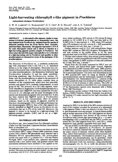
Proc. Natl. Acad. Sci. USA<br />
Vol. 91, pp. 679-683, January 1994<br />
Microbiology<br />
<strong>Light</strong>-<strong>harvest<strong>in</strong>g</strong> <strong>chlorophyll</strong> c-<strong>like</strong> <strong>pigment</strong> <strong>in</strong> <strong>Prochloron</strong><br />
(endosymbiosis/phylogeny/Prochlorophyta)<br />
A. W. D. LARKUM*, C. SCARAMUZZI*, G. C. Cox*, R. G. HILLERt, AND A. G. TURNERt<br />
*School of Biological Sciences and Electron Microscope Unit, University of Sydney, Sydney, NSW 2006, Australia; tSchool of Biological Sciences, Macquarie<br />
University, North Ryde, NSW 2113, Australia; and tBiological Sciences, Sydney Institute of Technology, Ultimo, NSW 2007, Australia<br />
Communicated by Andrew A. Benson, August 5, 1993<br />
ABSTRACT A <strong>chlorophyll</strong> c-<strong>like</strong> <strong>pigment</strong>, similar to magnesium-3,8-div<strong>in</strong>yl<br />
pheoporphyr<strong>in</strong> as monomethyl ester, has<br />
been isolated from <strong>Prochloron</strong> sp. obta<strong>in</strong>ed from five species of<br />
didemnid ascidians from the Great Barrier Reef, Australia,<br />
and from Palau, Micronesia. The <strong>pigment</strong> represents 4-15% of<br />
the total <strong>chlorophyll</strong> content and is shown to function <strong>in</strong> a<br />
light-<strong>harvest<strong>in</strong>g</strong> <strong>pigment</strong> prote<strong>in</strong> complex of <strong>Prochloron</strong>. The<br />
observation that all of the major <strong>chlorophyll</strong>s (a+b+c) function<br />
<strong>in</strong> a light-<strong>harvest<strong>in</strong>g</strong> role <strong>in</strong> <strong>Prochloron</strong> and possibly <strong>in</strong> other<br />
prochlorophytes is discussed <strong>in</strong> terms of the phylogeny of the<br />
prochlorophytes.<br />
The discovery of <strong>Prochloron</strong> sp., a symbiotic prokaryotic<br />
alga with <strong>chlorophyll</strong> (Chl) b and Chl a (1, 2), stimulated<br />
debate as to the orig<strong>in</strong> of chloroplasts. Discussion has cont<strong>in</strong>ued<br />
to the present time, fueled by the recent discoveries of<br />
two other prochlorophytes-the filamentous freshwater alga<br />
Prochlorothrix hollandica (3) and the small, unicellular,<br />
free-liv<strong>in</strong>g planktonic alga Prochlorococcus mar<strong>in</strong>us (4).<br />
Some view the prochlorophytes as allied to the ancestors of<br />
green chloroplasts (4, 5), while others consider them as<br />
members of the cyanobacteria, which have lost phycobiliprote<strong>in</strong>s<br />
and ga<strong>in</strong>ed Chl b, probably <strong>in</strong>dependently, <strong>in</strong> each<br />
group (6-10). The presence of a Chl c-<strong>like</strong> <strong>pigment</strong>, tentatively<br />
identified as magnesium 3,8-div<strong>in</strong>ylphaeoporphyr<strong>in</strong> a5<br />
monomethyl ester (MgDVP), has been claimed for Prochlorococcus<br />
(4, 11), although only the peak wavelengths of the<br />
natural and demetallated <strong>pigment</strong> were provided (11). It also<br />
has been claimed that MgDVP occurs <strong>in</strong> Prochlorothrix<br />
hollandica (11) <strong>in</strong> low concentrations. Here we present<br />
evidence, based on the spectra of <strong>pigment</strong>s resolved by<br />
th<strong>in</strong>-layer chromatography (TLC) and high-performance liquid<br />
chromatography (HPLC), that a Chl c-<strong>like</strong> <strong>pigment</strong> tentatively<br />
identified as MgDVP occurs <strong>in</strong> <strong>Prochloron</strong> <strong>in</strong> concentrations<br />
that are 4-15% of the total Chl. Moreover, the<br />
<strong>pigment</strong> is shown to be located <strong>in</strong> a light-<strong>harvest<strong>in</strong>g</strong> Chl<br />
<strong>pigment</strong>-prote<strong>in</strong> complex (LHC) and to act as a light<strong>harvest<strong>in</strong>g</strong><br />
<strong>pigment</strong>.<br />
MATERIALS AND METHODS<br />
<strong>Prochloron</strong> spp. were isolated from five didemnid ascidians<br />
(Table 1) at One Tree Reef, Capricorn Section, Great Barrier<br />
Reef, as described (15).<br />
Analysis of the <strong>pigment</strong>s of<strong>Prochloron</strong> was done on site by<br />
reverse-phase HPLC us<strong>in</strong>g a Waters 600E quaternary pump<br />
system with a Waters 990 photodiode array detector and a<br />
Shimadzu RF-551 spectrofluorimeter. The reverse-phase<br />
system was used with a C18 column (3.9 x 150 mm Waters<br />
Novapak 36975; 4-,um particle size, spherical end-capped<br />
material with a low carbon load), and gradient elution with<br />
acetonitrile/methanol/water (method 1) was applied as fol-<br />
The publication costs of this article were defrayed <strong>in</strong> part by page charge<br />
payment. This article must therefore be hereby marked "advertisement"<br />
<strong>in</strong> accordance with 18 U.S.C. §1734 solely to <strong>in</strong>dicate this fact.<br />
679<br />
lows: <strong>in</strong>itial conditions, 95% solvent A/5% solvent B; l<strong>in</strong>ear<br />
gradient to 1% A/99% B <strong>in</strong> 15 m<strong>in</strong>; and then held at 1%<br />
A/99%o B for 15 m<strong>in</strong> [solvent A = 50% water/25% acetonitrile/25%<br />
methanol (vol/vol); solvent B = 50% acetonitrile/<br />
50% methanol (vol/vol); flow rate, 2 ml m<strong>in</strong>-1].<br />
Further analyses used (i) TLC with polyethylene (Super<br />
Rylan 7007, sample no. PRX 1013, Koppers, Warr<strong>in</strong>gton, PA)<br />
and with acetone as the mobile phase or (ii) the same<br />
polyethylene used <strong>in</strong> an HPLC system (16) (hand-packed 4.6<br />
x 250 mm steel column) with a step solvent system (method<br />
2) as follows: system preequilibrated with 65% acetone/35%<br />
water; step gradient to 100% acetone at 0 m<strong>in</strong> and cont<strong>in</strong>ued<br />
for 15 m<strong>in</strong>; flow rate, 2 ml m<strong>in</strong>-1.<br />
Eluant from the Chl peaks separated by HPLC was collected,<br />
frozen, and freeze-dried under vacuum. Pigments<br />
were redissolved <strong>in</strong> 90% acetone/10% water, 100% acetone,<br />
or 100% ether, and their spectra were analyzed <strong>in</strong> a Beckman<br />
DU-8 spectrophotometer. Chl ratios were determ<strong>in</strong>ed on site<br />
<strong>in</strong> 90% acetone/10% water by us<strong>in</strong>g an Hitachi U-2000<br />
spectrophotometer and the trichromatic equation of Jeffrey<br />
and Humphrey (13). Demetallated Chl (i.e., removal of Mg<br />
from the porphyr<strong>in</strong> r<strong>in</strong>g) was produced by add<strong>in</strong>g 50 ,4 of 0.1<br />
M HCl to 2-4 ml of acetone or ether extracts. The LHC of<br />
<strong>Prochloron</strong> was isolated as described (15). Low-temperature<br />
(77 K) fluorescence spectra were measured with a Perk<strong>in</strong><br />
Elmer 44B Fluorimeter.<br />
Micromonas pusilla (culture CS-86 from the Culture Collection,<br />
CSIRO Fisheries, Hobart, Tasmania) was grown <strong>in</strong><br />
GP medium (17) under natural but low light (maximum, 200<br />
,uE-m-2s-1).<br />
RESULTS AND DISCUSSION<br />
Analysis by HPLC <strong>in</strong> the field of the 90% acetone extracts of<br />
each of five isolates revealed the presence of a <strong>chlorophyll</strong><br />
c-<strong>like</strong> <strong>pigment</strong> (Table 1) that was eluted at 14 m<strong>in</strong>; Chl a, Chl<br />
b, and carotenoids were also present. A typical elution profile<br />
and an on-l<strong>in</strong>e spectrum of the Chl c-<strong>like</strong> <strong>pigment</strong> are shown<br />
<strong>in</strong> Fig. 1. Comparisons (not shown) of the extracts from<br />
<strong>Prochloron</strong> with a brown alga (possess<strong>in</strong>g Chl cl + c2) and a<br />
d<strong>in</strong>oflagellate (Chl c2) <strong>in</strong>dicated that the HPLC system separated<br />
the Chl c-<strong>like</strong> <strong>pigment</strong> from Chl c1 and Chl c2, which<br />
themselves were partially separable from each other.<br />
The use of HPLC <strong>in</strong> the field was crucial for the discovery<br />
of the Chl c-<strong>like</strong> <strong>pigment</strong>(s) <strong>in</strong> <strong>Prochloron</strong>. At present<br />
<strong>Prochloron</strong> cannot be cultured (18), and didemnid ascidians,<br />
the major source of material, release acids when handled.<br />
Thus, unless fresh material is extracted and processed carefully<br />
and immediately, Chl breakdown products (e.g., <strong>chlorophyll</strong>ides<br />
and pheoporphyr<strong>in</strong>s) can be generated, which<br />
would obscure the presence of Chl c-<strong>like</strong> <strong>pigment</strong>s. However,<br />
further laboratory work was necessary, us<strong>in</strong>g polyethylene<br />
chromatography (14, 16, 19), to verify the presence of Chl<br />
Abbreviations: Chl, <strong>chlorophyll</strong>; BChl, bacterio<strong>chlorophyll</strong>; LHC,<br />
light-<strong>harvest<strong>in</strong>g</strong> Chl <strong>pigment</strong>-prote<strong>in</strong> complex; MgDVP, magnesium<br />
3,8-div<strong>in</strong>ylphaeoporphyr<strong>in</strong> a5 monomethyl ester.
680 Microbiology: Larkum et al.<br />
Table 1. The ratios of Chl a/b- and Chl a/c-<strong>like</strong> <strong>pigment</strong> found<br />
by reverse-phase HPLC analysis (method 1) for <strong>Prochloron</strong> sp.<br />
from five didemnid ascidians collected at One Tree Reef,<br />
Capricorn Section, Great Barrier Reef<br />
Species of ascidian host for <strong>Prochloron</strong> sp. Chl a/b Chl a/c-<strong>like</strong><br />
Didemnum molle (Herdmann, 1886) 3.1 15.8<br />
Diplosoma virens (Hartmeyer, 1908) 2.9 5.0<br />
Lissocl<strong>in</strong>um patella (Gottschaldt, 1898) 3.1 6.2<br />
Lissocl<strong>in</strong>um bistratum (Sluiter, 1905) 7.6 9.6<br />
Trididemnum paracyclops (Kott, 1980) 3.55 6.1<br />
Quantitation based on absorbance of <strong>in</strong>dividual peaks and ext<strong>in</strong>ction<br />
coefficients <strong>in</strong> methanol (12). Note that 90% acetone/10%o water<br />
extracts of the symbionts gave similar values for the Chl a/b ratio<br />
and, surpris<strong>in</strong>gly, also for the Chl a/c-<strong>like</strong> ratio (13), despite the<br />
difference <strong>in</strong> absorption maximum of Chl c and MgDVP <strong>in</strong> the red<br />
region of the spectrum (14).<br />
c-<strong>like</strong> <strong>pigment</strong>s. Thus, carefully prepared frozen or freezedried<br />
samples of <strong>Prochloron</strong> from L. patella were further<br />
analyzed <strong>in</strong> Sydney (the frozen samples came from a site at<br />
15-m depth near Lizard Island, Great Barrier Reef; the<br />
freeze-dried samples were a gift to G.C.C. from R. Lew<strong>in</strong> and<br />
were collected <strong>in</strong> Palau, Micronesia). TLC analysis (14, 19)<br />
revealed the presence of two Chl c-<strong>like</strong> compounds that had<br />
a significantly greater Rf value than those of either Chl c1 or<br />
Chl c2 (Fig. 2) and migrated <strong>in</strong> the relative position ofMgDVP<br />
(14, 19). There is also evidence from the fluorescence spectra<br />
of two related <strong>pigment</strong>s (see below). The presence of a Chl<br />
c-<strong>like</strong> <strong>pigment</strong> was also confirmed by reverse-phase HPLC<br />
with a polyethylene column, but only one broad peak was<br />
resolved (Fig. 3). The spectral peaks of the compound(s)<br />
eluted at 7.9 m<strong>in</strong> (Fig. 3 Lower, solid curve) (438 nm, 576 nm,<br />
and 626 nm-<strong>in</strong> 90% acetone/10% water) agree well with<br />
those of MgDVP (14, 19) <strong>in</strong> the same solvent. With extracts<br />
of brown algae or d<strong>in</strong>oflagellates, Chl c1 or Chl c2 or both<br />
were eluted later (not shown). The spectral peaks of the<br />
demetallated Chl c-<strong>like</strong> <strong>pigment</strong> (Table 2) (419 nm, 568 nm,<br />
and 588 nm) match well with those of demetallated MgDVP<br />
(11, 20). In addition, cells ofM. pusilla were extracted <strong>in</strong> 90%<br />
0<br />
0<br />
.0<br />
0<br />
.0<br />
0.2<br />
ol 1<br />
Time, m<strong>in</strong><br />
300 500<br />
Wavelength, nm<br />
700<br />
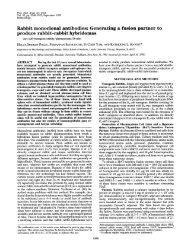
FIG. 1. (Upper) An elution profile (Adet = 440 mm) of the 90%o<br />
acetone extract of <strong>Prochloron</strong> sp. from Diplosoma virens separated<br />
by reverse-phase HPLC (method 1; see Table 1) on a Novapak C18<br />
column (Table 1). Bands: a, Chl a; b, Chl b; c, Chl c-<strong>like</strong> <strong>pigment</strong>;<br />
others, carotenoids. (Lower) On-l<strong>in</strong>e spectrum of Chl c-<strong>like</strong> <strong>pigment</strong><br />
eluted as band c at 14.4 m<strong>in</strong>. [This band was also detected by<br />
fluorescence emission <strong>in</strong> the red region of the spectrum (excitation,<br />
440 nm) with a maximum at 635 nm (not shown).]<br />
0<br />
co<br />
0 .0<br />
Q<br />
Proc. Natl. Acad. Sci. USA 91 (1994)<br />
O carotenoids<br />
* phaeophyt<strong>in</strong>s<br />
0 chi a(±b)<br />
@ chl c-<strong>like</strong><br />
e chl c-<strong>like</strong><br />
* chl c1<br />
0 chl c2<br />
* Orig<strong>in</strong><br />
A B C D E F<br />
o'<br />
. 0<br />
0 0 @ 0<br />
* 0 0 0 - -<br />
% a) 440 nm<br />
.07 . b)438 nm<br />
.05<br />
.05 .04 a<br />
.03b .03<br />
I 1 500 600<br />
Wavelength, nm<br />
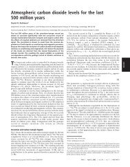
FIG. 2. (Upper) Diagram of a developed th<strong>in</strong>-layer chromatogram<br />
(14) of the follow<strong>in</strong>g 90% acetone extracts. Lanes: A, Endarachne<br />
b<strong>in</strong>ghamiae (Phaeophyta); B, Colpomenia s<strong>in</strong>uosa (Phaeophyta); C,<br />
<strong>Prochloron</strong> sp. (L. patella from Palau); D, <strong>Prochloron</strong> sp. (L. patella<br />
from Lizard Island); E, Pavlova lutheri (Prymnesiophyta); F, Amphid<strong>in</strong>ium<br />
carterae (Pyrrophyta). The develop<strong>in</strong>g solvent was acetone.<br />
In lane C only the lower Chl c-<strong>like</strong> <strong>pigment</strong> was clearly visible,<br />
s<strong>in</strong>ce the upper Chl c-<strong>like</strong> <strong>pigment</strong> was overla<strong>in</strong> by an orange<br />
<strong>pigment</strong>, probably a carotenoid. (Lower) Spectra of two Chl c-<strong>like</strong><br />
<strong>pigment</strong>s <strong>in</strong> 90% acetone from lane D <strong>in</strong> Upper. Spectra: a, spectrum<br />
of lower Chl c-<strong>like</strong> (stippled) <strong>pigment</strong>; b, spectrum ofupper Chl c-<strong>like</strong><br />
(hatched) <strong>pigment</strong>. (Amax for the major peak of a and b spectra are<br />
shown.)<br />
acetone/10% water and analyzed by HPLC with a C18<br />
column (method 1). A Chl c-<strong>like</strong> <strong>pigment</strong> was present, which<br />
was coeluted with the major Chl c-<strong>like</strong> <strong>pigment</strong> from <strong>Prochloron</strong>.<br />
It also had similar spectral properties to the Chl c-<strong>like</strong><br />
<strong>pigment</strong> of <strong>Prochloron</strong> (Table 2). The Chl c-<strong>like</strong> <strong>pigment</strong> <strong>in</strong><br />
Micromonas spp. has been tentatively identified as MgDVP<br />
(12, 14, 19) on the basis of its spectral properties and elution<br />
characteristics on TLC and HPLC.<br />
MgDVP has a fully unsaturated porphyr<strong>in</strong> macrocycle and<br />
does not carry a long-cha<strong>in</strong> esterify<strong>in</strong>g alcohol at C-17 and<br />
differs from Chl c2 only <strong>in</strong> the presence at C-17 of a propionic<br />
acid <strong>in</strong>stead of an acrylic acid (14). Evidence suggests that<br />
MgDVP is an <strong>in</strong>termediate <strong>in</strong> the pathway of Chl a [and<br />
bacterio<strong>chlorophyll</strong> (BChl)] biosynthesis (21). It was first<br />
documented <strong>in</strong> the photosynthetic bacterium Rhodobacter<br />
sphaeroides grown <strong>in</strong> the presence of 8-hydroxyqu<strong>in</strong>ol<strong>in</strong>e,<br />
but it probably occurs also <strong>in</strong> some mutants ofR. sphaeroides<br />
as a result of a lesion <strong>in</strong> the normal synthesis of BChl (21).<br />
MgDVP was also identified as a <strong>pigment</strong> ofunknown function<br />
<strong>in</strong> several mar<strong>in</strong>e flagellates (22) <strong>in</strong>clud<strong>in</strong>g the pras<strong>in</strong>ophyte<br />
M. pusilla, where it has s<strong>in</strong>ce been shown to be present <strong>in</strong> an<br />
LHC as a light-<strong>harvest<strong>in</strong>g</strong> <strong>pigment</strong> (23, 24). However, it<br />
should be po<strong>in</strong>ted out that the precise structure of the Chl<br />
c-<strong>like</strong> <strong>pigment</strong> <strong>in</strong> M. pusilla and the related alga Mantoniella<br />
squamata is debated (12, 20) [both algae are placed <strong>in</strong> the<br />
special group, the micromonadophytes (25)]. MgDVP is also<br />
probably present <strong>in</strong> the prochlorophyte Prochlorococcus<br />
mar<strong>in</strong>us (11), although full details have yet to be documented.<br />
On the basis of the present evidence, we conclude that a chl<br />
c-<strong>like</strong> <strong>pigment</strong>, similar to MgDVP, occurs <strong>in</strong> <strong>Prochloron</strong> sp.<br />
together with a closely related <strong>pigment</strong>; however, both <strong>pigment</strong>s<br />
need to be further exam<strong>in</strong>ed by nuclear magnetic
0<br />
c<br />
0.<br />
co °0.1<br />
CO 0.15<br />
Microbiology: Larkum et al.<br />
0.1<br />
0.05<br />
0 2 4 6<br />
Time, m<strong>in</strong><br />
a<br />
573<br />
Wavelength, nm<br />
8 12<br />
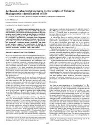
FIG. 3. (Upper) Profile of <strong>pigment</strong>s extracted <strong>in</strong> 90%o acetone<br />
from <strong>Prochloron</strong> sp. (L. patella from Palau) and separated by<br />
reverse-phase HPLC on a polyethylene column (17) (method 2)<br />
monitored at 440 nm (the eluant was also monitored by a fluorescence<br />
detector, which confirmed the fluorescence of Chl c-<strong>like</strong> at 635 nm<br />
<strong>in</strong> the band eluted at 7.9 m<strong>in</strong>). Bands: a, Chl a + b; c, Chl c-<strong>like</strong>;<br />
others, carotenoids. Extracts from a brown alga E. b<strong>in</strong>ghamiae and<br />
a d<strong>in</strong>oflagellate alga A. carterae showed that Chl ci was eluted at 9.2<br />
m<strong>in</strong> and Chl c2 was eluted at 11.8 m<strong>in</strong>. (profiles not shown). (Lower)<br />
On-l<strong>in</strong>e spectrum of the Chl c-<strong>like</strong> band (c) eluted at 7.9 m<strong>in</strong>. (solid<br />
curve) and of the <strong>pigment</strong> extracted and chromatographed <strong>in</strong> the<br />
same position from the LHC (dashed curve).<br />
resonance spectroscopy. It is possible that only one of the<br />
<strong>pigment</strong>s is native and that the other is a product of the<br />
preparative procedures. We reject the possibility that both<br />
<strong>pigment</strong>s are the result of contam<strong>in</strong>ation by another alga or<br />
photosynthetic bacterium. The preparations used were monitored<br />
microscopically and showed less than 0.3% contam<strong>in</strong>ation<br />
on a cell count basis (
682 Microbiology: Larkum et al.<br />
U)<br />
0 657<br />
a:<br />
600 ~~720<br />
_<br />
636 628<br />
_ _ _ _ _ _ _ _ _ _ _ _ _ _ _<br />
A, nm<br />
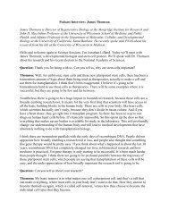
FIG. 4. Low-temperature (77 K) fluorescence emission spectra of the LHC isolated from <strong>Prochloron</strong> sp. (from L. patella found at 10-m depth<br />
on One Tree Reef). (A) Isolated LHC excited by light at 440 nm. (B) LHC extract <strong>in</strong> 95% acetone/10% water excited at 440 nm. (C) LHC extract<br />
<strong>in</strong> 95% acetone/5% water excited at 470 nm. (D) LHC <strong>in</strong> 2% SDS and heated to 70°C for 5 m<strong>in</strong>, excited at 440 nm. (E) LHC <strong>in</strong> 2% SDS and<br />
heated to 70°C for 5 m<strong>in</strong>, excited at 470 nm.<br />
procedure (15) as the LHC had no detectable levels of the Chl<br />
c-<strong>like</strong> <strong>pigment</strong>, although Chl a and b were present, as shown<br />
previously (15). This evidence suggests that the Chl c-<strong>like</strong><br />
<strong>pigment</strong> is not <strong>in</strong>timately connected to photosystem I. The<br />
nature and function of the major LHC <strong>in</strong> <strong>Prochloron</strong> sp. and<br />
the presence of other LHCs have yet to be fully elucidated.<br />
It may be similar to LHC II of chlorophytes and higher plants<br />
and therefore associated with photosystem II (15), but neither<br />
its molecular location nor its primary structure has been<br />
reported.<br />
The occurrence of Chls a, b, and c <strong>in</strong> a light-<strong>harvest<strong>in</strong>g</strong> role<br />
<strong>in</strong> <strong>Prochloron</strong> raises many <strong>in</strong>trigu<strong>in</strong>g questions. The presence<br />
of an identical or similar Chl c-<strong>like</strong> <strong>pigment</strong> to that <strong>in</strong><br />
<strong>Prochloron</strong> seems <strong>like</strong>ly <strong>in</strong> Prochlorococcus mar<strong>in</strong>us, although<br />
it has yet to be shown <strong>in</strong> the latter organism whether<br />
the <strong>pigment</strong> acts <strong>in</strong> a light-<strong>harvest<strong>in</strong>g</strong> capacity and is found <strong>in</strong><br />
an LHC. Is Prochlorothrix hollandica, the other prochlorophyte,<br />
similar? Prelim<strong>in</strong>ary <strong>pigment</strong> analysis of that organism<br />
<strong>in</strong>dicates that MgDVP may be present <strong>in</strong> low concentrations<br />
(11). If this is substantiated, then all three prochlorophytes<br />
are <strong>like</strong>ly to conta<strong>in</strong> the Chl c-<strong>like</strong> <strong>pigment</strong>, although it would<br />
still be necessary to show that it acts <strong>in</strong> a light-<strong>harvest<strong>in</strong>g</strong><br />
capacity. At the moment it is possible to say that <strong>Prochloron</strong><br />
sp. and possibly each of the three prochlorophytes are more<br />
closely allied to the micromonadophyte chloroplast l<strong>in</strong>e (25,<br />
28) than to any other chloroplast l<strong>in</strong>e on the evidence that<br />
they all share a Chl c-<strong>like</strong> <strong>pigment</strong>. However, the dist<strong>in</strong>ct<br />
differences <strong>in</strong> the LHCs between <strong>Prochloron</strong> (15) and<br />
Prochlorothrix (29) on the one hand (the type of LHC <strong>in</strong><br />
Prochlorococcus is not known) and the micromonadophytes<br />
on the other hand (20, 30) (where the LHC is of the chromophytic<br />
type) stand aga<strong>in</strong>st a close aff<strong>in</strong>ity between these<br />
two groups. The recent f<strong>in</strong>d<strong>in</strong>g of div<strong>in</strong>yl Chl a and b <strong>in</strong><br />
Prochlorococcus (4, 11) and the evidence that both <strong>Prochloron</strong><br />
and Prochlorococcus share the presence of a Chl c-<strong>like</strong><br />
<strong>pigment</strong> raise the possibility that div<strong>in</strong>yl <strong>chlorophyll</strong>s a and b<br />
also exist <strong>in</strong> <strong>Prochloron</strong>.<br />
The occurrence of a Chl c-<strong>like</strong> <strong>pigment</strong> and Chl b [or<br />
div<strong>in</strong>yl-Chl b (11)] <strong>in</strong> all of the prochlorophytes would<br />
weaken the argument that these organisms are merely cyanobacteria,<br />
where Chl b orig<strong>in</strong>ated <strong>in</strong>dependently <strong>in</strong> each of<br />
the three groups (7-10). We believe that the phylogenetic tree<br />
analysis on which that argument was based is flawed (31-33).<br />
Another explanation is that the prochlorophytes are an<br />
anciently diverged group (34) widely separated from the<br />
Proc. Natl. Acad. Sci. USA 91 (1994)<br />
Cyanobacteria, as orig<strong>in</strong>ally suggested (2). Thus, all three<br />
known prochlorophytes may be only very distantly related to<br />
the green chloroplast and may have diverged considerably<br />
from each other. F<strong>in</strong>ally, the present evidence lends support<br />
to the recent suggestion that Chl c-<strong>like</strong> <strong>pigment</strong>s were present<br />
<strong>in</strong> the earliest photosynthetic prokaryotes (28).<br />
We are grateful to Dr. S. W. Jeffrey for the supply of M. pusilla.<br />
This work was supported by the Australian Research Council (Small<br />
Grants).<br />
1. Lew<strong>in</strong>, R. A. (1975) Phycologia 14, 153-160.<br />
2. Lew<strong>in</strong>, R. A. (1977) Phycologia 16, 217.<br />
3. Burger-Wiersma, T., Veenhuis, M., Korthals, H. J., Van de<br />
Wiel, C. C. M. & Mur, R. (1986) Nature (London) 320, 262-<br />
264.<br />
4. Chisholm, S. W., Olson, R. J., Zettler, E. R., Goericke, R.,<br />
Waterbury, J. B. & Welschmeyer, N. A. (1988) Nature (London)<br />
334, 340-343.<br />
5. Palmer, J. (1985) Annu. Rev. Genet. 19, 325-354.<br />
6. Meyer, T. E., Cusanovich, M. A. & Kamen, M. D. (1986)<br />
Proc. Natl. Acad. Sci. USA 83, 217-220.<br />
7. Turner, S., Burger-Wiersma, T., Giovannoni, J., Mur, L. R. &<br />
Pace, N. R. (1989) Nature (London) 337, 380-385.<br />
8. Morden, C. W. & Golden, S. S. (1991) J. Mol. Evol. 32,<br />
379-395.<br />
9. Palenik, B. & Haselkorn, R. (1992) Nature (London) 355,<br />
265-267.<br />
10. Urbach, E., Robertson, D. L. & Chisholm, S. (1992) Nature<br />
(London) 355, 267-270.<br />
11. Goericke, R. & Repeta, D. J. (1992) Limnol. Oceanogr. 37,<br />
425-433.<br />
12. Wright, S. W., Jeffrey, S. W., Mantoura, R. F. C., Llewellyn,<br />
C. A., Bjornland, T., Repeta, D. & Welschmeyer, N. (1991)<br />
Mar. Ecol. Prog. Ser. 77, 183-196.<br />
13. Jeffrey, S. W. & Humphrey, G. P. (1975) Biochem. Physiol.<br />
Pflanzen (BPP) 167, 191-194.<br />
14. Jeffrey, S. W. & Wright, S. W. (1987) Biochim. Biophys. Acta<br />
894, 180-188.<br />
15. Hiller, R. G. & Larkum, A. W. D. (1985) Biochim. Biophys.<br />
Acta 806, 107-115.<br />
16. Shioi, Y. & Beale, S. 1. (1987) Anal. Biochem. 162, 493-499.<br />
17. Loeblich, A. R. & Smith, V. E. (1968) Lipids 9, 5-13.<br />
18. Lew<strong>in</strong>, R. A. & Cheng, L. (1989) <strong>Prochloron</strong>: A Microbial<br />
Enigma (Chapman Hall, New York).<br />
19. Jeffrey, S. W. (1989) <strong>in</strong> The Chromophytic Algae: Problems<br />
and Perspectives, eds. Green, J. C., Leadbetter, B. S. C. &<br />
Diver, W. L. (Clarendon, Oxford, U.K.), pp. 13-36.<br />
20. Wilhelm, C. (1987) Biochim. Biophys. Acta 892, 23-29.
21.<br />
22.<br />
23.<br />
24.<br />
25.<br />
26.<br />
27.<br />
28.<br />
Microbiology: Larkum et al.<br />
Jones, 0. T. G. (1979) <strong>in</strong> The Porphyr<strong>in</strong>s, ed. Dolph<strong>in</strong>, D.<br />
(Academic, New York), Vol. 6, pp. 179-230.<br />
Ricketts, T. R. (1966) Phytochemistry 5, 223-229.<br />
Brown, J. (1985) Biochim. Biophys. Acta 807, 1143-1146.<br />
Wilhelm, C., Lenartz-Weiler, I., Wiedemann, I. & Wild, A.<br />
(1986) Phycologia 25, 304-312.<br />
Fawley, M. W., Stewart, K. D. & Mattox, K. R. (1986) J. Mol.<br />
Evol. 23, 168-176.<br />
Larkum, A. W. D. & Barrett, J. (1983) Adv. Bot. Res. 10,<br />
1-219.<br />
Hiller, R. G., Larkum, A. W. D. & Wrench, P. (1988) Biochim.<br />
Biophys. Acta 932, 223-231.<br />
Larkum, A. W. D. (1991) <strong>in</strong> Chlorophylls, ed. Scheer, H.<br />
(CRC, Boca Raton, FL), pp. 367-383.<br />
Proc. Natl. Acad. Sci. USA 91(1994) 683<br />
29. Bullerjahn, G. S., Matthijs, H. C. P., Mur, L. R. & Sherman,<br />
L. A. (1987) Eur. J. Biochem. 168, 295-300.<br />
30. Herold, A., Schmitt, A., Wilhelm, C. & Wild, A. (1991)<br />
Photosynthetica 25, 645-653.<br />
31. Lockhart, P. J., Beanland, T. J., Howe, C. J. & Larkum,<br />
A. W. D. (1992) Proc. Natl. Acad. Sci. USA 89, 2742-2746.<br />
32. Lockhart, P. J., Penny, D., Hendy, M. D., Howe, C. J., Beanland,<br />
T. J. & Larkum, A. W. D. (1992) FEBS Lett. 301, 127-131.<br />
33. Lockhart, P. J. & Penny, D. (1993) <strong>in</strong> Research <strong>in</strong> Photosynthesis,<br />
ed. Murata, M. (Kluwer, Dordrecht, The Netherlands),<br />
Vol. 3, pp. 499-505.<br />
34. Larkum, A. W. D. (1993) <strong>in</strong> Research <strong>in</strong> Photosynthesis, ed.<br />
Murata, M. (Kluwer, Dordrecht, The Netherlands), Vol. 3, pp.<br />
475-482.