Xylem and Phloem
Plants don’t have blood vessels - instead they have xylem and phloem vessels. Xylem transports water whereas phloem transports glucose and dissolved amino acids. They are both specialised to carry out their function and arranged in such a way to give the plant as much structure and support as possible.
Vascular Bundles
Plants contain vessels which function to transport water and sugars from one part of the plant to another. Xylem vessels transport water and dissolved mineral ions from the roots to the rest of the plant and also provide structural support. Phloem vessels transport dissolved substances, such as sucrose and amino acids, from the leaves to the rest of the plant. Xylem and phloem vessels are grouped together within the plant stem and form vascular bundles. Sclerenchyma fibres are also found within vascular bundles and provide support to the stem.
Within the plant stem, xylem vessels are found right on the inside. Phloem tissue is located in the middle of the vascular bundle and sclerenchyma fibres are found on the outside. Having the stronger xylem vessels in the centre provides strength to the stem and acts like an internal ‘scaffolding’ to support the stem and prevent it from bending in the wind.
In the root, the xylem forms a cross-like structure in the centre which is surrounded by phloem vessels. This arrangement adds strength to the root as it pushed through the soil.
Within the leaf, the xylem vessels are found towards the top of the vascular bundle with the phloem vessels found underneath.
Xylem
Xylem vessels transport water and mineral ions from the roots to the rest of the plant. They are made up of dead, hollow cells with no end cell walls. This forms one continuous tube when the xylem cells are stacked on top of each other. The cells have no organelles or cytoplasm, which creates more space inside the vessel for transporting water. The cell walls contain pits which allows water and mineral ions to move into and out of the vessel. The cell wall also contains a tough, woody substance called lignin, which strengthens the xylem vessel and provides structure and support to the plant.
Phloem
Phloem vessels transport dissolved substances, such as sucrose and amino acids from parts of the plant where they are made (sources) to the parts of the plant where they are used (sinks). Leaves are sources because they produce glucose from photosynthesis and parts of the plant where sugar is stored, such as roots and bulbs, act as sinks.
Phloem vessels are made up of two types of cell - sieve tube elements and companion cells. The sieve tube elements are living cells and are joined end-to-end to form sieve tubes. The ends of each cell consist of a ‘sieve plate’ which contains lots of holes to allow solutes to move from one cell to the next. The sieve tube cells contain no organelles and very little cytoplasm to create more space for solutes to be transported. The absence of a nucleus and other organelles means that these cells cannot survive on their own, so each sieve tube element is associated with a companion cell, which contains a nucleus and is packed full of mitochondria. The mitochondria provide lots of energy for the active loading of sucrose into the sieve tube element. The sieve tube element and the companion cell are connected through plasmodesmata (channels in the cell wall) which allows the two cells to communicate.
Sclerenchyma
Together with xylem and phloem vessels, sclerenchyma fibres are also found within vascular bundles and provide structural support to the plant. They are made up of bundles of long, dead cells. The cells have a hollow lumen and the cell walls are thickened with lignin. The cell walls also contain more cellulose than a typical plant cell which makes sclerenchyma fibres particularly strong.
Dissecting Plant Stems
To view vascular bundles under the microscope, you first need to dissect the plant stem and prepare a tissue sample. You would do this by following the method below:
Cut a thin section of the plant stem using a scalpel. Take care when using the sample and remember to cut away from you.
Place the tissue sample into water to prevent it from drying out.
Place the tissue sample into a small dish containing the stain. A common stain that is used to view vascular bundles is toluidine blue O (TBO) which stains lignin blue/green which will enable you to visualise the xylem and sclerenchyma fibres. The phloem cells and remaining tissue will appear a pink/purple colour.
Rinse the tissue samples in water and place each one onto a microscope slide.
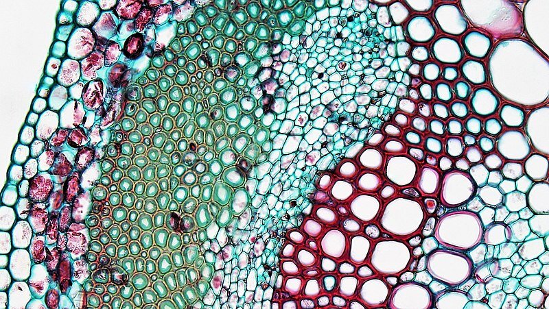
Vascular bundle in a clover leaf viewed under the microscope. Image credit: Berkshire Community College Bioscience Image Library.

Did you know…
Russian scientists have been able to revive a plant which had died 32,000 years ago after its fruit was stored by an arctic ground squirrel. The fruit was of the narrow-leafed campion plant (pictured) which had been preserved for thousands of years by the cold temperatures in the Siberian tundra.
Next Page: Plant Fibres and Sustainability