
Dandelion Seed

Dandelion Seed - Head - 7X LM EDF

Dandelion Seed - Parachute Top - 4000X SE

Dandelion Seed - Parachute Top - 750X SE

Dandelion Seed - Parachute Top - 100X SE

Dandelion Seed - 150X SE

Dandelion Seed Dispersal
Product Description
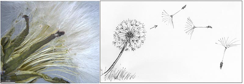
This dandelion species (Taraxacum officinale) is a native of Europe and Asia but is now found throughout temperate regions of the world. It is a flowering plant that is considered an irritating lawn and garden weed as it has very successfully adapted reproductively. This dandelion propagates through its root system and asexually by seeds that are produced without pollination. The seeds have the same genetic make-up as the plant; thus, it essentially clones itself.
The dandelion seed structure is complex. It consists of a bristly seed fruit called the achene; a thin hollow stalk called the beak, and a parachute end called the pappus. The pappus is a tuft of approximately 180 thin rigid “hairs” that spread out radially. Each hair is 6 – 7.5 mm long and consists of a bundle of hollow branching pipes that decrease in number toward the tip making it very light and strong. The hairs also have feather-like projections that increase the surface area. The seed is blown by the wind from the seed head ball. The force of gravity does not prevent it from floating easily through turbulent air using the drag forces of its pappus parachute. Thus, the dandelion has perfected seed dispersal in the air over long distances.
References:
https://en.wikipedia.org/wiki/Taraxacum
Morphological Design Of Dandelion – Sem-Proceedings.Com
https://fliphtml5.com/sibd/jiba/basic
PURCHASE 5 OR MORE IMAGES AND GET 20% OFF YOUR ENTIRE ORDER!
Please contact us for custom images.
The image store is a collection of organisms that have been examined under a stereo light microscope (LM) and or scanning electron microscope (SEM). Each group of organisms has a short description and a longer more detailed description or story about the organism. Clicking on the product group shows the individual images. Each series takes the observer from macro to micro or nano on a particular organism, starting with a macro photographic image(s) for perspective, micro images taken by the light microscope, and most have micro to nano scanning electron microscope images. The SEM images will appear in black and white as a beam of electrons is used to illuminate the specimen rather than light. A few SEM images are colorized (lotus leaf). More information about the labeling and techniques used is below.
For the curious:
The light microscope images are labeled LM and a Z is included if it is a vertical composite of images effectively extending the depth of field or EDF of the microscope.
SEM images are labeled by the type of detector use:
SE (secondary electron)
LSE (Low vacuum secondary electron)
BES (backscattered electron shadow mode)
BEC (backscattered electron compositional mode)
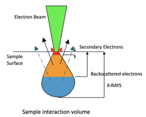
The SEM instrument works by producing a beam of electrons under a vacuum that interacts with the sample surface and subsurface producing different signals, as shown in the diagram at right. Secondary electrons, backscattered electrons and x-rays are detected using different instrument modes. In addition to morphological information to produce an image the SEM can determine elemental composition by energy dispersive x-ray spectroscopy (EDS).
Personal/Educational License:
For personal or educational use only. Images cannot be given or resold to others.
Commercial License:
For Profit or Non-profit business use within the organization, in presentations and publications with image credit. No resale of images.
An image license does not grant exclusive rights to an image. Lichen Labs retains the copywrite to all images.