Anatomy of the Phalanges
Updated:
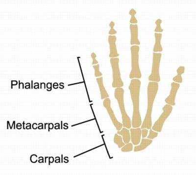
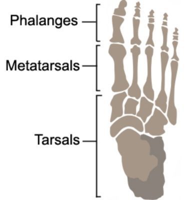
The phalanges are the bones that make up the fingers and toes (Figures 1,2). There are 14 phalanges in each hand and foot, with three phalanges in each finger and toe except for the thumb and big toe, which have two. Understanding the anatomy of the phalanges is essential for diagnosing and treating injuries to the fingers and toes. This article will cover the bony landmarks, muscular attachments, and common injuries associated with the phalanges.
Phalanges Anatomy: Bony Landmarks
The phalanges are long bones that make up the finger and toe skeleton. Each finger and toe consists of a proximal, middle, and distal phalanx (with the exception of the thumb and great toe which only has a proximal and distal phalanx). The proximal phalanx is the bone closest to the metacarpal or metatarsal bone. The middle phalanx is the bone in the middle, and the distal phalanx is the bone closest to the fingertip or toe. Each phalanx has a head, base, shaft, and several bony landmarks, including tuberosities, fossae, and ridges. These landmarks serve as attachment points for ligaments and tendons and provide stability to the finger and toe joints.
Phalanges Anatomy: Muscular Attachments
The phalanges have numerous muscular attachments that allow for movement and flexibility of the fingers and toes. The flexor digitorum superficialis muscle attaches to the middle phalanges in the hand and allows for flexion of the fingers. The extensor digitorum muscle attaches to the distal phalanges and allows for extension of the fingers. Additionally, the interossei muscles, located between the metacarpals or metatarsals, attach to the proximal and middle phalanges and allow for abduction and adduction (spreading the fingers apart and bringing them together) of the fingers and toes.
Common Injuries affecting the Phalanges
Injuries to the phalanges are common and can result from trauma, repetitive use, or overuse. Fractures, dislocations, sprains and tendon injuries are the most common injuries associated with the phalanges. Fractures can occur at any point along the bone and can be displaced or nondisplaced. Dislocations occur when the bone is forced out of its normal position, often resulting in severe pain and swelling. Tendon injuries, such as mallet finger or trigger finger, can result from repetitive use or overuse and can cause limited range of motion and discomfort.
In conclusion, understanding the anatomy of the phalanges is essential for diagnosing and treating injuries to the fingers and toes. The bony landmarks, muscular attachments, and common injuries associated with the phalanges provide a framework for identifying and treating these injuries. If you experience pain or discomfort in your fingers or toes, consult with a Physiotherapist or healthcare professional to determine the cause and best course of treatment.
References:
- Netter, F. H. (2014). Atlas of human anatomy. Elsevier Health Sciences.
- Standring, S. (Ed.). (2016). Gray’s anatomy: the anatomical basis of clinical practice. Elsevier Health Sciences.
- Wilkins, K. E., & Beaty, J. H. (2014). Fractures and dislocations of the hand and carpus. In Rockwood and Wilkins’ fractures in children (pp. 696-739). Wolters Kluwer Health/Lippincott Williams & Wilkins.
- Osterman, A. L. (2017). The hand: fundamentals of therapy. Elsevier Health Sciences.
- Varacallo, M., & Mair, S. D. (2020). Phalangeal Fractures. In StatPearls [Internet]. StatPearls Publishing.
- Phalanges of the Hand by radiopaedia – https://radiopaedia.org/articles/phalanges-of-the-hands

Link to this Page
If you would like to link to this article on your website, simply copy the code below and add it to your page:
<a href="https://physioadvisor.com.au/phalanges-anatomy”>Anatomy of the Phalanges – PhysioAdvisor.com</a><br/>Learn about Phalanges anatomy including bony landmarks, muscular attachments and common injuries on PhysioAdvisor.
Return to the top of Anatomy of the Phalanges.