Nail Bed Repair
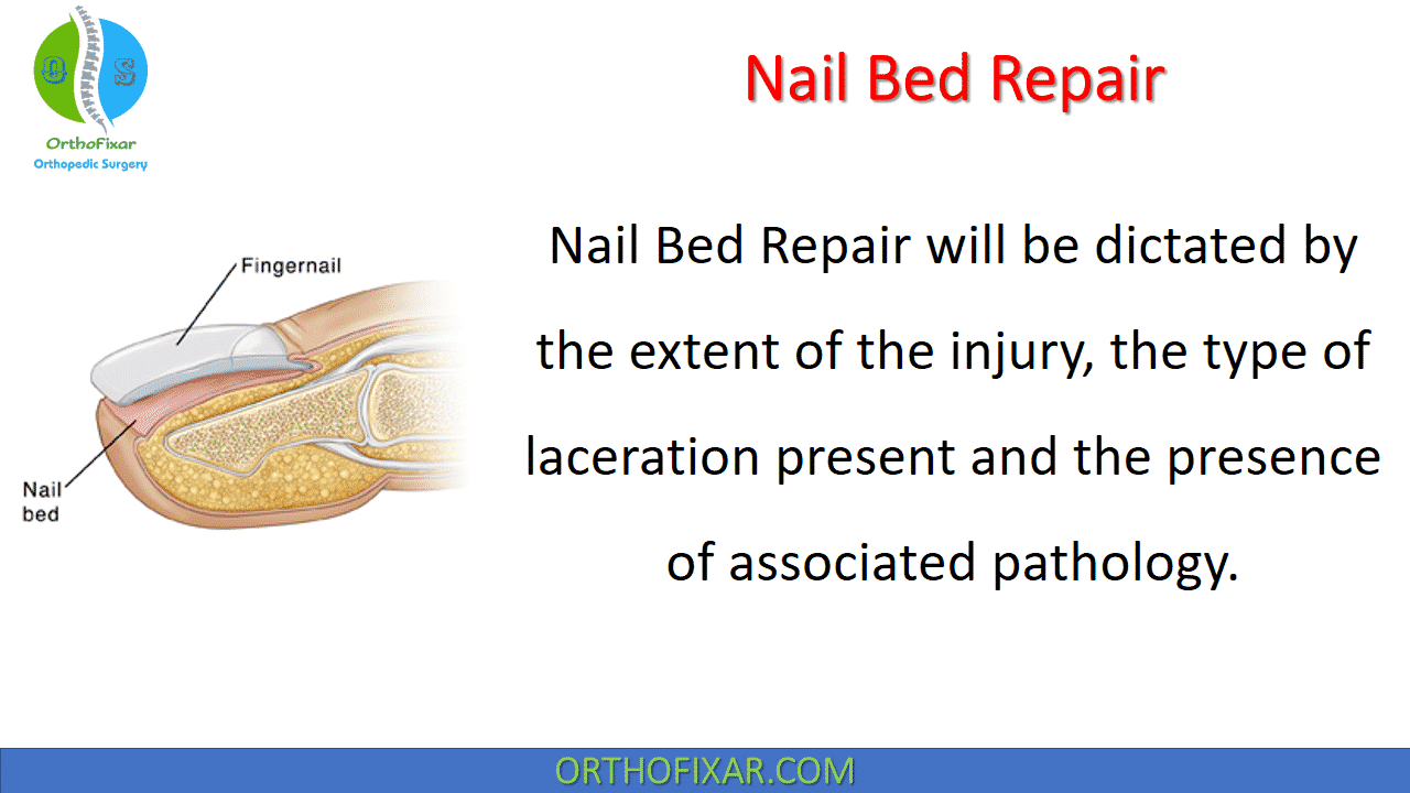
Nail Bed Repair will be dictated by the extent of the injury, the type of laceration present and the presence of associated pathology.
Small subungual hematomas (those involving <25% of the nail bed) can be managed with observation or trephination, if the area is painful.
For subungual hematomas involving more than 25% of the nail bed, nail plate removal is recommended for examination of the nail bed and repair of any laceration that may be present.
Treatment options for both simple and stellate nail bed lacerations include suture repair or the use of 2-octylcyanoacrylate (Dermabond).
See Also: Hand Anatomy

Necessary Supplies for Nail Bed Repair
Materials required for nail bed repair are described as follows:
- 1% lidocaine without epinephrine for digital nerve block .
- 10 cc syringe .
- 25- or 27-gauge needle for the digital nerve block .
- 0.5” or 1” penrose drain to act as a finger tourniquet .
- Povidone-iodine solution .
- Kidney basin.
- Sterile drapes.
- Laceration tray—necessary instruments include iris scissors, small needle driver, forceps and a small tissue elevator.
- 6-0 absorbable monofilament suture .
- 6-0 nylon suture (keep the foil package to use as an interposition underneath the eponychial fold if the patient’s nail is unavailable or unusable) .
- 2-octylcyanoacrylate (Dermabond, if this treatment approach is utilized) .
- Xeroform non-adherent dressing .
- Sterile dressings (gauze and kling wrap) .
- Finger splint.
- Loupe magnification (optional).

Nail Bed Repair Technique
Suture Repair and 2-Octylcyanoacrylate Repair:
With the patient in supine position, the affected hand is placed palm down onto an arm extension or small mayo stand type table. The injured finger is copiously irrigated with saline removing any debris or contamination from the region of the nail. The finger is then prepped using povidone-iodine solution and the area is draped using a sterile extremity drape.
A digital nerve block is then provided making sure to cover all four digital nerves that supply the injured finger. The penrose drain is then applied as a finger tourniquet, wrapped from distal to proximal and anchored around the base of the finger. The nail plate is then removed and the nature of the nail bed laceration is assessed.
See Also: Nerve Block For Hand Surgery
Nail Plate Removal
- Using the iris scissors placed between the nail bed and the nail plate (hyponchium), the nail plate is systematically released, opening and closing the tips of the scissors to provide tissue
separation. - The iris scissors are advanced proximally until they reach the nail fold—making sure to free the nail plate proximally, radially and ulnarly.
- Once removed from the underlying nail bed, the nail plate should be saved for later interposition underneath the eponychial fold (place in kidney basin soaking in povidone-iodine).

Suture Repair
- Irrigate the nail bed removing any hematoma or debris before Nail Bed Repair.
- Classify the laceration as either simple or stellate.
- Debride the laceration edges removing any irregularities that may be present with the iris scissors to ensure a smooth repair without tension.
- Using the small tissue elevator, the edges of the nail bed laceration can be slightly undermined from the underlying periosteum, mobilizing this tissue for repair.
- Repair the laceration using 6-0 absorbable monofilament sutures tied in simple fashion.

2-Octylcyanoacrylate Repair
- Once the laceration site has been irrigated, debrided and prepared, a single layer of 2-octylcyanoacrylate is applied to the laceration site.
- The edges of the laceration are held reapproximated manually as the tissue adhesive dries (approximately 60–90 seconds).
- Once the first layer is dry, a second layer is applied and allowed to dry completely.

Eponychial Interposition
- Following Nail Bed Repair of the laceration, either the removed nail plate or a piece of foil from the suture packaging can be used as a stent underneath the eponychial fold to provide space for new nail growth and prevent the development of adhesions and later nail plate deformity.
- This can trim/shape the edge of the removed nail plate can be trimmed to easily fit underneath the eponychial fold.
- Either interposition choice can be held in place using either 6-0 nylon suture or leftover 2-octylcyanoacrylate.
- For cases in which 6-0 nylon suture is used to hold the nail plate in place, the first pass is antegrade through the skin of the dorsum of the finger 5 mm proximal to the nail fold with the needle exiting between the eponychial fold and the nail bed. The next suture pass is through the prepared nail plate from posterior to anterior followed by another pass through the nail plate from anterior to posterior. The final suture pass is retrograde starting between the eponychial fold and the nail bed exiting through the skin of the dorsum of the finger approximately 5 mm proximal to the nail fold. Tension on the sutures will pull the nail plate into position underneath the eponychial fold. The sutures are then tied creating a horizontal mattress type configuration.
Dressings and Follow-up Care
- Xeroform, non-adherent gauze is then applied covering the nail bed repair.
- The finger is then wrapped in a kling dressing.
- The repaired nail bed is then protected in a finger splint, immobilizing the DIP joint.
- Patients are discharged home with a 5-day course of oral antibiotics for prophylaxis against infection (cephalexin 250 mg PO qid).
- The repair site should be re-evaluated in 5 to 7 days.
- The interposed nail or suture packet foil can be removed from underneath the eponychial fold at three weeks post treatment.
- Educate the patient that new nail growth may take 3 to 6 months to occur and often some cosmetic deformity is present after Nail Bed Repair.

References
- Brown RE. Acute nail bed injuries. Hand Clin. 2002 Nov;18(4):561-75. doi: 10.1016/s0749-0712(02)00075-6. PMID: 12516973.
- Elbeshbeshy BR, Rettig ME. Nail bed repair and reconstruction. Tech Hand Up Extrem Surg. 2002 Jun;6(2):50-5. doi: 10.1097/00130911-200206000-00002. PMID: 16520617.
- Hart RG, Kleinert HE. Fingertip and nail bed injuries. Emerg Med Clin North Am. 1993 Aug;11(3):755-65. PMID: 8359141.
- Strauss EJ, Weil WM, Jordan C, Paksima N. A prospective, randomized, controlled trial of 2-octylcyanoacrylate versus suture repair for nail bed injuries. J Hand Surg Am. 2008;33(2):250-3.
- Van Beek AL, Kassan MA, Adson MH, Dale V. Management of acute fingernail injuries. Hand Clin. 1990;6(1):23-35; discussion 37-8.
- Zook EG, Guy RJ, Russell RC. A study of nail bed injuries: causes, treatment, and prognosis. J Hand Surg Am. 1984;9(2):247-52.
- Lifetime product updates
- Install on one device
- Lifetime product support
- Lifetime product updates
- Install on one device
- Lifetime product support
- Lifetime product updates
- Install on one device
- Lifetime product support
- Lifetime product updates
- Install on one device
- Lifetime product support