
MAGGOT INFESTATION
Maggot infestation is a condition in which the fly maggots feed off and develop in the tissues of living organisms. True myiasis results from flies deliberately laying eggs in or on the tissues. There are two forms of myiasis: obligate, in which it is necessary for the maggots to feed on living tissues and facultative, where flies opportunistically take advantage of wounds or degenerative necrotic conditions as a site in which to incubate their larvae.In general obligate myiasis of humans is topical in origin, whereas facultative myiasis can occur anywhere in the world. Majority of flies that are likely to cause myiasis in humans belong either to the blowfly group, family calliphoridae, or the housefly group, family muscidae. Most species causing facultative myiasis in humans are not pathogenic which is why some are used in larval therapy, while obligate parasites range from the essentially begin to the potentially lethal. Life cycle of maggot Female flies may visit wounds to feed or to lay eggs. They generally lay 50–300 eggs at a time and at skin temperature these hatch around 8–12 hours later. The eggs are about 1.7 mm long and the emerging larvae are about the same length but less easy to detect. Once emerged they grow rapidly, within 24 hours at human skin temperature they grows up to 7–8.5 mm long and in only 50–60 hours they attain full growth. They then stop feeding and migrate from the tissue to seek a dry crevice or soil in which to pupate (life stage in which it attains maturation). In all cases this is self-linking, determine only by the temperature and the availability of food. Insects in this group normally only take necrotic tissue and slough and it is rare to find them debriding viable tissue. Symptoms The symptoms of myiasis depend on the area of the body that is infested. Cutaneous myiasis: in which the maggot penetrates the skin and develops in the tissue under the skin, is probably the most commonly observed form of myiasis. The most common infestation sites are exposed areas such as the extremities, back, and scalp. Within 24 hours, a papule resembling an insect bite will swell into a boil-like lesion ranging anywhere from 10 to 35 mm in diameter. Often, there is a small (2–3 mm diameter) pore at the center of the boil which allows the larvae to breathe. The patient may experience pain, and some have reported feeling the larvae moving around within the tissues. This phenomenon is probably more common with D. hominis, which have relatively large larvae possessing outer layers of spikes. Creeping myiasis: occurs with parasitic maggots which are not able to develop in humans. Man serves as an accidental host for these flies, which include several species of Hypoderma. The primary symptom is a painful swelling that “creeps” throughout the body as the first in star larvae migrate and look for suitable sites for its development. Wound myiasis: occurs as a result of egg deposition on decaying flesh or pus-discharging wounds. If the maggots invade rather than staying on superficial layers of exposed tissue, subcutaneous nodules can result. Myiasis of body cavities: results from maggot infestation on the eye, nasal passages, ear canal, or mouth. It is usually caused by D. hominis and the screw worms. If the maggots penetrate into the base of the brain, meningitis and death can result. Ophthalmomyiasis is commonly a result of O. ovis infestation. In rare cases, there could be blindness due to invasion into the optic nerve. Accidental myiasis: results from ingestion of eggs or existing maggots into the gastrointestinal tract. Local irritation, vomiting, and diarrhea are the usual symptoms. The low oxygen levels in the gut usually will kill the maggots, but some survive intact because their outer layers are resistant to digestive enzymes. Management therapy The treatment of myiasis, forcible removal of larva from the host tissue is not possible because of the larva's tapered shape and many rows of spines and hooks that it uses to grip the tissue cavity. While myiasis is self-limiting and, in many cases, not dangerous to the host, several authors suggest that the psychological distress associated with maggot infestations alone is sufficient reason to treat even the most harmless cutaneous myiasis. Surgical debridement Surgical incision and extraction of the larva is usually done under local anesthesia. Care must be taken to prevent laceration of the larva; any portion of the larva remaining in the tissue cavity will produce an undesirable inflammatory response, a bacterial infection, or the formation of a granuloma. Surgery may be unnecessary except in cases in which the larva has died inside the lesion. The surgical treatment is accompanied by systemic administration of antimicrobials to control secondary infection. Innovative alternative treatment strategies An alternative to both surgical and suffocation techniques is the injection of lidocaine at the base of the tissue cavity in which the larva inhibits. The local swelling forces the larva to the surface, where it is easily grasped and removed. This technique may be of limited use in cases involving multiple larvae, as necessary doses lidocaine or other anesthetic could prove toxic.
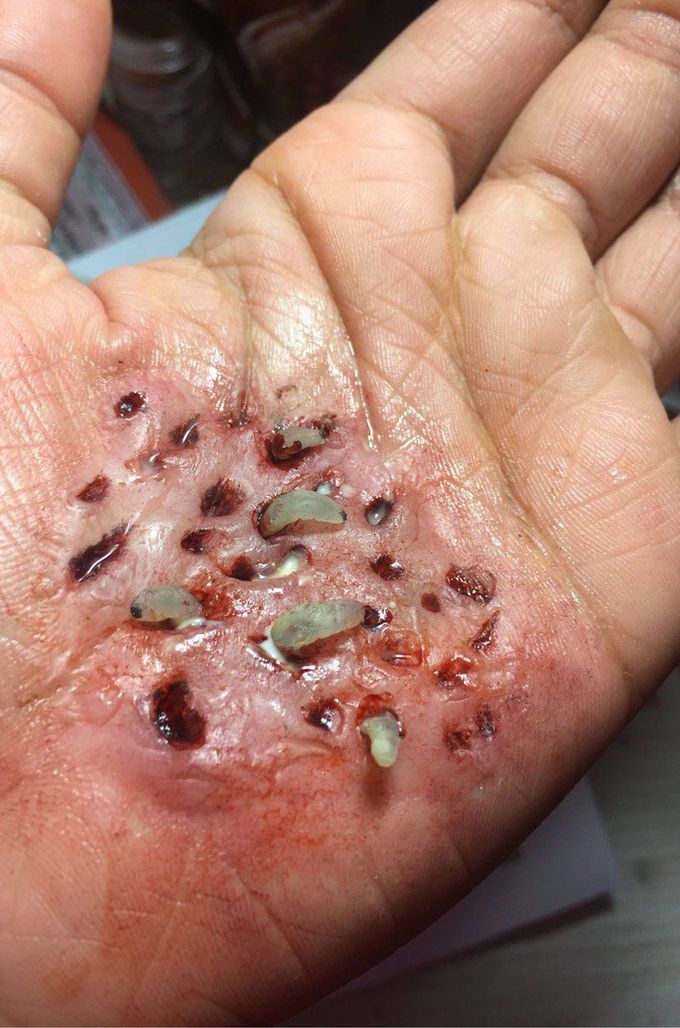
I can see maggot infestation in a patient with wounds and not sanitary conditions . In the picture shown can you tell me how this may happen . My first impression just from picture would be bot fly . Thank you for any information.
Now that I think about it if it was botfly this would be maggot infestation . Would I be correct thinking botfly?