One of the more amazing animals I observed in class is the sea urchin. There are over 200 species of sea urchins, which belong to the Class Echinoidea and Phylum Echinodermata. The name “Echinoidea” is derived from the Greek “echinus”, which means “hedgehog”. An appropriate comparison since the spines covering the urchins dorsal surface make them resemble an underwater hedgehog.
Several interesting things distinguish Echinoidea from other Echinodermata from a morphological standpoint. At first glance, it appears that sea urchins lack the characteristic five arms. Yet, penta-radial symmetry is still present because sea urchins have 5 plates, which are joined together to form a ball. Tube feet run up the dorsal surface of these plates, from the sea urchin’s mouth on the bottom to it’s anus on the top (aboral surface). Areas without tube feet are called interambulacral zones (Pechenik, 2010).

Figure 1: Sea urchin test (endoskeleton)
(Adapted from Brock, 2010)
Echinodea also have an exclusive feeding apparatus, called Aristotle’s lantern, which is located on the oral surface. It is made of five plates formed into a lantern-like structure with a tooth at the end of each plate. The plates are attached to muscles, which allow the lantern to be moved in various directions to bite, scrape food, or eat molluscs. Food is forwarded into the esophagus and digested in the intestine before the waste is expelled out of the anus on the dorsal surface of the urchin (Carnevali et al, 1993).
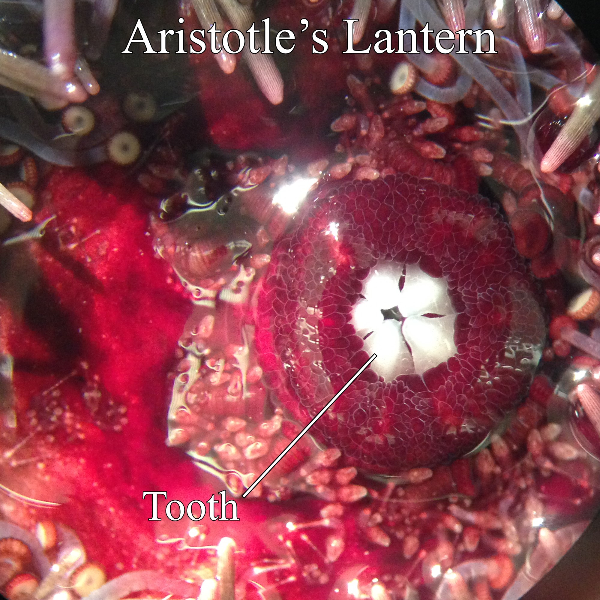
Figure 2: Aristotle’s Lantern . Image Credit: Justin Bermudez
A Sea Urchin moves using several tube feet that project through holes in its endoskeleton and are associated with its water-vascular system. It can “pump-up” the foot by contracting muscles on either side of the tube. Discs at the end of the tube feet also have muscles that can squeeze water back into the tube and lengthen it, while causing the disc to hold onto a surface like a suction cup. In addition, muscles on one side can contract allowing the foot and the urchin to pull itself in any direction (Helms, 1989).
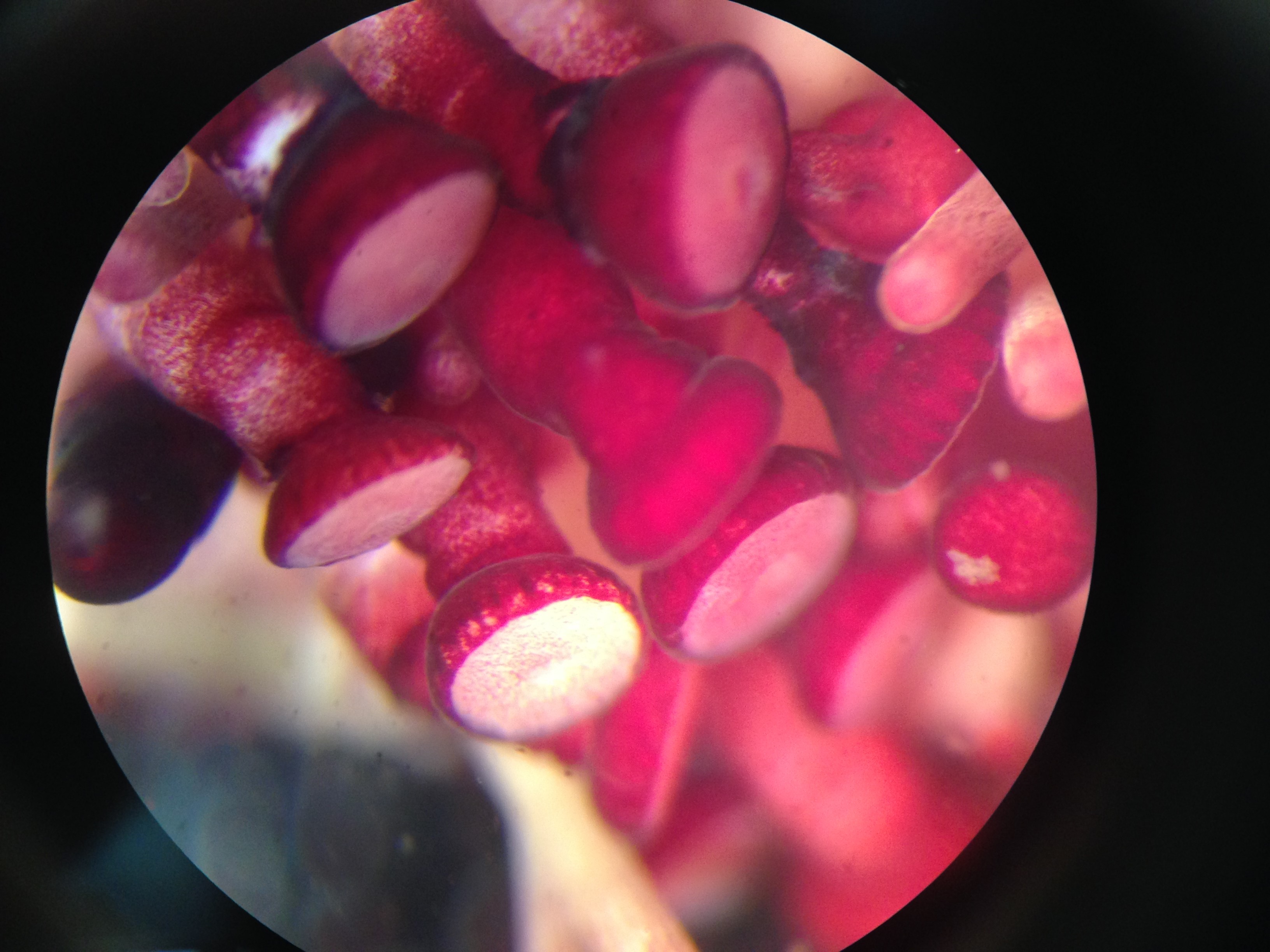
Figure 3: Tube feet. Image Credit: Justin Bermudez
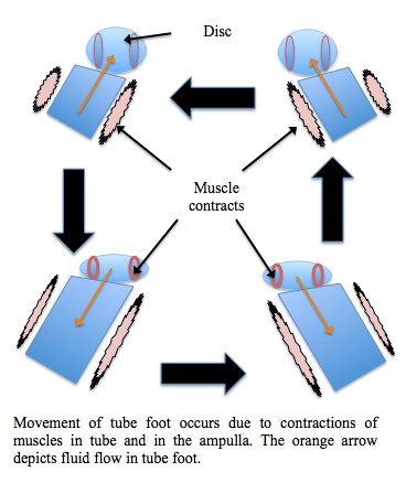
Figure 4: Tube foot motion
Like a tiny-armed soldier, the sea urchin has four means of defense. Most obvious are the spines protruding from its body. The spines are indented at the base and sit on spherical mounds on the shell, (see Figure 1), forming something like a ball and socket joint so that the muscles can move them around. The spines contain toxins in some sea urchins, that can be lethal to certain species. Second, they have the hard calcium based endoskeleton, which supports and defends its internal organs. Third, the urchin has little stealth attackers called pedicellaria. The pedicellaria, are 3-jawed pinchers, that extend from the urchin’s surface via stalks. They are distributed among the spines, but are much shorter and harder to see. When provoked, the little pinchers can bite and some can inject venom as well (Carefoot, 2010). They can also be used for catching or crushing prey or cleaning the urchin. The pedicellaria are controlled by muscles, which allow them to change their direction of attack (Pechenik, 2010).
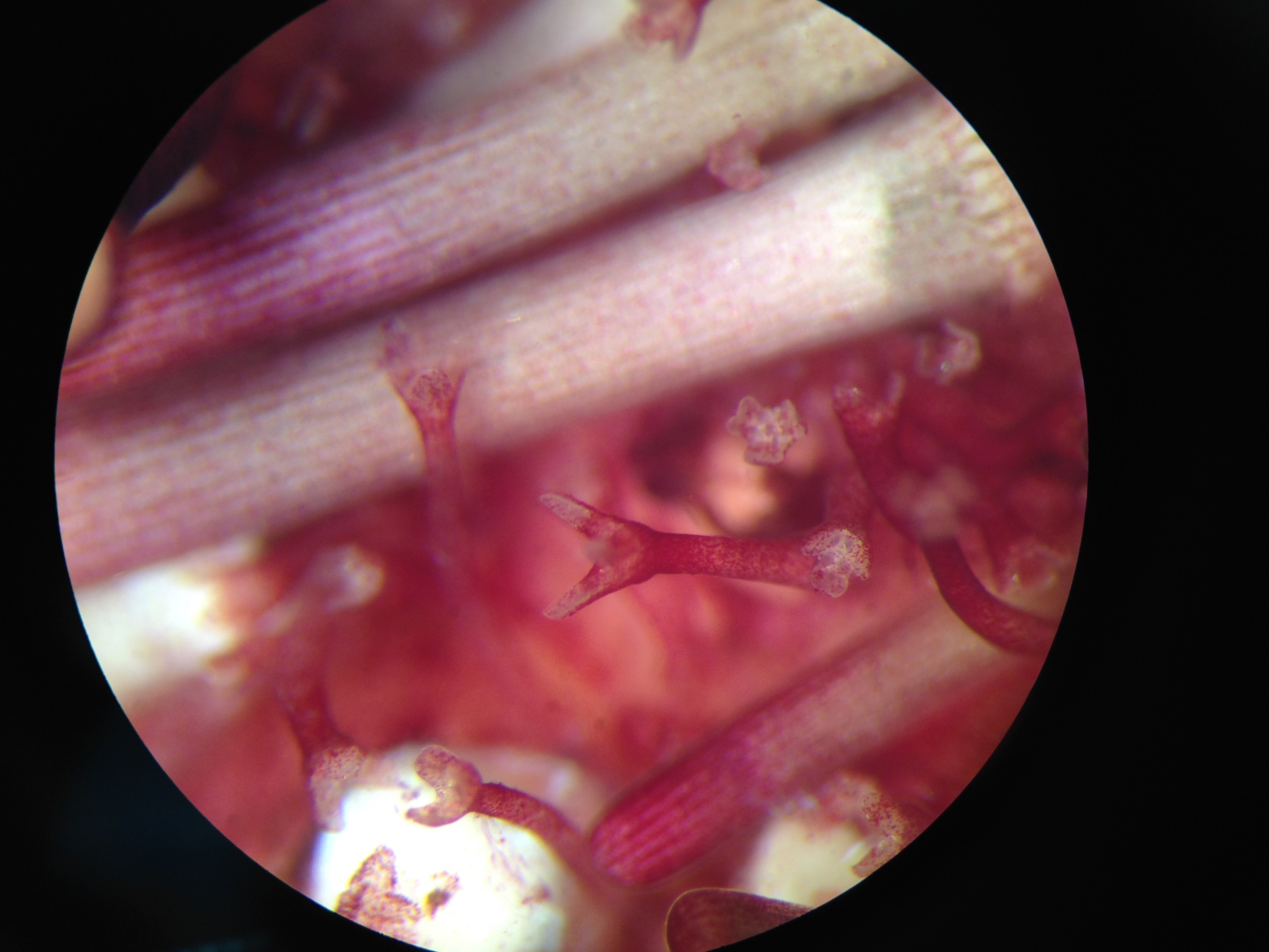
Figure 5: Open 3-pronged pedicellaria. Image Credit: Justin Bermudez
Their fourth and most fascinating defense is camouflage. Using their tube feet, they can pick up shells or algae and use them to cover their body in order to blend into the ocean floor (Encyclopedia Aquatic World, 2004). Unfortunately, due to pollution, they also pick up bottle caps and other trash, which inadvertently turns then into little underwater advertisements.
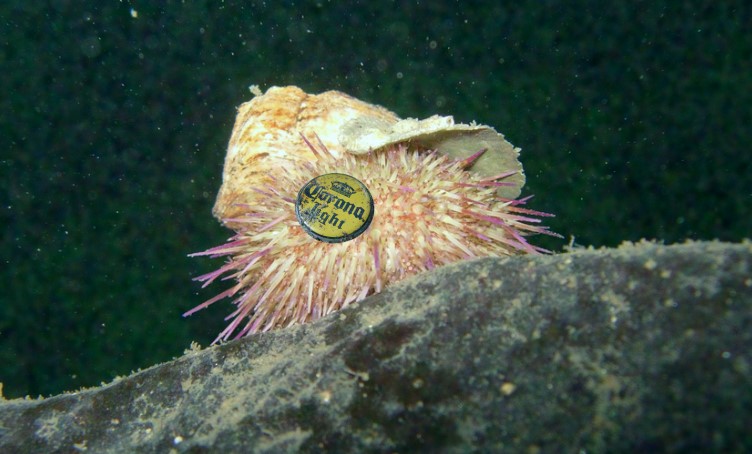
Figure 6: Sea urchin camouflage
(Adapted from Alfaro, 2009 & Milligan, 2010)
References
Alfaro, Alan. Out of Place. 08 Mar 2009, Online image. Flickr. 14 Mar 2014. <http://www.flickr.com/photos/ajalfaro/3338024313/>
Brock, Vicky. Shetland sea urchin. 04 July 2010, Online image. Flickr. 14 Mar 2014. <http://www.flickr.com/photos/vickyb/4777278268/>
Carefoot, Tom, (2010) Sea Urchin. Learn About Sea Urchins: Feeding, nutrition, & growth. www.asnailsodyssey.com. Bamfield Marine Sciences Center, Web. 6, Mar. 2014.
Carefoot, Tom, (2010) Sea Urchin. Learn About Sea Urchins: Predators & Defenses. www.asnailsodyssey.com. Bamfield Marine Sciences Center, Web. 6, Mar. 2014.
Carnevali, Candia et al (1993) The Aristotle’s lantern of the sea-urchin Stylocidaris affinis: functional morphology of the musculo-skeletal system. Zoomorphology 113: 173-189
Encyclopedia of the Aquatic World. (2004) Marshall Cavendish Corp. Tarrytown, NY.
Helms, Doris et al. (1989) More biology in the laboratory. W. H. Freeman; First Edition, New York, NY.
Milligan, Gordon. Camouflaged Sea Urchin. 22 Oct 2012, Online image. Flickr. 14 Mar 2014. <https://www.flickr.com/photos/el-milligano/8109912145/>
Pechenik, Jan. (2010) Biology of Invertebrates. McGraw Hill; Sixth Edition, New York, NY.
