


Filters

Stock royalty-free photos and images of Maxillary sinus
Discover unlimited high resolution images of Maxillary sinus and stock visuals for commercial use.

Sinuses anatomy
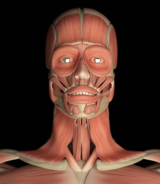
Human face anatomy model, 3D illustration
Human face anatomy model icon, 3D illustration
Human face anatomy model icon, 3D illustration
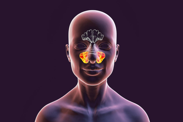
Anatomy of paranasal sinuses. 3D illustration showing female with highlighted maxillary sinuses, also known as antrum of Highmore, front view

Anatomy of paranasal sinuses. 3D illustration showing male with highlighted paranasal sinuses, frontal, maxillary, ethmoid, and sphenoid

Anatomy of paranasal sinuses. 3D illustration showing male with highlighted paranasal sinuses, frontal, maxillary, ethmoid, and sphenoid

Anatomy of paranasal sinuses. 3D illustration showing male with highlighted maxillary sinuses, also known as antrum of Highmore, front view

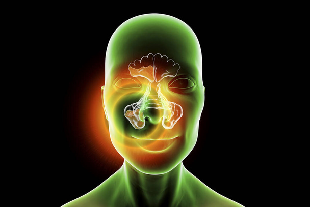
Anatomy of paranasal sinuses. 3D illustration showing human body with highlighted maxillary sinuses, also known as antrum of Highmore
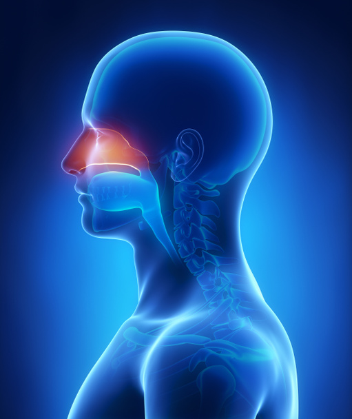
Nasal cavity

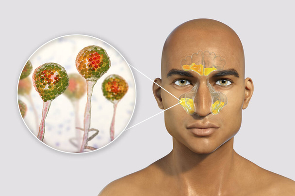
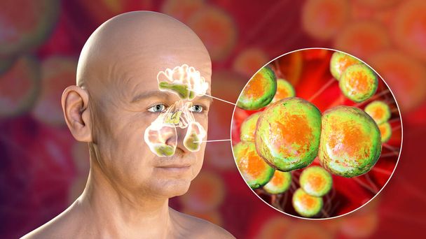
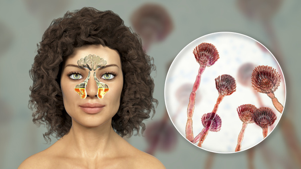
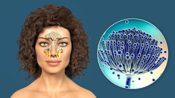
Aspergillus fungi as a cause of sinusitis. 3D illustration showing inflammation of maxillary sinuses in a female and close-up view of Aspergillus fungus. Chronic fungal rhinosinusitis

Mucor fungi as a cause of sinusitis, 3D illustration. Inflammation of maxillary sinuses and close-up view of fungi Mucor. Mucormycosis, Covid-19 complication, sinusitis in immunocompromised patients

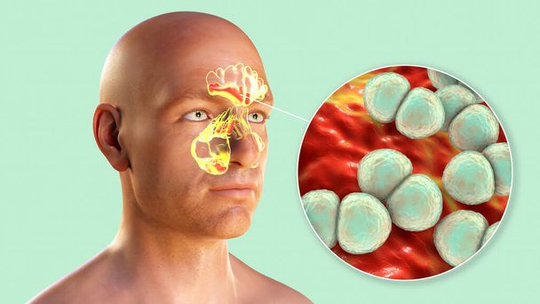
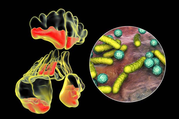
Staphylococcus aureus bacteria as a cause of sinusitis. 3D illustration showing purulent inflammation of frontal sinuses in an African man and close-up view of staphylococci bacteria
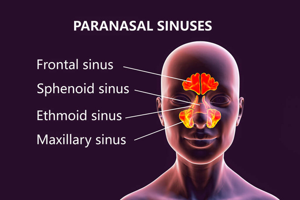
Anatomy of paranasal sinuses. 3D illustration showing male with highlighted paranasal sinuses, frontal, maxillary, ethmoid, and sphenoid. Labelled image
Sinusitis, inflammation of paranasal cavities. 3D illustration showing purulent inflammation of frontal and maxillary sinuses
Streptococcus pneumoniae bacteria as a cause of sinusitis. 3D illustration showing purulent inflammation of frontal, maxillary, and ethmoid sinuses and close-up view of pneumococci bacteria
Moraxella catarrhalis bacteria as a cause of sinusitis. 3D illustration showing purulent inflammation of frontal sinuses in an African man and close-up view of Moraxella bacteria
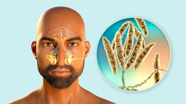
Maxillary rhinosinusitis due to fungi Fusarium, 3D illustration. Fungal rhinosinusitis in immunocompromised patients
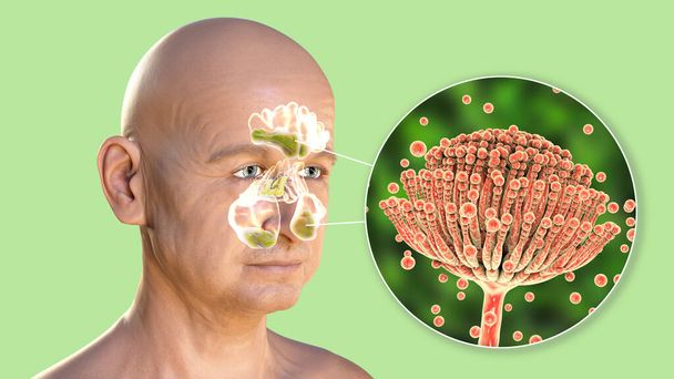
Aspergillus fungi as a cause of sinusitis. 3D illustration showing inflammation of frontal, maxillary and ethmoid sinuses in a man and close-up view of Aspergillus fungus. Chronic fungal rhinosinusitis
Anatomy of paranasal sinuses. 3D illustration showing female with highlighted paranasal sinuses, frontal, maxillary, ethmoid, and sphenoid. Labelled image
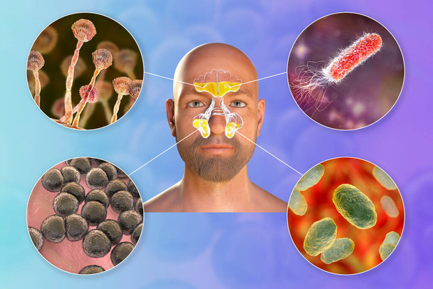
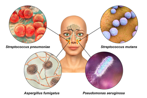
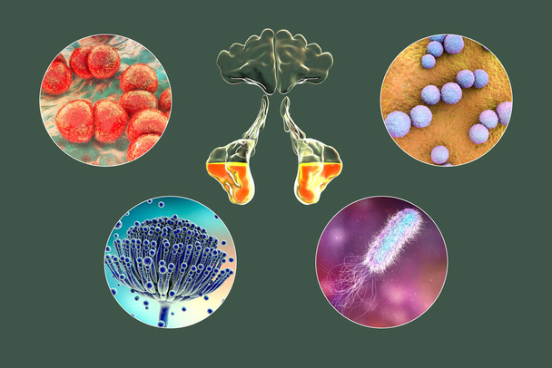
Rhinosinusitis and microorganisms that cause sinusitis, fungi Aspergillus, bacteria Pseudomonas aeruginosa, Streptococcus pneumoniae and Haemophilus influenzae, 3D illustration
Streptococcus pneumoniae bacteria as a cause of sinusitis. 3D illustration showing purulent inflammation of frontal sinuses in an African man and close-up view of streptococci bacteria
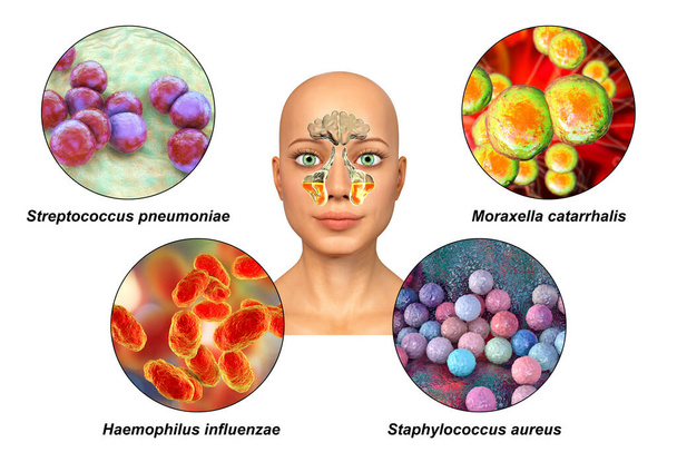
Anatomy of rhinosinusitis and bacteria that cause sinusitis Streptococcus pneumoniae, Moraxella catarrhalis, Haemophilus influenzae, and Staphylococcus aureus, labelled 3D illustration

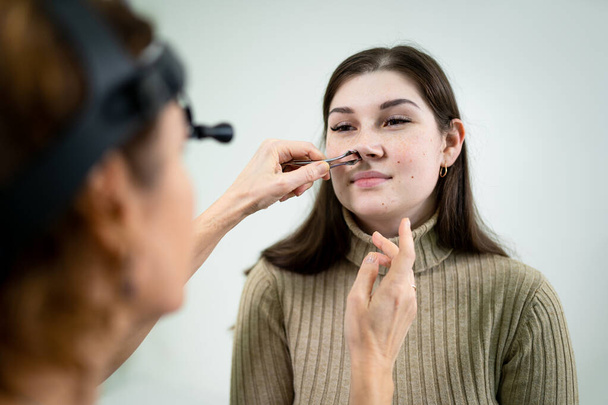
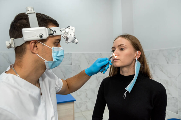
Professional female doctor otorhinolaryngologist doing nose examination with otoscope in modern hospital. Nasal congestion, sinusitis, allergy concept. Woman ENT doctor and patient in office clinics.

Streptococcus pyogenes bacteria as a cause of sinusitis. 3D illustration showing purulent inflammation of frontal and maxillary sinuses and close-up view of streptococci bacteria
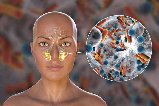
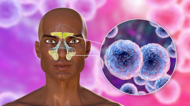
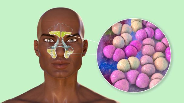
Sinusitis, inflammation of paranasal cavities. 3D illustration showing inflammation of maxillary sinuses in a female person and close-up view of bacteria that cause sinusitis
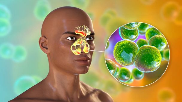
Moraxella catarrhalis bacteria as a cause of sinusitis. 3D illustration showing purulent inflammation of frontal sinuses in an African man and close-up view of Moraxella bacteria
Streptococcus pyogenes bacteria and other streptococci as a cause of sinusitis. 3D illustration showing inflammation of frontal sinuses in an African man and close-up view of streptococcal bacteria

Otolaryngology concept. Positive woman otorhinolaryngologist checking nose with otoscope of his patient at hospital. Nasal congestion, sinusitis, allergy concept. Female patient at modern ENT clinic.
Pseudomonas aeruginosa bacteria as a cause of sinusitis. 3D illustration showing inflammation of maxillary sinuses and close-up view of Pseudomonas bacteria
Film mandible showed Righr fracture of body and angle of Mandible.

Anatomy of rhinosinusitis and bacteria that cause sinusitis Staphylococcus aureus, Streptococcus pneumoniae, Haemophilus influenzae, and Moraxella catarrhalis, 3D illustration
Mucor fungi as a cause of sinusitis, 3D illustration. Inflammation of maxillary sinuses and close-up view of fungi Mucor. Mucormycosis, Covid-19 complication, black fungus, yellow fungus
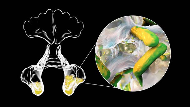
Anatomy of rhinosinusitis and microorganisms that cause sinusitis Streptococcus pneumoniae, Streptococcus mutans, Aspergillus fumigatus, and Pseudomonas aeruginosa, labelled 3D illustration
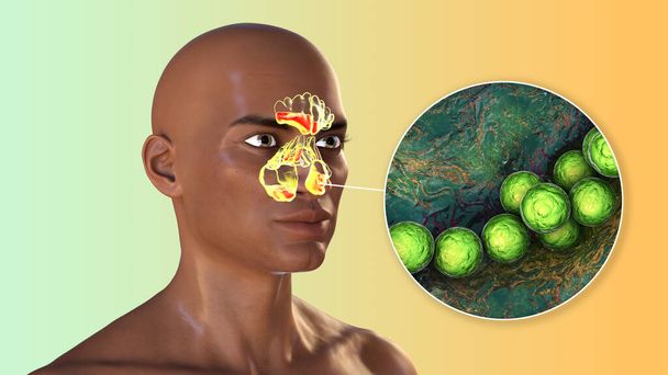
Moraxella catarrhalis bacteria as a cause of sinusitis. 3D illustration showing purulent inflammation of frontal, maxillary, and ethmoid sinuses and close-up view of Moraxella bacteria
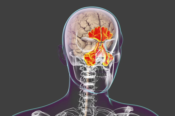
Anatomy of paranasal sinuses. 3D illustration showing male body with skeleton, brain and highlighted paranasal sinuses

Sinusitis, inflammation of paranasal cavities. 3D illustration showing purulent inflammation of maxillary sinuses in a man and close-up view of bacteria that cause sinusitis
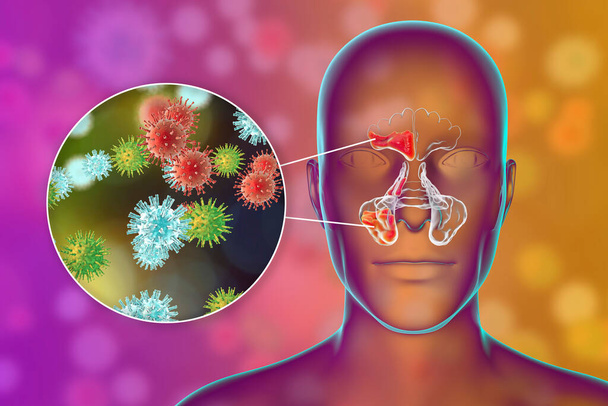
Viral sinusitis, inflammation of paranasal cavities. 3D illustration showing inflammation of frontal sinus and close-up view of viruses that cause sinusitis
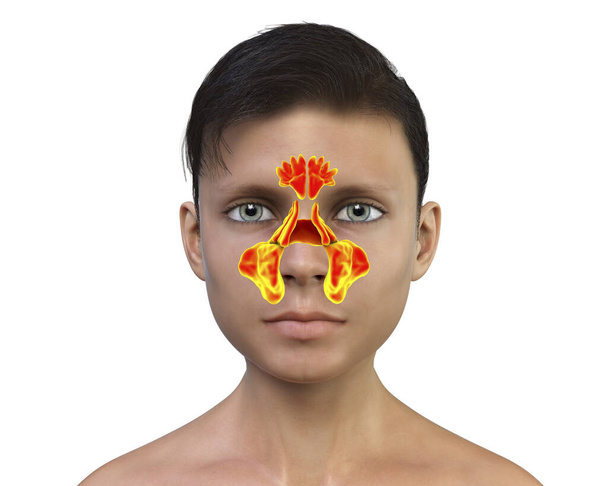
Anatomy of paranasal sinuses. 3D illustration showing teenager boy with highlighted paranasal sinuses, frontal, maxillary, ethmoid, and sphenoid
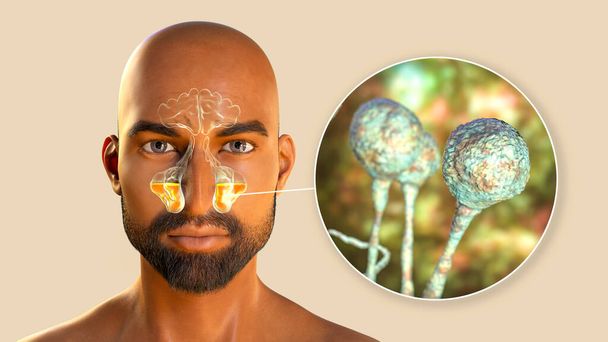
Mucor fungi as a cause of sinusitis, 3D illustration. Inflammation of maxillary sinuses and close-up view of fungi Mucor. Mucormycosis, Covid-19 complication, sinusitis in immunocompromised patients
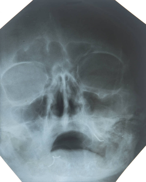
X-ray photo of breathing disorders in the sinuses of the nose
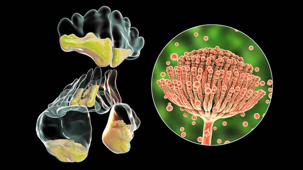
Aspergillus fungi as a cause of sinusitis. 3D illustration showing inflammation of frontal, maxillary, and ethmoid sinuses and close-up view of Aspergillus fungus. Chronic fungal diffuse rhinosinusitis
Otolaryngology concept. Positive woman otorhinolaryngologist checking nose with otoscope of his patient at hospital. Nasal congestion, sinusitis, allergy concept. Female patient at modern ENT clinic.
Sinusitis, inflammation of paranasal cavities. 3D illustration showing purulent inflammation of frontal, maxillary, and ethmoid sinuses and close-up view of bacteria that cause sinusitis
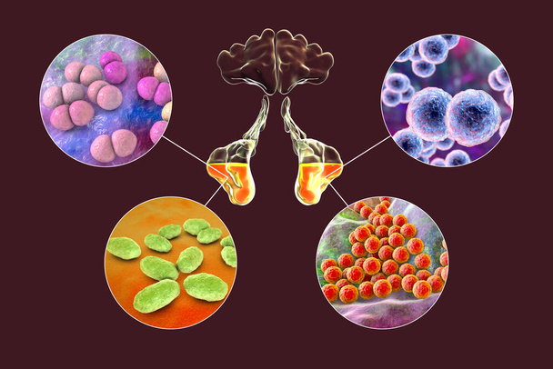
Anatomy of rhinosinusitis and bacteria that cause sinusitis Streptococcus pneumoniae, Moraxella catarrhalis, Haemophilus influenzae, and Staphylococcus aureus, 3D illustration
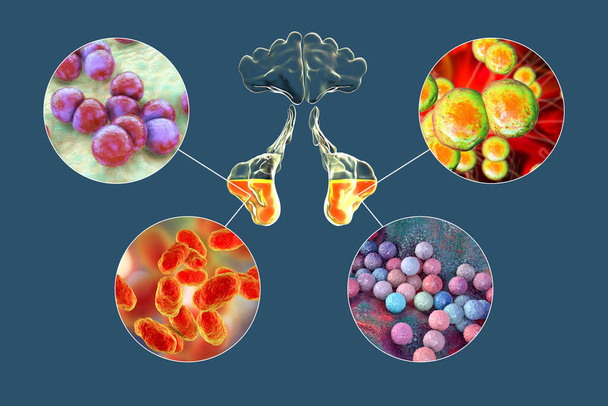
Anatomy of rhinosinusitis and bacteria that cause sinusitis Streptococcus pneumoniae, Moraxella catarrhalis, Haemophilus influenzae, and Staphylococcus aureus, 3D illustration
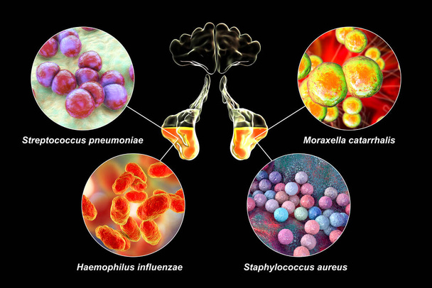
Anatomy of rhinosinusitis and bacteria that cause sinusitis Streptococcus pneumoniae, Moraxella catarrhalis, Haemophilus influenzae, and Staphylococcus aureus, 3D labelled illustration
Sinusitis, inflammation of paranasal cavities. 3D illustration showing purulent inflammation of frontal, maxillary, and ethmoid sinuses, close-up view

Sinusitis, medicines as concept of ordinary treatment, conceptual image
Aspergillus fungi as a cause of sinusitis. 3D illustration showing inflammation of maxillary sinuses in a female person and close-up view of Aspergillus fungus. Chronic fungal rhinosinusitis
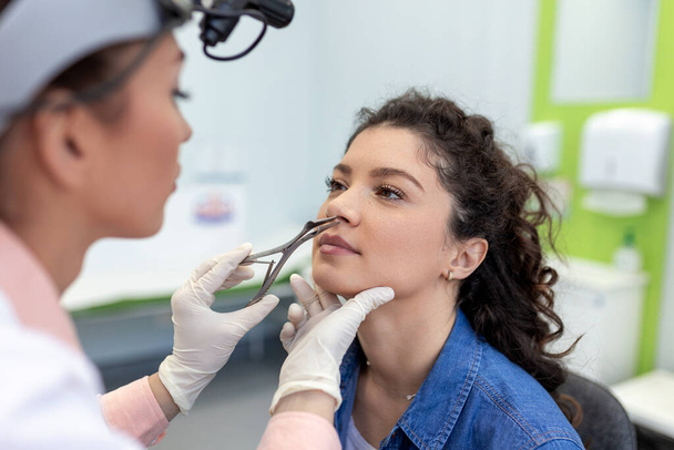
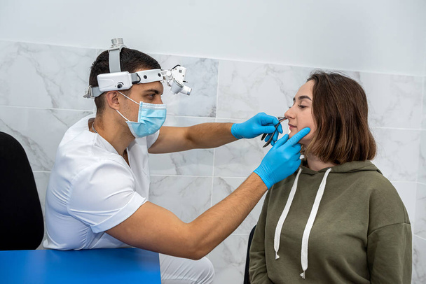
Female ENT doctor examines patient's sinuses with medical instrument in modern clinic. Otorhinolaryngologist checks the patient's girl's nose for diseases, allergies and curvature of the nasal septum.
Positive male otorhinolaryngologist is checking nose with otoscope of his patient girl in hospital. Nasal congestion. A patient in a modern ENT clinic. The concept of otolaryngology.
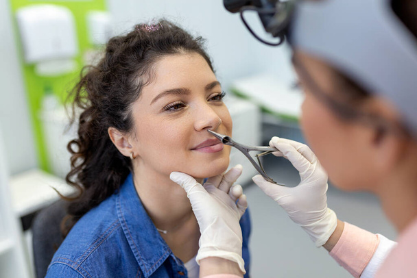
Portrait of an otolaryngologist while working with a girl patient on an examination of the sinuses. nasal congestion medicine
Anatomy of rhinosinusitis and microorganisms that cause sinusitis Streptococcus pneumoniae, Streptococcus mutans, Aspergillus fumigatus, and Pseudomonas aeruginosa, 3D illustration
Otolaryngology concept. Positive woman otorhinolaryngologist checking nose with otoscope of his patient at hospital. Nasal congestion, sinusitis, allergy concept. Female patient at modern ENT clinic.

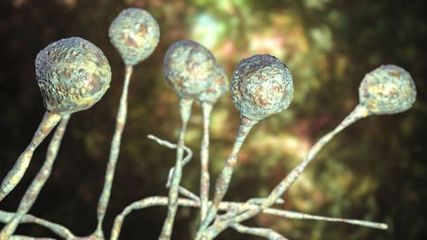
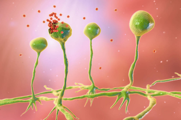
Mucor mold, black fungus, bread mold fungi, 3D illustration. Opportunistic fungi that cause mucormycosis involving skin, nasal sinuses, brain and lungs. Complication of Covid-19
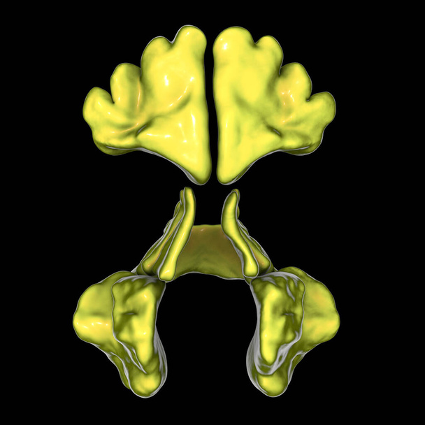
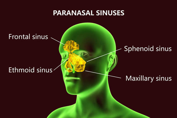
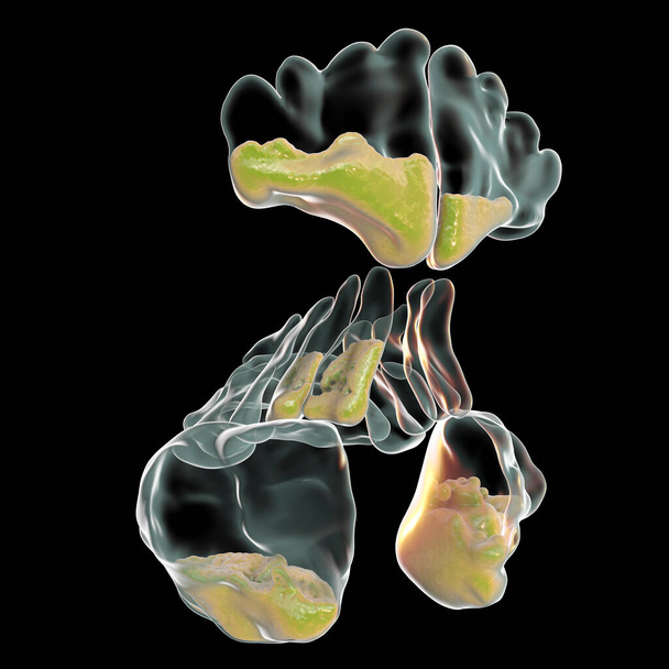
Anatomy of paranasal sinuses. 3D illustration showing frontal, maxillary, ethmoid, and sphenoid sinuses
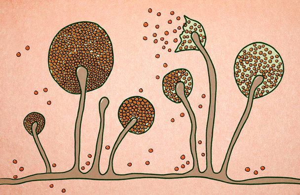
Mucor mold, bread mold fungi, found in soil, digestive systems, 3D illustration. Opportunistic fungi that cause zygomycosis, and also involved in nose infections, chronic fungal rhinosinusitis
Aspergillus fungi as a cause of sinusitis. 3D illustration showing inflammation of maxillary sinuses in a female person and close-up view of Aspergillus fungus. Chronic fungal rhinosinusitis

ENT-Doctor Scientist Examine Ear,Nose,Throat.Tonsilis,Sore Throat,Otitis,Larynx,Sinusitis Digital Treatment.Online Smartphone Consultation. Medical Internet Diagnostics. ORL Clinic.Vector Illustration

ENT-Doctors Scientists Examine Ear,Nose,Throat.Tonsilis,Sore Throat,Otitis,Larynx,Sinusitis Digital Treatment.Research Trial.Clinical Investigation.ORL Clinic. Medical Council Diagnostics Illustration
Structure of Mucor mold, black fungus, yellow fungus, 3D illustration. Opportunistic fungi that cause mucormycosis involving skin, nasal sinuses, brain and lungs. Complication of Covid-19

Side view of woman with symptoms of frontal sinusitis, feeling of pressure between the eyes sitting on the sofa in the living room
Structure of Mucor mold, black fungus, yellow fungus, illustration. Opportunistic fungi that cause mucormycosis involving skin, nasal sinuses, brain and lungs. Complication of Covid-19
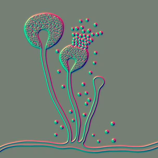
Rhizopus mold, also known as bread mold and black fungus, 3D illustration. Opportunistic fungi that cause mucormycosis involving skin, nasal sinuses, brain and lungs. Complication of Covid-19
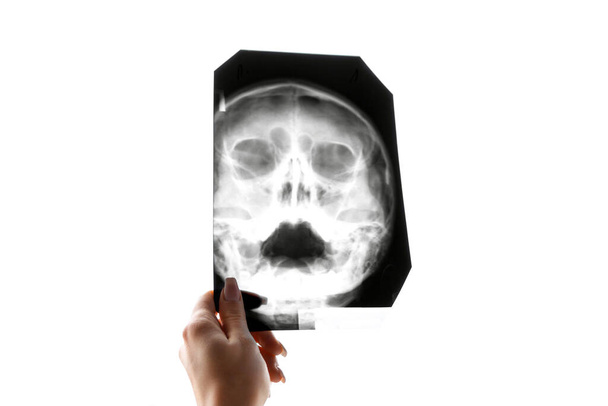
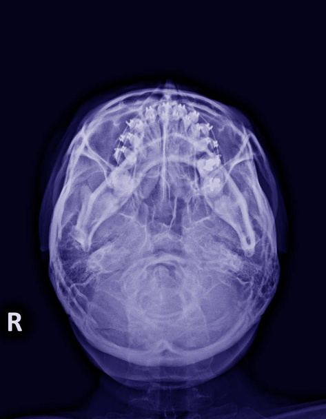
X-ray of the head. Fluorography of maxillary sinuses. Concept of medicine.

Sinusitis, inflammation of paranasal cavities. 3D illustration showing purulent inflammation of frontal and maxillary sinuses in a man and close-up view of bacteria that cause sinusitis