Introduction
Skin tears are a common presentation, where even minor trauma can create a problematic wound with a high risk of evolving into a complex, chronic wound. The International Skin Tear Advisory Panel (ISTAP) defines skin tears as traumatic wounds caused by shearing/mechanical forces (including removal of adhesives) where severity may vary by depth.1
Skin tears typically occur in the elderly population who undergo histological changes to the architecture of their skin. Contributing factors include thinning of the epidermis, atrophy and contracture of the dermis, elastosis (loss of elasticity and strength), reduction in sebaceous gland oil production (causing dryer skin), fragile blood vessels and thinning of the subcutaneous tissue, which typically provides a cushioning layer.1 Elderly patients are often frail with a reduced physiological reserve and a preponderance of comorbid medical conditions, which places them at high risk of blunt trauma injury post mechanical fall or syncopal events.2
Treatment aims to minimise infection risk, promote wound healing and achieve wound coverage to allow complete functional recovery. Cosmetic appearance is a secondary, albeit important, consideration, particularly in the elderly patient population.
Skin tear classification systems include the Skin Tear Audit Research (STAR) classification and the ISTAP classification, which categorises wounds according to whether the skin edges can be realigned to anatomical position, the amount of skin flap loss and the quality of the remaining skin.3 While these systems are helpful for research and audits, our experience suggests skin tears are described in clinical practice according to estimated size, the colour of skin flaps, degree of tissue loss and presence of visible contamination.
A thorough assessment of skin tears requires consideration of:
-
anatomical location:
- typically occurs on the upper or lower limbs
-
time to presentation:
-
skin tears are often overlooked, initially managed with steristrip/dressing application or not considered significant enough to warrant treatment
-
considerable delay in management increases the risk of infection
-
-
size of wound:
- small defects may be amenable to re-laying of skin and dressing
-
surface area of skin remaining:
- the amount of remaining skin determines if coverage is possible with a mesh graft
-
quality of remaining skin:
- degree of necrosis, underlying haematoma
-
patient factors:
-
comorbidities and medications
-
anticoagulated patients should have compression bandages and strict limb elevation.
-
Skin tears that are small in size have minimal tissue loss, and are easily reapproximated can typically be managed with wound irrigation, approximation of wound edges, fixation with tissue glue or adhesive dressing and coverage with an overlying padded dressing until a review in three to seven days.
Large skin tears that have tissue loss, gross contamination or have failed treatment by reapproximation and dressing typically undergo formal debridement and skin grafting. This carries risk and morbidity associated with anaesthesia and surgery, including prolonged fasting, general anaesthesia, delay to definitive care, graft donor site morbidity, as well as the financial costs associated with such procedures, including operative theatre time, hospital stay (pre- and post-procedure) and cost of inpatient services (such as physiotherapy etc.).
Royal North Shore Hospital (RNSH, Sydney, Australia) studied the use of a mesh protocol where skin flaps that were pale/dusky or unamenable to realignment underwent debridement, amputation, meshing and reapproximation under local anaesthesia in the emergency department (ED).4 Our study aimed to validate the reproducibility of the results in a different hospital network and identify avenues to streamline the process further.
Methods
A prospective review was undertaken from July 2021 to January 2022 of all patients who underwent management of skin tears via the mesh protocol at Northern Beaches Hospital (NBH, NSW, Australia). The study cohort comprised patients treated in either the ED or ward. There were no exclusion criteria.
Data were collected prospectively from a consecutive patient group, including patient age, comorbidities, mechanism of injury, time from injury to presentation, location, size of the injury, dressing interface used, percentage of graft take at first review (days five–seven post grafting) and any complications.
The national hospital cost data collection report, produced by the Independent Hospital Pricing Authority (IHPA), was used for analysis of operative and admission costs. Costs of donor site dressings and theatre time were based on the average wound size in this cohort. Costs of graft site dressings are unchanged between the mesh protocol and formal grafting, so these were not calculated.
Descriptive statistics were used for the majority of variables.
Procedure for mesh protocol
All patients with skin tears in the ED or ward were referred to the plastics registrar for an opinion on management. In collaboration with a plastic surgeon, a decision on conservative management versus mesh grafting was made following an in-person review. For a patient to be considered to be managed with the mesh protocol review by the NBH plastics registrar was required within 48 hours from the time of injury.
Following informed consent from the patient and/or guardian, the mesh protocol was undertaken in the ED or on the ward by the plastics registrar under local anaesthesia (including adrenaline in the context of minimal access to diathermy), with betadine preparation and sterile draping. All areas of devascularised and/or detached skin were amputated and either had mesh applied 1.5:1 or were hand fenestrated with a blade, depending on the wound coverage required. The skin was re-layed, and inset with adhesive glue, then dressed with a non-adhesive layer with an outer dressing of compression bandage or negative pressure wound therapy (NPWT).
Patients were then either discharged after mobility review by a physiotherapist or admitted if further medical treatment was necessary. Typically, no mobility restrictions were placed, with the advice to elevate the affected limb when resting.
Graft check was performed on days five and seven days, either in person if the patient was an inpatient or via photographs from the community nurses or nursing home facility nurse. Further management in the form of dressing advice was then initiated, with patients usually discharged with ongoing community nursing.
Results
Fifty-three consecutive patients who sustained 64 discrete skin tears underwent the mesh protocol over a seven-month period. The average patient age was 86 years. Of 53 patients, 28 were discharged to home/nursing home, 19 patients were admitted under a medical team for other reasons, and six patients were admitted under the plastic and reconstructive surgery department for five days for social/self-care reasons. Twenty-seven patients were given anticoagulants. Twenty-four patients were seen within 0–12 hours of injury, 19 within 12–24 hours of injury and 10 within 24–36 hours of injury.
Mechanisms of injury included mechanical fall (43), traumatic wound (8), two dog scratch wounds (2) and one haematoma evacuation. The average wound size was 50 cm2. Eleven patients had more than one involved location. Twenty-one wounds were pretibial, 18 on the forearm/hand, 10 on the knee, eight on the leg, six on the arm/elbow and one on the forehead (Table 1).
Following graft review on days five–seven, 94 per cent (60/64) of all grafts achieved full graft take. A total of four sites on four different patients did not achieve this. One patient required formal debridement and delayed grafting in theatre after mesh graft failure, requiring a total 12-day admission. Another patient required silver dressings and intravenous antibiotics for methicillin-resistant staphylococcus aureus (MRSA) and pseudomonas growth (while admitted under the geriatric team for pneumonia). The remaining two patients achieved wounding healing with an extended period of dressings in the community.
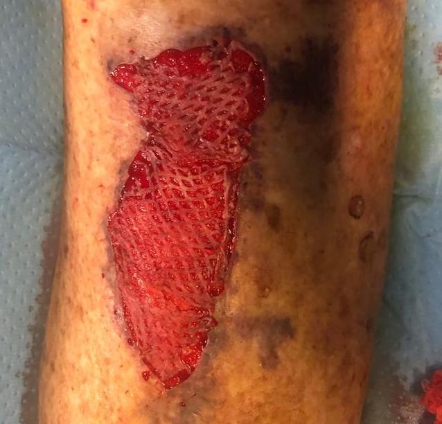
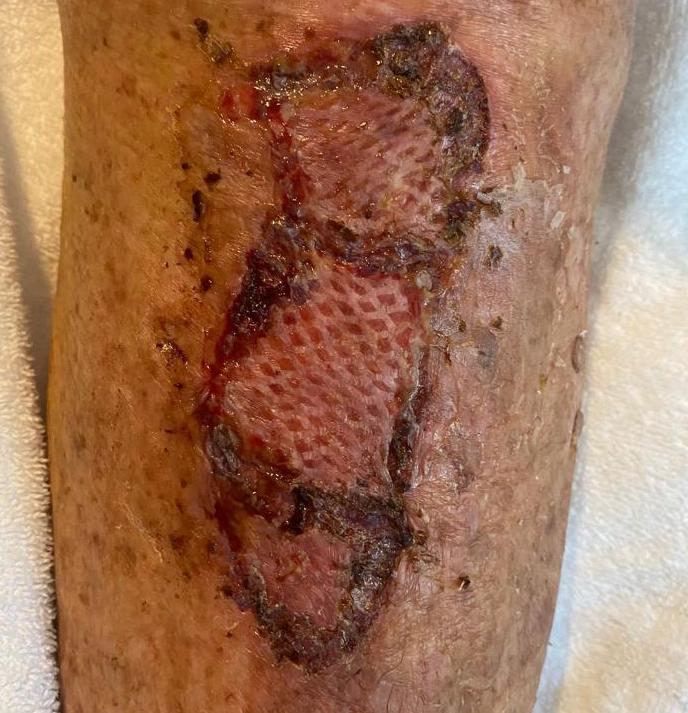
Case example
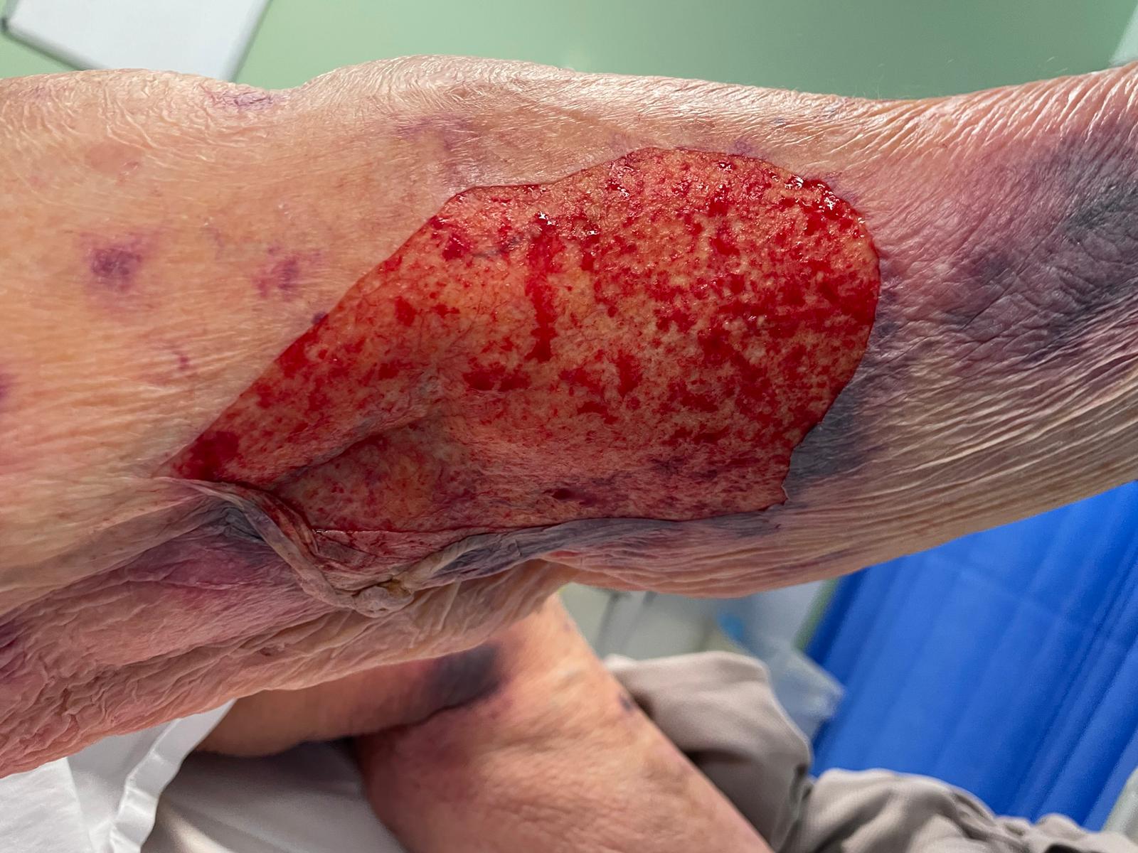
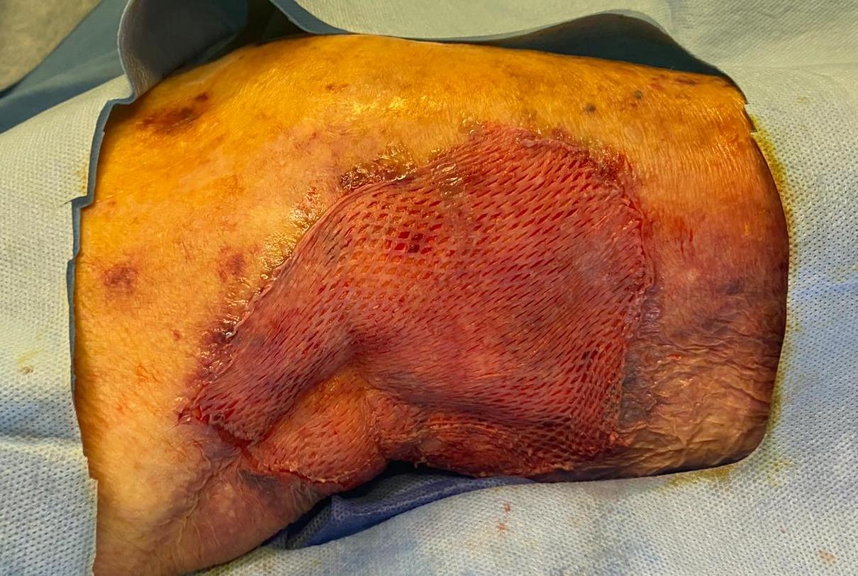
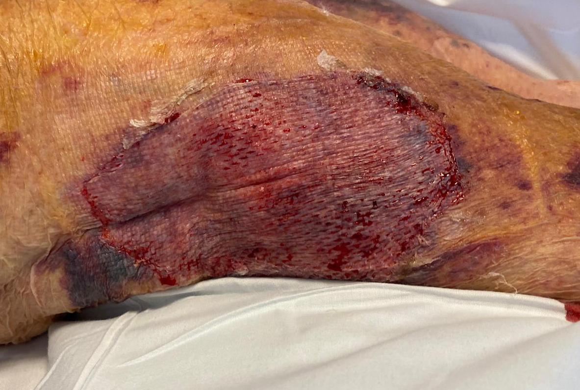
84-year-old woman with a 25 x 15 cm skin tear to her right posterolateral thigh/leg, which had 100 per cent graft taken on day five (figures 1, 2 and 3)


Cost analysis
Given that 83 per cent of patients whose primary reason for presentation to the ED was due to their skin tear were able to be discharged after undergoing mesh protocol, this scenario was used as the base case cost analysis. The base case for comparison was a patient requiring day surgery for formal debridement and skin grafting.
Following a presentation to the ED in New South Wales (NSW), the average cost before discharge is A$385 and if subsequently admitted, is A$1663 per bed day. The cost per admitted bed day includes standard emergency operative costs. Donor site dressings were estimated at A$67 (uncomplicated) to A$175 (complicated/delayed healing) per patient. The cost of a mesh protocol in a patient discharged from ED is therefore calculated as A$385. The cost of formal debridement and skin grafting is estimated as A$1663 + A$67 or A$1663 + A$175 = A$1730 or A$1838. Subsequently, estimated cost saving ranges from A$1345–A$1453 with an additional cost of $1663 for each subsequent day of admission. A five-day admission following formal grafting (as is conventional practice) would increase costs by over A$8000.
Discussion
Performing this minor procedure as a part of an established referral pathway for acute skin tears will likely reduce the under-management of skin tears leading to a reduction in the number of chronic wounds. A recent NSW Health analysis reports chronic wounds to account for over 5000 patient admissions, 6000 additional ED presentations, and half a million outpatient visits each year in the NSW public hospital system.5 Each patient has an average of three admissions for each wound. Admitted patients are usually older and two-thirds have multiple comorbidities. These patients are more likely to suffer nosocomial diseases such as venous thromboembolism (VTE), pneumonia, urinary tract infection (UTI) and pressure sores.6 This has a profound impact on the health system and is projected to cost over A$3 billion to the public system alone over the next decade. This projected cost is independent of the cost of wound care in the general practitioner (GP) setting and private sector where a large proportion of treatment takes place. Given the distinct lack of reimbursement for wound care services and associated consumables, minimising the cost burden to the patient and healthcare system is of crucial importance.7
There are several reports in the literature describing the use of traumatically avulsed skin or a cutaneous skin flap overlying haematoma as surrogate skin grafts to avoid the creation of a new wound in the form of a donor site.8–10 Reports of such techniques formed the basis for Vandervord and colleagues’ pilot study, which was reproduced by our department in a separate hospital network to validate the protocol.
The results presented in this study are superior to those reported in the literature regarding operative split-thickness skin grafting.
Llanos and colleagues described a cohort of 60 patients who underwent split-thickness grafting for burns with patients having an average stay of 10 days from grafting to discharge, and with 17 of 30 patients requiring a second coverage procedure.11 Aerdan and colleagues studied 87 wounds of mixed aetiology with a mean graft take of 88 per cent and five complete graft failures, despite all patients having NPWT attached and hospital admission for five days post grafting.12
Comparison to the RNSH pilot study confirms the reproducibility of the pilot study’s cost-benefit and the positive clinical outcomes reported. This is an estimated saving between A$1345–A$1453 for each patient who avoided formal debridement and grafting compared to A$1588.29–A$1879.29. The estimated saving per additional bed day of A$1663 is also comparable to A$1422 in the original study. The original study did not describe the cost of each individual mesh protocol.
Our protocol differs from the RNSH pilot study in that most graft reviews were performed in the outpatient setting, with clinical images communicated via telehealth appointments. This is mainly in the context where no specialist wound dressing clinics are available, which is likely to be the case at most hospitals other than tertiary referral centres. The community nursing model is currently the most common wound care system; even in the setting of complex chronic wounds, evidence in the literature shows no difference in patient satisfaction, health-related quality of life or healing time in specialist clinic care versus home-based care.13,14
The decision to treat patients as semi-ambulatory discharge patients or as inpatients after lower limb split skin grafting remains controversial. Beyond instances of social or medical indications for admission, there is robust evidence to support discharge home following formal operative skin grafting as long as proper counselling regarding timing of mobility and maximising leg elevation is undertaken.15 Our study supports this notion given our experience of reliable outcomes following grafting using tissue from a skin tear. It is also important to note that patients in our study received either simple layered dressings or NPWT, and simple dressings in combination with semi-ambulatory discharge status remained effective.
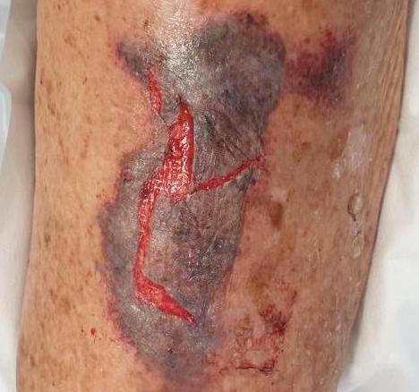
Typical management following uncomplicated mesh protocol or formal skin graft includes weeks of ongoing wound dressings until complete epithelialisation has occurred. While a mesh graft may not necessarily cover the entirety of the wound bed, our experience still suggests that small areas left ungrafted due to lack of available tissue can re-epithelialise.
Figures 4, 5 and 6 show piecemeal and dusky skin flaps and thin epidermis with sections between the mesh grafts that could not be covered. Graft take at day five shows ingrowth and epithelialisation occurring (figures 4, 5 and 6).


Even in partial graft loss, a sloughy wound base or minor cellulitis, further treatment involves a wound swab and ongoing antimicrobial dressings with oral antibiotic cover, all of which can typically be initiated and managed in the outpatient or telehealth setting. Any concerns regarding fulminant infection, wound breakdown and so on, should prompt review in the ED or the next available specialist wound clinic, whichever is more appropriate based on a multidisciplinary discussion about the clinical context.
Practical considerations of the mesh protocol include the simplicity of the procedure, making it possible even for junior members of the plastic surgical team to perform the protocol after initiation and training. In the context of multiple anatomical sites, the total aggregate of skin remaining can be divided and redistributed to provide the largest amount of wound coverage to facilitate epithelialisation.
Conclusion
The mesh protocol is an adaptable and robust method for the acute management of skin tears. It can be reproduced successfully across other units where significant benefits are maintained to patients and the healthcare system alike. Wound care is particularly amenable to safe and effective management in the telehealth setting, which further enhances these important benefits.
Patient consent
Patients/guardians have given informed consent to the publication of images and/or data.
Conflict of interest
The authors have no conflicts of interest to disclose.
Funding declaration
The authors received no financial support for the research, authorship and/or publication of this article.
Revised: 21 July 2022